

- •Contents
- •Preface to the first edition
- •Flagella
- •Cell walls and mucilages
- •Plastids
- •Mitochondria and peroxisomes
- •Division of chloroplasts and mitochondria
- •Storage products
- •Contractile vacuoles
- •Nutrition
- •Gene sequencing and algal systematics
- •Classification
- •Algae and the fossil record
- •REFERENCES
- •CYANOPHYCEAE
- •Morphology
- •Cell wall and gliding
- •Pili and twitching
- •Sheaths
- •Protoplasmic structure
- •Gas vacuoles
- •Pigments and photosynthesis
- •Akinetes
- •Heterocysts
- •Nitrogen fixation
- •Asexual reproduction
- •Growth and metabolism
- •Lack of feedback control of enzyme biosynthesis
- •Symbiosis
- •Extracellular associations
- •Ecology of cyanobacteria
- •Freshwater environment
- •Terrestrial environment
- •Adaption to silting and salinity
- •Cyanotoxins
- •Cyanobacteria and the quality of drinking water
- •Utilization of cyanobacteria as food
- •Cyanophages
- •Secretion of antibiotics and siderophores
- •Calcium carbonate deposition and fossil record
- •Chroococcales
- •Classification
- •Oscillatoriales
- •Nostocales
- •REFERENCES
- •REFERENCES
- •REFERENCES
- •RHODOPHYCEAE
- •Cell structure
- •Cell walls
- •Chloroplasts and storage products
- •Pit connections
- •Calcification
- •Secretory cells
- •Iridescence
- •Epiphytes and parasites
- •Defense mechanisms of the red algae
- •Commercial utilization of red algal mucilages
- •Reproductive structures
- •Carpogonium
- •Spermatium
- •Fertilization
- •Meiosporangia and meiospores
- •Asexual spores
- •Spore motility
- •Classification
- •Cyanidiales
- •Porphyridiales
- •Bangiales
- •Acrochaetiales
- •Batrachospermales
- •Nemaliales
- •Corallinales
- •Gelidiales
- •Gracilariales
- •Ceramiales
- •REFERENCES
- •Cell structure
- •Phototaxis and eyespots
- •Asexual reproduction
- •Sexual reproduction
- •Classification
- •Position of flagella in cells
- •Flagellar roots
- •Multilayered structure
- •Occurrence of scales or a wall on the motile cells
- •Cell division
- •Superoxide dismutase
- •Prasinophyceae
- •Charophyceae
- •Classification
- •Klebsormidiales
- •Zygnematales
- •Coleochaetales
- •Charales
- •Ulvophyceae
- •Classification
- •Ulotrichales
- •Ulvales
- •Cladophorales
- •Dasycladales
- •Caulerpales
- •Siphonocladales
- •Chlorophyceae
- •Classification
- •Volvocales
- •Tetrasporales
- •Prasiolales
- •Chlorellales
- •Trebouxiales
- •Sphaeropleales
- •Chlorosarcinales
- •Chaetophorales
- •Oedogoniales
- •REFERENCES
- •REFERENCES
- •EUGLENOPHYCEAE
- •Nucleus and nuclear division
- •Eyespot, paraflagellar swelling, and phototaxis
- •Muciferous bodies and extracellular structures
- •Chloroplasts and storage products
- •Nutrition
- •Classification
- •Heteronematales
- •Eutreptiales
- •Euglenales
- •REFERENCES
- •DINOPHYCEAE
- •Cell structure
- •Theca
- •Scales
- •Flagella
- •Pusule
- •Chloroplasts and pigments
- •Phototaxis and eyespots
- •Nucleus
- •Projectiles
- •Accumulation body
- •Resting spores or cysts or hypnospores and fossil Dinophyceae
- •Toxins
- •Dinoflagellates and oil and coal deposits
- •Bioluminescence
- •Rhythms
- •Heterotrophic dinoflagellates
- •Direct engulfment of prey
- •Peduncle feeding
- •Symbiotic dinoflagellates
- •Classification
- •Prorocentrales
- •Dinophysiales
- •Peridiniales
- •Gymnodiniales
- •REFERENCES
- •REFERENCES
- •Chlorarachniophyta
- •REFERENCES
- •CRYPTOPHYCEAE
- •Cell structure
- •Ecology
- •Symbiotic associations
- •Classification
- •Goniomonadales
- •Cryptomonadales
- •Chroomonadales
- •REFERENCES
- •CHRYSOPHYCEAE
- •Cell structure
- •Flagella and eyespot
- •Internal organelles
- •Extracellular deposits
- •Statospores
- •Nutrition
- •Ecology
- •Classification
- •Chromulinales
- •Parmales
- •Chrysomeridales
- •REFERENCES
- •SYNUROPHYCEAE
- •Classification
- •REFERENCES
- •EUSTIGMATOPHYCEAE
- •REFERENCES
- •PINGUIOPHYCEAE
- •REFERENCES
- •DICTYOCHOPHYCEAE
- •Classification
- •Rhizochromulinales
- •Pedinellales
- •Dictyocales
- •REFERENCES
- •PELAGOPHYCEAE
- •REFERENCES
- •BOLIDOPHYCEAE
- •REFERENCE
- •BACILLARIOPHYCEAE
- •Cell structure
- •Cell wall
- •Cell division and the formation of the new wall
- •Extracellular mucilage, biolfouling, and gliding
- •Motility
- •Plastids and storage products
- •Resting spores and resting cells
- •Auxospores
- •Rhythmic phenomena
- •Physiology
- •Chemical defense against predation
- •Ecology
- •Marine environment
- •Freshwater environment
- •Fossil diatoms
- •Classification
- •Biddulphiales
- •Bacillariales
- •REFERENCES
- •RAPHIDOPHYCEAE
- •REFERENCES
- •XANTHOPHYCEAE
- •Cell structure
- •Cell wall
- •Chloroplasts and food reserves
- •Asexual reproduction
- •Sexual reproduction
- •Mischococcales
- •Tribonematales
- •Botrydiales
- •Vaucheriales
- •REFERENCES
- •PHAEOTHAMNIOPHYCEAE
- •REFERENCES
- •PHAEOPHYCEAE
- •Cell structure
- •Cell walls
- •Flagella and eyespot
- •Chloroplasts and photosynthesis
- •Phlorotannins and physodes
- •Life history
- •Classification
- •Dictyotales
- •Sphacelariales
- •Cutleriales
- •Desmarestiales
- •Ectocarpales
- •Laminariales
- •Fucales
- •REFERENCES
- •PRYMNESIOPHYCEAE
- •Cell structure
- •Flagella
- •Haptonema
- •Chloroplasts
- •Other cytoplasmic structures
- •Scales and coccoliths
- •Toxins
- •Classification
- •Prymnesiales
- •Pavlovales
- •REFERENCES
- •Toxic algae
- •Toxic algae and the end-Permian extinction
- •Cooling of the Earth, cloud condensation nuclei, and DMSP
- •Chemical defense mechanisms of algae
- •The Antarctic and Southern Ocean
- •The grand experiment
- •Antarctic lakes as a model for life on the planet Mars or Jupiter’s moon Europa
- •Ultraviolet radiation, the ozone hole, and sunscreens produced by algae
- •Hydrogen fuel cells and hydrogen gas production by algae
- •REFERENCES
- •Glossary
- •Index

Chapter 14
Heterokontophyta
DICTYOCHOPHYCEAE
These golden-brown algae are characterized by tentacles or rhizopodia on basically amoeboid vegetative cells (Moestrup, 1995; Preisig, 1995). Amoeboid cells are relatively rare among the algae, being mostly restricted to the Dictyochophyceae and the Xanthophyceae (Hibberd and Chretiennot-Dinet, 1979). The algae in the Dictyochophyceae have been previously classified in the Chrysophyceae, although molecular evidence shows them to be most closely related to the Pelagophyceae (CavalierSmith et al., 1995) or Eustigmatophyceae (Daugbjerg and Andersen, 1997).
Classification
The Dictyochophyceae can be divided into three orders (Preisig, 1995):
Order 1 Rhizochromulinales: marine and freshwater unicells with tentacles.
Order 2 Pedinellales: unicells with a long anterior flagellum and a second flagellum reduced to a basal body, usually three to six chloroplasts (if chloroplasts are present), marine and freshwater.
Order 3 Dictyocales: marine unicells with an external silicified skeleton.
Rhizochromulinales
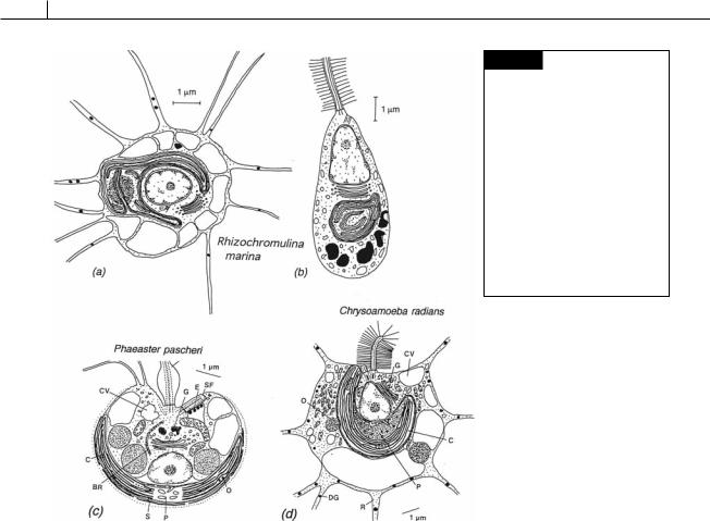
This order contains the more primitive organisms in the order (O’Kelly and Wujek, 1995). Rhizochromulina (Fig. 14.1(a), (b)) has amoeboid
non-flagellated vegetative cells with many fine beaded-filipodia and a single golden-brown chloroplast (Hibberd and Chretiennot-Dinet, 1979). The fusiform zoospore has a single tinsel flagellum with a second basal body in the protoplasm (Fig. 14.1(b)). Chrysoamoeba (Fig. 14.1(d)) lives as a solitary amoeba for the greater part of its life cycle, transforming into swimming cells with a single long flagellum only for short periods. In Phaeaster (Fig. 14.1(c)), the anterior portion of the cell is drawn out into rhizopodia.
Pedinellales
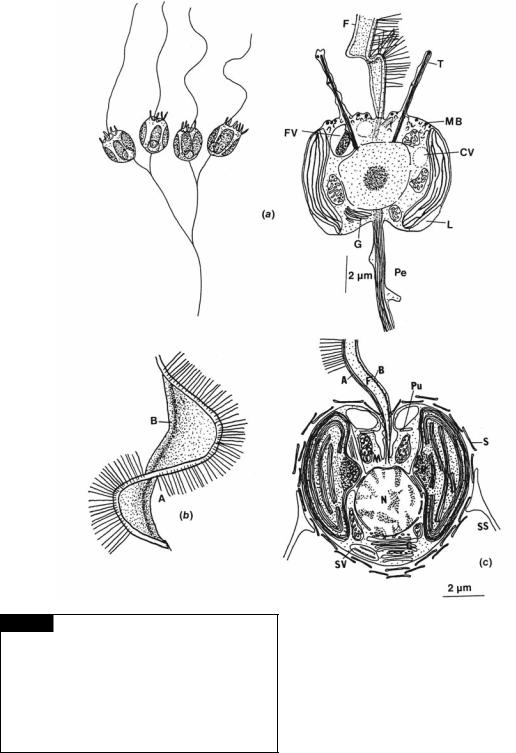
The pedinellids are unique in containing genera that are phototrophic (Apedinella (Fig. 14.2(c)), Pseudopedinella), mixotrophic (able to photosynthesize and take up organic compounds) (Pedinella) (Figs. 14.2(a), (b), 14.3, 14.4), and phagotrophic (Pteridomonas, Ciliophrys). The phagotrophic genera have vestigial chloroplasts and evolved from genera with chloroplasts (Sekiguchi et al., 2002). The organisms in this order have three interconnected microtubules (triads) that course from the nuclear envelope through tentacles (if they are present) to the plasma membrane (Fig. 14.2(a)) (Daugbjerg, 1996). A long apical flagellum is extended into a lateral wing by a paraxonemal rod (Figs. 14.2, 14.3(b)). The apical flagellum is inserted in a pit and there is a second flagellum that is reduced to a basal body. The basal bodies are at a slight angle to each other. The cells are radially symmetrical with a large central nucleus and a posterior Golgi apparatus. There are usually three to six chloroplasts present
360 CHLOROPLAST E.R.: EVOLUTION OF TWO MEMBRANES
Fig. 14.1 (a), (b) Rhizochromulina marina. Vegetative cell (a) and zoospore (b). (c) Phaeaster pascheri.
(d) Chrysoamoeba radians. (BR) Basal root; (C) chloroplast; (CV) contractile vacuole; (DG) dense granule; (E) eyespot; (G) Golgi body; (LF) long flagellum; (O) oil;
(P) pyrenoid; (R) rhizopodium;
(S) scale; (SF) short flagellum. ((a), (b) Redrawn from Hibberd and Chretiennot-Dinet, 1979; (c) redrawn from Belcher and Swale, 1971; (d) redrawn from Hibberd, 1971.)
if the organism is phototrophic (whereas in the Chrysophyceae there are usually only one or two chloroplasts). Some genera, such as Pedinella (Fig. 14.4(d), (e)) and Apedinella (Fig. 14.2(c)), have scales attached to the plasma membrane by microligaments (Koutoulis et al., 1988).
A posterior trailing stalk, associated with a system of vacuoles, can be present. Pedinella (Figs. 14.2(a), 14.4) cells rotate while swimming, trailing the stalk behind. The stalk appears to be sticky, and swimming cells will often adhere to a substrate by means of the stalk. Pedinella can undergo phagotrophy and ingest other small cells. Bacteria are passed down the flagellum and adhere to the plasma membrane, just outside the row of tentacles. Secretions of the muciferous bodies in this region provide an adhesive that sticks the cells to the plasmalemma. Usually within a minute, the bacteria become enveloped in a sheet of cytoplasm that is extruded from the cell.
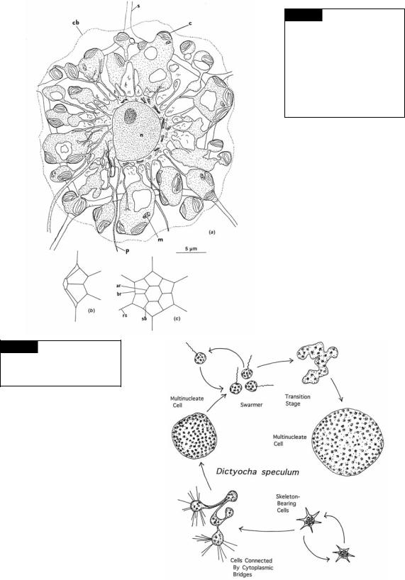
Dictyocales
The Dictyocales or silicoflagellates are a group of cosmopolitan marine flagellates, presently represented by only one extant genus Dictyocha, although an abundance of taxa have been described from fossil siliceous skeletons (Henriksen et al., 1993).
Silicoflagellate cells have one emergent flagellum and a skeleton of hollow siliceous rods outside of the protoplasm. The skeleton can be a simple ring, an ellipse, or a triangle, but it is often much more complete. In Dictyocha fibula (Fig. 14.5), the skeleton is composed of a series of peripheral polygons surrounding a central hexagon. The nucleus is in the center of the protoplasm with a number of cytoplasmic processes extending from the central mass. The chloroplasts are usually in the cytoplasmic processes (van Valkenburg, 1971a, b). Most species have chloroplasts that are derived from secondary endosymbioses, although
HETEROKONTOPHYTA, DICTOCHOPHYCEAE |
361 |
|
|
Fig. 14.2 (a) Pedinella hexacostata in the light and electron microscope. (b) Structure of the winged flagellum of P. hexacostata. (c) Apedinella spinifera. (A) Axoneme of flagellum;
(B) paraxonemal rod of flagellum; (CV) contractile vacuole;
(F) flagellum with hairs; (FV) food vacuole; (G) Golgi body;
(L) leucosin; (M) mitochondrion; (MB) muciferous body;
(N) nucleus; (Pe) peduncle; (Pu) pusule; (S) scale; (SS) spined scale; (SV) scale vesicle; (T) tentacle. ((a), (b) after Swale, 1969; (c) after Throndsen, 1971.)
there has been a report of chloroplasts derived from haptophytes through tertiary endosymbioses (Daugbjerg and Henriksen, 2001).
In Dictyocha speculum, the skeleton-bearing cells multiply vegetatively by mitotic division (Fig. 14.6) (Hendriksen et al., 1993). Cells connected by bridges develop and give rise to large spherical cells without skeletons that become multinucleate. Uninucleate swarmers with a single flagellum develop in the large spherical cells. The swarmers are released and grow into large multinucleate

362 CHLOROPLAST E.R.: EVOLUTION OF TWO MEMBRANES
Fig. 14.3 Pedinella squamata.
Transmission electron micrograph.
(C) Chloroplast; (M) mitochondrion;
(N) nucleus; (Py) pyrenoid. (From
Sekiguchi et al., 2003.)
Fig. 14.4 Pedinella squamata. Transmission electron micrographs. (a) Whole mount of a cell showing the flagellum and mastigonemes. (b) Higher magnification of a flagellum showing a paraxonemal rod (arrowhead). (c) Higher magnification of mastigonemes with no lateral filaments on the shafts. (d) Plate scales scattered about a whole mount of a cell. (e) A single plate scale with fibers (arrow) and the marginal rim (arrowhead). (From Sekiguchi et al., 2003.)
HETEROKONTOPHYTA, DICTOCHOPHYCEAE |
363 |
|
|
Fig. 14.6 Growth stages of the
silicoflagellate Dictyocha speculum.
(Adapted from Henriksen et al.,
1993.)
Fig. 14.5 (a) A drawing of the fine structure of Dictyocha fibula. (b), (c) Side and front views of the skeleton of Dictyocha. (ar) Apical ring; (br) basal ring; (c) chloroplast; (cb) cell boundary; (m) mitochondrion; (n) nucleus;
(p) pseudopodium; (rs) radial spine;
(s) silica skeleton; (sb) supporting bar. (After van Valkenburg, 1971a,b.)