


BASIC CHARACTERISTICS OF THE ALGAE |
19 |
|
|
Fig. 1.23 Drawing of a phycobilisome from the cyanobacterium Synechococcus. (Adapted from Grossman et al., 1993.)
phycobilisomes (cyanobacteria, cryptophytes, and red algae) an important ecological advantage in the open ocean, which is predominantly nitrogen limited (Vergara and Niell, 1993).
Mitochondria and peroxisomes
There are two types of mitochondria in algal cells (Leipe et al., 1994). Mitochondria with flat lamellae cristae occur in the red algae, green algae, euglenoids, and cryptophytes (Fig. 1.26). Mitochondria with tubular cristae occur in heterokonts and haptophytes.
Glycolate, the major substrate of photorespiration, can be broken down by either glycolate dehydrogenase in the mitochondria, or by glycolate oxidase in peroxisomes, single membranebounded bodies in the cytoplasm (for the reactions, see the chapter on Chlorophyta). The distribution of the two enzymes is as follows (Betsche et al., 1992; Iwamoto et al., 1996):
Fig. 1.24 Chromatic adaptation in a phycobilisome of a
cyanobacterium.
Fig. 1.25 Phycobilisome breakdown under conditions of nitrogen deprivation. (Adapted from Grossman et al., 1993.)
(a) |
|
(b) |
Fig. 1.26 Drawings of the two types of mitochondria that occur in the algae. (a) Mitochondrion with flat lamellar cristae. (b) Mitochondrion with tubular cristae.
20 INTRODUCTION
1Glycolate dehydrogenase occurs in the cyanobacteria, cryptophytes, euglenoids,
diatoms, and the green algae with the exception of the Charophyceae.
2Glycolate oxidase occurs in the glaucophytes, red algae, brown algae, and the Charophyceae in the green algae and higher plants.
Division of chloroplasts and mitochondria
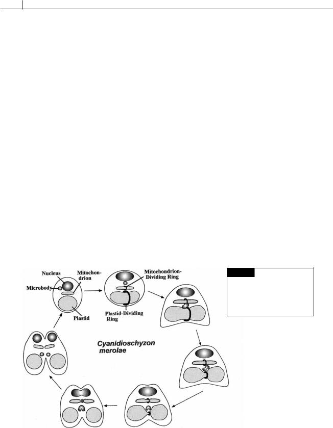
Chloroplasts and mitochondria divide by pinching in half to form two new organelles. A plastiddividing (PD) ring or mitochondrion-dividing (MD) ring surrounds the organelle in the area of fission (Fig. 1.27) (Miyagishima et al., 2003; Osteryoung and Nunnari, 2003). Each ring is composed of two parts, an outer ring in the protoplasm outside of the chloroplast and an inner ring in the stroma inside the inner membrane of the chloroplast. These rings are also called FtsZ (filamentous temperature-sensitive) rings after a counterpart that is present when bacteria divide. The similarity is indicative of the endosymbiotic origin of chloroplasts and mitochondria from bacteria. The plastid-dividing ring appears in the area of division and begins to contract after a microbody has migrated to the plas- tid-dividing ring (Fig. 1.27). The plastid-dividing
ring contracts around the area of plastid fission in association with GTPase proteins called dynamins. The PD ring disappears after fission is completed.
Storage products
The storage products that occur in the algae are as follows:
High-molecular-weight compounds
1-1,4 Linked glucans
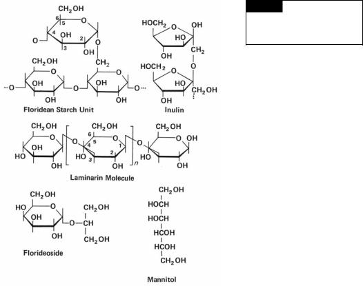
a Floridean starch (Fig. 1.28): This substance occurs in the Rhodophyta and is similar to the amylopectin of higher plants. It stains red-violet with iodine, giving a color similar to that of the stain reaction of animal glycogen. Floridean starch occurs as bowlshaped grains from 0.5 to 25 m outside the chloroplast, inferring the host in the original endosymbiosis took over formation of storage product. This differs from the Chlorophyta where starch is produced in the chloroplast. Despite the differing locations of starch synthesis, the Rhodophyta and Chlorophyta use a common pathway in the synthesis of starch (Patron and Keeling, 2005).
Fig. 1.27 Diagrammatic representation of the behavior of the plastid-dividing ring and mitochondrion-dividing ring in the unicellular red alga Cyanidioschyzon merolae.
BASIC CHARACTERISTICS OF THE ALGAE |
21 |
|
|
Fig. 1.28 The structure of
floridean starch, inulin, laminarin,
and floridoside. (After Percival and
McDowell, 1967.)
bMyxophycean starch: Found in the Cyanophyta, myxophycean starch has a similar structure to glycogen. This reserve product occurs as granules ( -granules), the shape varying between species from rod-shaped granules to 25-nm particles to elongate 31to 67-nm bodies.
cStarch: In the Chlorophyta, starch is composed of amylose and amylopectin. It occurs inside the chloroplast in the form of starch grains (Fig. 1.12(e)). In the Cryptophyta, starch has an unusually high content of amylose and occurs as grains between the chloroplast envelope and the chloroplast E.R. (Fig. 1.12(b)). In the Dinophyta also, starch occurs in the cytoplasm outside of the
chloroplast, but its structure is not known.
2-1,3 Linked glucans
a Laminarin (Fig. 1.28): In the Phaeophyceae, laminarin consists of a related group of predominantly -1,3 linked glucans containing 16 to 31 residues. Variation in the molecule
is introduced by the number of 1 → 6 linkages, the degree of branching, and the occurrence of a terminal mannitol molecule. The presence of a high proportion of C-6 interresidue linkages and of branch points seems to determine the solubility of the polysaccharide in cold water: the greater
the number of linkages, the higher the solubility. Laminarin occurs as an oil-like liquid outside of the chloroplasts, commonly in a vesicle surrounding the pyrenoid.
bChrysolaminarin (leucosin): In the Chrysophyceae, Prymnesiophyta, and Bacillario-
phyceae, chrysolaminarin consists of -1,3 linked D-glucose residues with two 1 → 6
glycosidic bonds per molecule. Chrysolaminarin occurs in vesicles outside of the chloroplast and has more glucose residues per molecule than laminarin.
cParamylon: In the Euglenophyta, Xanthophyceae, and Prymnesiophyta
(Pavlova mesolychnon), paramylon occurs as

22 INTRODUCTION
water-soluble, single-membrane-bounded inclusions of various shapes and dimensions outside of the chloroplast (Fig. 6.2). Paramylon consists solely of -1,3 linked glucose residues, and the molecule is about as large as that of chrysolaminarin.
3Fructosans: Acetabularia (Chlorophyta) has an inulin-like storage product consisting of a series of 1,2 linked fructose units terminated by a glucose end group (Fig. 1.28).
Low-molecular-weight compounds
1Sugars: Chlorophyta and Euglenophyta form sucrose as a reserve product; trehalose is found in the Cyanophyta and at low levels in
the Rhodophyta.
2 Glycosides: The glycerol glycosides, floridoside (Fig. 1.28) and isofloridoside, are widely distributed in the Rhodophyta.
3Polyols: Mannitol (Figs. 1.28, 4.4) occurs in Rhodophyta and Phaeophyceae. It is also present in lower green algae, where it replaces sucrose as a photosynthetic product. Free glycerol occurs widely in the algae and is an important photosynthetic product in several zooxanthellae (endosymbiotic algae in animals) and in some marine Volvocales, especially Dunaliella.
Contractile vacuoles
The ability of algal cells to adjust to changes in the salinity of the medium is an important aspect of the physiology of these cells. In cells with walls, this osmoregulation is accomplished with the aid of turgor pressure, whereas in naked cells it is accomplished by means of contractile vacuoles and/or regulation of the solutes present in the cells. In the latter case, cells increase the internal concentration of osmotically active molecules and ions when the concentration of dissolved solutes increases in the external medium. Likewise, the internal concentration of such molecules decreases when the concentration of dissolved salts in the external medium decreases.
Most algal flagellates have two contractile vacuoles in the anterior end of a cell (Fig. 1.1). A contractile vacuole will fill with an aqueous solution (diastole) and then expel the solution outside of
the cell and contract (systole). The contractile vacuole rhythmically repeats this procedure. If there are two contractile vacuoles, they usually fill and empty alternately. Contractile vacuoles occur more frequently in freshwater than marine algae, a phenomenon that gives credence to the theory that the contractile vacuoles maintain a water balance in the cells. The algal cells in freshwater have a higher concentration of dissolved substances in their protoplasm than in the surrounding medium so that there is a net increase of water in the cells. The contractile vacuoles act to expel this excess water. An alternate theory on the function of the contractile vacuoles is that they remove waste products from the cells. The Dinophyta have a structure similar to a contractile vacuole, called a pusule, which may have a similar function but is more complex.
The contractile vacuoles of the Cryptophyta are characteristic of the algae (Fig. 1.29). In the Cryptophyta, the contractile vacuole occurs in a fixed anterior position next to the flagellar depression (Patterson and Hausmann, 1981). At the beginning of the filling phase (diastole), there is no distinct contractile vacuole, only a region filled with small (ca. 0.5- m diameter) contributory vacuoles. These vacuoles fuse to form a large irregular vacuole which subsequently rounds up. The contributory vacuoles destined to form the next contractile vacuole now appear around the rounded contractile vacuole. The contractile vacuole fuses with the plasma membrane of the flagellar pocket and discharges its contents outside the cell. The area of the plasma membrane that fuses with the contractile vacuole does not have a periplast (specialized plates within the plasma membrane). This area is, instead, bounded by microtubules. The membrane of the contractile vacuole is recovered by the cell as small vesicles with an electron-dense coat, and the membrane components are reutilized by the cell. These vesicles plus the contractile vacuole occur in the spongiome or area around the contractile vacuole. In freshwater algae the contractile vacuole cycle lasts for 4 to 16 seconds, whereas in marine species the cycle can last for up to 40 seconds.
Algal flagellates use a combination of contractile vacuoles and osmoregulation to control the water content of their cells. In the chrysophyte