

BASIC CHARACTERISTICS OF THE ALGAE |
9 |
|
|
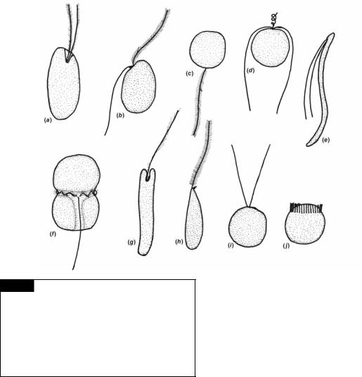
Fig. 1.9 The shape of eukaryotic motile algal cells and their flagella. The drawings represent the common arrangement of flagella in the groups. There are a number of modifications in structure that are not included here. (a) Cryptophyta; (b) most of the Heterokontophyta; (c) Bacillariophyceae of the Heterokontophyta; (d) Prymnesiophyta; (e) Chlorophyta; (f) Dinophyta; (g) Euglenophyta; (h) Eustigmatophyceae of the
Heterokontophyta; (i, j) Chlorophyta.
they are called isokont flagella; if they are of unequal length, they are called anisokont flagella; and if they form a ring at one end of the cell, they are called stephanokont flagella. Heterokont refers to an organism with a hairy and a smooth flagellum (Moestrup, 1982).
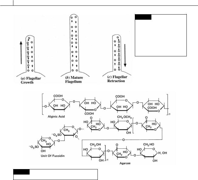
Flagella can be of different length in the same cell. This is controlled by intraflagellar transport, defined as the bi-directional movement of particles along the length of the flagellum between the axoneme and the flagellar membrane (Beech, 2003). A mature flagellum that is not elongating has a steady disassembly of the flagellum that is countered by an equally steady assembly provided by intraflagellar transport (Fig. 1.10). A change in length of the
flagellum is produced by an imbalance in the assembly or disassembly of flagellar components (Rosenbaum and Witman, 2002). Thus, disassembly occurs faster than assembly in flagellar retraction. The opposite occurs during flagellar growth. The differences in length of flagella arise from the shorter flagellum being delayed in the initial stages of construction. The assembly rate of the shorter flagellum is the same as the longer flagellum. There may be a gate at the base of the flagellum that regulates the passage of flagellar precursors into the basal body and the flagellum (Schoppmeier and Lechtreck, 2003).
Cell walls and mucilages
In general, algal cell walls are made up of two components: (1) the fibrillar component, which forms the skeleton of the wall, and (2) the amorphous component, which forms a matrix within which the fibrillar component is embedded.
The most common type of fibrillar component is cellulose, a polymer of 1,4 linked -D-glucose. Cellulose is replaced by a mannan, a polymer of 1,4 linked -D-mannose, in some siphonaceous
10 INTRODUCTION
Fig. 1.11 Structural units of alginic acid, fucoidin, and
agarose. (After Percival and McDowell, 1967.)
greens, and in Porphyra and Bangia in the Rhodophyta. In some siphonaceous green algae and some Rhodophyta (Porphyra, Rhodochorton, Laurencia, and Rhodymenia), fibrillar xylans of different polymers occur.
The amorphous mucilaginous components occur in the greatest amounts in the Phaeophyceae and Rhodophyta, the polysaccharides of which are commercially exploited. Alginic acid (Fig. 1.11) is a polymer composed mostly of -1,4 linked D- mannuronic acid residues with variable amounts of L-guluronic acid. Alginic acid is present in the intercellular spaces and cell walls of the
Fig. 1.10 (a) Intraflagellar transport results in more assembly of flagellar subunits than disassembly during flagellar growth. (b) A mature flagellum has an equal amount of assembly and disassembly of flagellar subunits. (c) There is more disassembly of flagellar subunits during flagellar retraction.
Phaeophyceae. Fucoidin (Fig. 1.11) also occurs in the Phaeophyceae and is a polymer of -1, 2, -1, 3, and -1, 4 linked residues of L-fucose sulfated at C- 4. In the Rhodophyta the amorphous component of the wall is composed of galactans or polymers of galactose, which are alternatively -1,3 and -1,4 linked. These galactans include agar (made up of agaropectin and agarose, Fig. 1.11) and carrageenan (Fig. 4.15).
Plastids
The basic type of plastid in the algae is a chloro- plast, a plastid capable of photosynthesis. Chromoplast is synonymous with chloroplast; in the older literature a chloroplast that has a color other than green is often called a chromoplast. A proplastid is a reduced plastid with few if any
BASIC CHARACTERISTICS OF THE ALGAE |
11 |
|
|
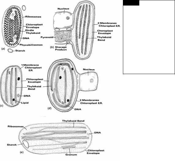
Fig. 1.12 Types of chloroplast structure in eukaryotic algae. (a) One thylakoid per band , no chloroplast endoplasmic reticulum (Rhodophyta). (b) Two thylakoids per band, two membranes of chloroplast E.R. (Cryptophyta). (c) Three thylakoids per band, one membrane of chloroplast E.R. (Dinophyta, Euglenophyta). (d) Three thylakoids per band, two membranes of chloroplast E.R. (Prymnesiophyta and Heterokontophyta). (e) Two to six
thylakoids per band, no chloroplast E.R. (Chlorophyta).
thylakoids. A proplastid will usually develop into |
to the chloroplasts in a secondary endosymbio- |
a chloroplast although in some heterotrophic |
sis. In the Euglenophyta and Dinophyta, there is |
algae it remains a proplastid. A leucoplast or amy- |
one membrane of chloroplast E.R. (Fig. 1.12(c). In |
loplast is a colorless plastid that has become |
the Cryptophyta, Prymnesiophyta, and Heterokon- |
adapted for the accumulation of storage product. |
tophyta, there are two membranes of chloroplast |
In the Rhodophyta and Chlorophyta, the |
E.R., with the outer membrane of chloroplast E.R. |
chloroplasts are bounded by the double membrane |
usually continuous with the outer membrane of |
of the chloroplast envelope (Fig. 1.12(a), (e)). In the |
the nuclear envelope, especially if the chloroplast |
other eukaryotic algae, the chloroplast envelope |
number is low (Fig. 1.12 (b), (d )). |
is surrounded by one of two membranes of |
The basic structure of the photosynthetic appa- |
chloroplast endoplasmic reticulum (chloroplast |
ratus in a plastid consists of a series of flattened |
E.R.), which has ribosomes attached to the outer |
membranous vesicles called thylakoids or discs, |
face of the membrane adjacent to the cytoplasm. |
and a surrounding matrix or stroma. The thylak- |
The chloroplast E.R. is the remnant of the food |
oids contain the chlorophylls and are the sites |
vacuole membrane and/or the plasma membrane |
of the photochemical reactions; carbon dioxide |
involved in the original endosymbiosis leading |
fixation occurs in the stroma. The thylakoids can |

12 INTRODUCTION
be free from one another or grouped to form thylakoid bands. In the cyanobacteria and Rhodophyta (Fig. 1.12(a)), the thylakoids are usually free from one another, with phycobilisomes (containing the phycobiliproteins) on the surface of the thylakoids. The phycobilisomes on the surface of one thylakoid alternate with those on the surface of an adjacent thylakoid. The phycobilisomes appear as 35-nm granules when phycoerythin predominates, or as discs when phycocyanin predominates. In the more primitive members of the Rhodophyta the thylakoids terminate close to the chloroplast envelope, whereas in advanced members of the Rhodophyta peripheral thylakoids are present, which enclose the rest of the thylakoids. In the Cryptophyta, the chloroplasts contain bands of two thylakoids (Fig. 1.12(b)); the phycobiliproteins are dispersed within the thylakoids. In the Euglenophyta and Heterokontophyta the thylakoids are grouped in bands of three with a girdle or peripheral band running parallel to the chloroplast envelope. In the Dinophyta, Prymnesiophyta, and Eustigmatophyceae, the thylakoids are also in bands of three, but there is no girdle band (Fig. 1.12(c), (d)). In the Chlorophyta, the thylakoids occur in bands of two to six, with thylakoids running from one band to the next. The above grouping of algal thylakoids into bands occurs under normal growth conditions. Abnormal growth conditions commonly cause lumping of thylakoids and other variations in structure.
A pyrenoid (Fig. 1.12(b)) is a differentiated region within the chloroplast that is denser than the surrounding stroma and may or may not be traversed by thylakoids. A pyrenoid is frequently associated with storage product. Pyrenoids contain
Fig. 1.13 The structure of Form I variation of Rubisco
showing the eight large subunits and eight small subunits.
ribulose-1, 5-bisphosphate carboxylase/oxygenase (Rubisco), the enzyme that fixes carbon dioxide (Jenks and Gibbs, 2000; Nagasato et al., 2003). Consequently, the size of the pyrenoid will vary depending on how much Rubisco is present.
Rubisco exists in two forms (Jenks and Gibbs, 2000; Zhang and Lin, 2003):
1Form I occurs in some bacteria, the cyanobacteria, in all green plants and nongreen plants. Form I is composed of eight large subunits and eight small subunits (Fig. 1.13).
Form I has a high affinity for CO2 and a low catalytic efficiency (low rate of CO2 fixation). In green algae, euglenoids, and green plants,
the large subunit is coded by chloroplast DNA and the small subunit by nuclear DNA. In the cyanelle (endosymbiotic cyanobacterium) of
Cyanophora paradoxa and in some non-green algae, both subunits are coded by chloroplast
DNA.
2Form II occurs in some eubacteria and in the dinoflagellates and is composed of two large
subunits. Form II has a low affinity for CO2 and a high catalytic efficiency.
The common ancestor of all ribulose-1,5-bisphos- phate carboxylase was probably similar to Form II and was adapted to the anaerobic conditions and high CO2 concentrations prevailing in the ancient earth (Haygood, 1996). Form I evolved as the earth’s atmosphere became oxygenated, and CO2 concentration declined and with it the need for a greater affinity for CO2. The greater affinity for CO2 in Form I, however, came at the price of reduced catalytic efficiency.
Chloroplasts contain small (30–100 nm), spherical lipid droplets between the thylakoids (Fig. 1.12 (c), (d)). These lipid droplets serve as a pool of lipid reserve within the chloroplast.
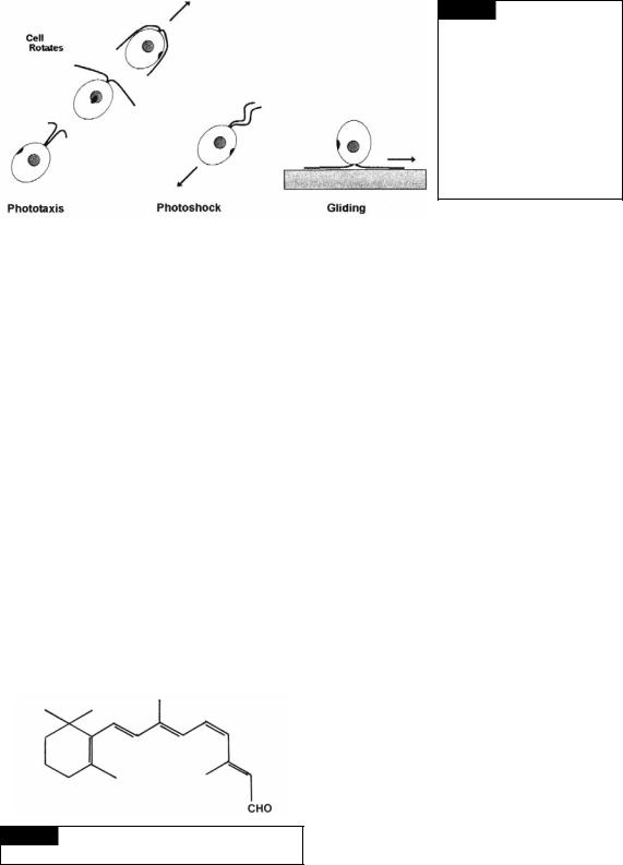
Many motile algae have groups of tightly packed carotenoid lipid-globules that constitute an orange-red eyespot or stigma (Fig. 5.2) that is involved in response to light. Motile algae exhibit three types of responses to light (Kawai and Kreimer, 2000): phototaxis, photophobia, and gliding (Fig. 1.14).
1Phototaxis. In phototaxis, the orientation of cell movement is effected by the direction and
BASIC CHARACTERISTICS OF THE ALGAE |
13 |
|
|
Fig. 1.14 Three types of flagellar orientation in Chlamydomonas. In phototaxis, the cells swim forward and rotate. Phototaxis requires that cells swim forward in a spiral path that causes rotation of the symmetrically placed eyespot. In photoshock, the cell has a transient avoidance response that causes the cell to swim backwards. In gliding, the leading flagellum and passive flagellum are 180° apart.
intensity of light. The cells move toward the light in positive phototaxis and away from the light in negative phototaxis. The photoreceptor in the green alga Chlamydomonas is chlamyrhodopsin (Fig. 1.15) in the plasma membrane over the eyespot. Chlamyrhodopsin contains an all-trans, 6-S-trans retinal chromophore that undergoes a 13-trans to cis isomerization during illumination (Hegemann, 1997). The eyespot periodically shades the photoreceptor as the cell rotates during swimming. The eyespot has a different structure in the different groups of algae and will be covered in the appropriate chapters. Eyespots have certain basic characteristics (Kawai and Kreimer, 2000):
(1) Eyespots usually have carotenoid-rich lipid globules packed in a highly ordered hexagonal arrangement. (2) Eyespots are usually single structures in peripheral positions, most often oriented perpendicular to the axis of the swimming path.
Phototaxis in Chlamydomonas is controlled by the beating of each flagellum. The flagellum closest to the eyespot is the cis flagellum while
Fig. 1.15 The structure of chlamyrhodopsin, the
photoreceptor in Chlamydomonas.
the trans flagellum is furthest from the eyespot. The light is received by the photoreceptor which controls the opening and closing of calcium channels, and the level of intraflagellar calcium concentration. The calcium concentration within the flagellum effects the interactions of the radial spokes with the central pair of microtubules (Mitchell, 2000). When the plasma membrane of Chlamydomonas is made permeable, Chlamydomonas cells swim normally at 10 8 M calcium in the medium. Decreasing the calcium to 10 9 M reduces the stroke velocity of the trans flagellum, while increasing the calcium to 10 7 M reduces the stroke velocity of the cis flagellum.
2Photophobia (photoshock). Photophobia is a change in direction of movement of the cell caused by a rapid change in light intensity, irrespective of the direction of the light. Swimming cells stop and change the beat pattern from the normal asymmetric flagellar stroke to a symmetrical stroke that propels the cell backward (Fig. 1.14). At the end of the photophobic response, the cells tumble and resume swimming in a new direction. Laboratory experiments with
Chlamydomonas link photophobic responses to increases in calcium above 10 6 M (Mitchell, 2000). Unlike phototaxis, interactions between radial spokes and central-pair microtubules are not necessary for a photophobic reaction.
3Gliding (quiesence). In gliding, the flagella stop beating and adhere to a surface or an

14 INTRODUCTION
Fig. 1.16 Transmission electron
micrograph of DNA in the
chloroplast of the dinoflagellate
Prorocentrum micans. (From Laatsch
et al., 2004.)
Fig. 1.17 Semidiagrammatic drawing of the two types of distribution of DNA in algal chloroplasts. Side and face views of the plastids are drawn. (Adapted from Coleman, 1985.)
air/water interface (Mitchell, 2000). The cells can glide over the surface with one flagellum actively leading and the other passively trailing (Fig. 1.14). Cells may switch direction by changing which flagellum is active. Gliding motility may be a common phenomenon among organisms that live in the thin film of water on soil particles.
In the Chlorophyta (Fig. 5.2), Cryptophyta (Fig. 9.4) and most of the Heterokontophyta (Fig. 10.1), the eyespot occurs as lipid droplets in the chloroplast. In the Euglenophyta (Fig. 6.2), Eustigmatophyceae (Fig. 12.1), and Dinophyta (Figs. 7.21, 7.22, 7.23), the eyespot occurs as a group of membrane-bounded lipid droplets, free of the chloroplast.
Most chloroplasts contain prokaryotic DNA in an area of the chloroplast devoid of 70S ribosomes (Figs. 1.16 and 1.17). The DNA is an evolutionary remnant of the cyanobacterium involved in the endosymbiosis leading to the chloroplast. The individual DNA microfibrils are circular, are attached to the chloroplast membranes, and lack basic proteins (histones). The algae can be divided into two general groups according to the distribution of DNA in the plastids (Coleman, 1985). In the first group, the clumps of DNA (nucleoids) are scattered throughout the plastids. This group includes the Cryptophyta, Dinophyta, Prymnesiophyta, Eustigmatophyceae, Rhodophyta, and Chlorophyta. In the second group, the DNA occurs in a ring just within the girdle lamella. This group includes the Chrysophyceae, Bacillariophyceae, Raphidophyceae, and Xanthophyceae (with the exception of Vaucheria and three genera known to lack girdle lamellae – Bumilleria, Bumilleriopsis, and Pseudobumilleriopsis). The Euglenophyta fit into neither group, showing a variable distribution of chloroplast DNA.
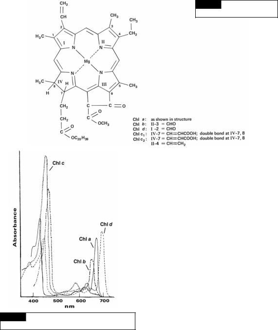
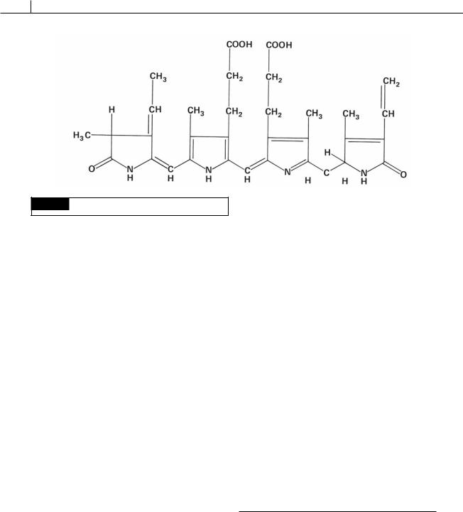
The photosynthetic algae have chlorophyll in their chloroplasts. Chlorophyll is composed of a
BASIC CHARACTERISTICS OF THE ALGAE |
15 |
|
|
Fig. 1.18 The structure of the
chlorophylls. (From Meeks, 1974.)
Fig. 1.19 The absorption spectra of chlorophylls a, b, c,
and d.
porphyrin-ring system that is very similar to that of hemogloblin but has a magnesium atom instead of an iron atom (Fig. 1.18). The algae have four types of chlorophyll, a, b, c (c1 and c2), and d. Chlorophyll a is the primary photosynthetic pigment (the light receptor in photosystem I of the
light reaction) in all photosynthetic algae and ranges from 0.3% to 3.0% of the dry weight. Chlorophyll a is insoluble in water and petroleum ether but soluble in alcohol, diethyl ether, benzene, and acetone. The pigment has two main absorption bands in vitro, one band in the red light region at 663 nm and the other at 430 nm (Fig. 1.19).
Whereas chlorophyll a is found in all photosynthetic algae, the other algal chlorophylls have a more limited distribution and function as accessory photosynthetic pigments. Chlorophyll b is found in the Euglenophyta and Chlorophyta (Fig. 1.18). Chlorophyll b functions photosynthetically as a light-harvesting pigment transferring absorbed light energy to chlorophyll a. The ratio of chlorophyll a to chlorophyll b varies from 2:1 to 3:1. The solubility characteristics of chlorophyll a are similar to chlorophyll b, and in vitro chlorophyll b has two main absorption maxima in acetone or methanol, one at 645 nm and the other at 435 nm (Fig. 1.19).
Chlorophyll c (Fig. 1.18) is found in the Dinophyta, Cryptophyta, and most of the Heterokontophyta. Chlorophyll c has two spectrally different components: chlorophyll c1 and c2. Chlorophyll c2 is always present, but chlorophyll c1 is absent in the Dinophyta and Cryptophyta. The ratio of chlorophyll a to chlorophyll c ranges from
16 INTRODUCTION
1.2:2 to 5.5:1. Chlorophyll c probably functions as an accessory pigment to photosystem II. The pigment is soluble in ether, acetone, methanol, and ethyl acetate, but is insoluble in water and petroleum ether. Extracted chlorophyll c1 has main absorption maxima at 634, 583, and 440 nm in methanol, whereas chlorophyll c2 has maxima at 635, 586, and 452 nm.
Chlorophyll d (Fig. 1.18) occurs in some cyanobacteria (Murakami et al., 2004). It has three main absorption bands at 696, 456, and 400 nm.
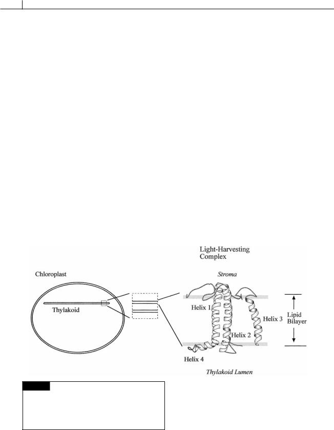
The photosynthetically active pigments of algae are gathered in discrete pigment–protein complexes which can be divided functionally into two groups (Grossman et al., 1990):
1the photochemical reaction center containing chlorophyll a, where light energy
is converted into chemical energy;
2the light-harvesting complexes that serve as antennae to collect and transfer available light energy to the reaction center.
The light-harvesting complexes use different antennae pigment complexes to capture light energy. All of the light-harvesting complexes are
composed of three-membrane spanning helices (Fig. 1.20).
1Green algae and higher plants use chlorophyll a/b binding proteins.
2Brown and golden-brown algae, (diatoms, chrysophytes, dinoflagellates, brown algae, and related groups) use a fucoxanthin chlorophyll a/c complex that is an integral part of the thylakoid membrane. The ratio of fucoxanthin to chlorophyll in this complex is approximately 2 : 1 and the characteristic brown or goldenbrown color of these algae is due to the high level of fucoxanthin in these cells. Due to chlorophyll c and special xanthophylls, these organisms are especially suited to harvest blue and green light, which are the most abundant at increasing ocean depths. This lightharvesting complex also is composed of three membrane-spanning helices and is closely related to the light-harvesting complex in the
first group (Caron et al., 1996).
3Cyanobacteria, cryptophytes and red algae use the phycobilisome as the major lightharvesting complex.
Fig. 1.20 The basic structure of the light-harvesting complex in all eukaryotic plants. Three transmembrane helices traverse the membrane. The similarity of the lightharvesting complex in all eukaryotic plants is an argument for the chloroplast arising from a single endosymbiotic event. (Modified from Kuhlbrandt et al., 1994.)
BASIC CHARACTERISTICS OF THE ALGAE |
17 |
|
|
Carotenoids are yellow, orange, or red pigments that usually occur inside the plastid but may be outside in certain cases. In general, naturally occurring carotenoids can be divided into two classes:
(1)oxygen-free hydrocarbons, the carotenes; and
(2)their oxygenated derivatives, the xanthophylls. The most widespread carotene in the algae is-carotene (Fig. 1.21). There are a large number of different xanthophylls, with the Chlorophyta having xanthophylls that most closely resemble those in higher plants. Fucoxanthin (Fig. 1.21) is the principal xanthophyll in the golden-brown algae (Chrysophyceae, Bacillariophyceae, Prymnesiophyceae, and Phaeophyceae), giving these algae their characteristic color. Like the chlorophylls, the carotenoids are soluble in alcohols, benzene, and acetone but insoluble in water.
The cyanobacteria and chloroplasts of the Rhodophyta and Cryptophyta have evolved mem- brane-peripheral antenna complexes containing phycobiliproteins that transfer light energy to photosytem II reaction centers. Like chlorophyll b/c/d, the phycobiliproteins expand the range of light energy that can be utilized in photosynthesis. Light tends to become blue-green as it courses down the water column, and this light is better absorbed by the biliproteins than chlorophyll a.
Phycobiliproteins are water-soluble blue or red pigments located on (Cyanophyta, Rhodophyta) or inside (Cryptophyta) thylakoids of algal
chloroplasts (Glazer, 1982). They are described as chromoproteins (colored proteins) in which the prosthetic group (non-protein part of the molecule) or chromophore is a tetrapyrole (bile pigment) known as phycobilin. The prosthetic group is tightly bound by covalent linkages to its apoprotein (protein part of the molecule) (see Fig. 1.21). Because it is difficult to separate the pigment from the apoprotein, the term phycobiliprotein is used. There are two different apoproteins, and , which together form the basic unit of the phycobiliproteins. To either or are attached the colored chromophores. The major “blue” chromophore occurring in phycocyanin and allophycocyanin is phycocyanobilin, and the major “red” chromophore occurring in phycoerythrin is phycoerythrobilin (Fig. 1.22).
The general classification of phycobiliproteins is based on their absorption spectra. There are three types of phycoerythrin: R-phycoerythrin and B-phycoerythrin in the Rhodophyta, and C- phycoerythrin in the Cyanophyta. There are also three types of phycocyanin: R-phycocyanin from the Rhodophyta and C-phycocyanin and allophycocyanin from the Cyanophyta. In addition, in the Cryptophyta there are three spectral types of phycoerythrin and three spectral types of phycocyanin.
The basic subunit of a phycobilisome consists of apoproteins and , each of which is attached to a chromophore (Anderson and Toole,
Fig. 1.21 The structure of -carotene and fucoxanthin.
18 INTRODUCTION
Fig. 1.22 The structure of phycoerythrobilin.
1998; Samsonoff and MacColl, 2001) (Fig. 1.23). In the core of the phycobilisome and are attached to allophycocyanins, which are closest to chlorophyll in the energy transfer pathway. In the outer rods, and are attached to phycocyanin or phycocyanin. In the core of the phycobilisome and are attached to allophycocyanin. The , molecules are assembled into hexamers ( 1, 1) cylindrical in shape. The hexamers that make up the core of the phycobilisome are assembled in pairs, with the hexamers of the rods radiating from the core. The hexamers are joined together by linker polypeptides. The linker polypeptides are basic whereas the hexamers are acidic; this suggests that electrostatic interactions are important in assembling phycobiliproteins. There are high-molecular-weight polypeptides that anchor the phycobilisome to the area of the thylakoid membrane that contains the reaction center and associated chlorophylls.
The pathway of energy transfer (Glazer et al., 1985) is
In intact cells, the overall efficiency of energy transfer from the phycobilisome to chlorophyll a in the thylakoids exceeds 90% (Porter et al., 1978).
Chromatic adapters change their pigment components under different light wavelengths (Fig. 1.24). For example, the cyanobacterium Synechocystis grown in green light produces phycoerythrin (red in color), phycocyanin (blue), and allophycocyanin (blue-green) in a molar ratio of about 2 : 2 : 1; when it is grown in red light, the ratio is about 0.4 : 2 : 1. The phycobilisome structure changes appropriately, with the peripheral rods having more phycoerythrin hexamers under green light, and less phycocyanin hexamers. The allophycocyanin core hexamers stay the same.
Depriving cells of nitrogen results in an ordered degradation of phycobilisomes (Fig. 1.25). There is a progressive degradation of hexamer rod and linker polypeptides followed by the core peptides. New phycobilisomes are rapidly synthesized on the addition of nitrogen to the medium. Phycobilisomes are, thus, an important source of internal nitrogen and offer the algae that have
phycoerythrin |
|
|
allophycocyanin B |
|
( max 565) |
→ phycocyanin → |
allophycocyanin → |
( max 670) |
→ chlorophyll a |
or |
or |
|||
phycoerythrocyanin |
( max 620–638) |
( max 650) |
high-molecular- |
|
( max 568) |
|
|
weight polypeptide |
|
|
|
|
( max 665) |
|