


3. Management of Colorectal Cancer by “Hot Biopsy” and Snare Resection
YOSHIHIRO SAKAI
1. Introduction
Cancer can sometimes be diagnosed on endoscopy, but small carcinomas in adenomas are difficult to detect. When cancer is suspected, resection of the lesion is required either endoscopically or surgically, depending on the estimated depth of invasion in cases of cancer. Endoscopic treatment must be supported by clear-cut evidence that the lesion is superficial and can be completely resected by endoscopic techniques. Endoscopic procedures can be used only when tumor invasion is confined to the mucosa or is estimated to be within 1000 mm from the muscularis mucosae. Magnification and dye techniques are often used to estimate the depth of invasion. Endoscopic ultrasonography (EUS) is also used. With the former technique, lesions invading the submucosa that are exposed and have lost their glandular structure are not indicated for endoscopic therapy.With EUS, lesions in which the third layer is interrupted or the fourth layer is displaced also cannot be treated endoscopically. For convenience, physiological saline solution can be locally injected immediately below the lesion just before endoscopic treatment. The endoscopist then determines whether or not the lesion can be lifted. However, this technique can lead to error because lesions associated with fibrosis resulting from previous biopsy or other causes cannot be lifted. Another potential source of error is that whether or not a lesion can be lifted is often related to the technique and subjective judgment of the endoscopist. Even if the lesion is lifted, sometimes the cancer has invaded deeply in the submucosa close to the proper muscle layer.
2. “Hot Biopsy” for Colorectal Cancer
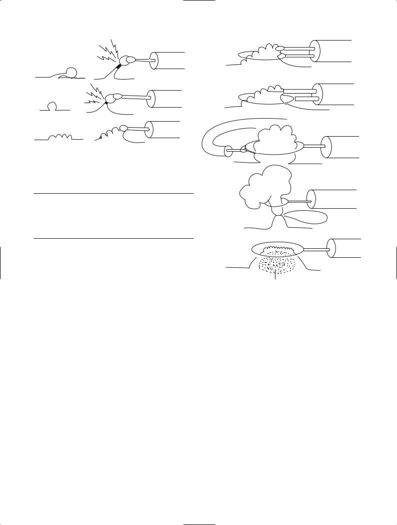
“Hot biopsy” [1] is used in the management of polyplike lesions. Part of the lesion is grasped with a hotbiopsy forceps. The lesion is then pulled into the center of the lumen and a high-frequency current, particularly coagulation waves, is applied. In terms of size, lesions indicated for this technique must be able to be grasped by the hot-biopsy forceps and elevated. The shape of the
lesion should facilitate concentration of high-frequency current, thereby producing heat. Clinically, pulling the lesion into the center of the lumen causes it to become pendunculated or semi-pendunculated, allowing current to be efficiently applied to the lesion base and the tissue to be retrieved. Even if only small fragments of tissue are collected in the cup of the forceps, the part of the lesion remaining after heat-induced coagulation of the base sloughs off (Fig. 1). Hot biopsy therefore cannot be used to treat sessile lesions or lesions more than 1 cm in diameter. This technique is therefore indicated for small pedunculated lesions less than 5 mm in diameter or small nonpedunculated lesions that have a narrow base.
An advantage of hot biopsy is the ability to repeat treatment within a short period. However, whether the target lesion has been adequately treated cannot be confirmed, leading to an increased risk of recurrence. Lesions containing carcinoma can be evaluated if completely retrieved in the cup of the hot-biopsy forceps. Many lesions cannot fit into the cup of the forceps. In some cases, carcinoma tissue is collected in the cup, whereas in others only the surrounding adenomatous components are collected, increasing the risk of misdiagnosis. In patients with carcinoma, it is an urgent task to estimate the depth of invasion and determine whether the lesion has sloughed off or remains, but histological techniques of verifying the outcome are lacking. If no cancer tissue is retrieved, it is unclear whether the diagnosis of cancer before hot biopsy was incorrect or whether the site grasped by the forceps did not contain cancer tissue. Whether treatment was adequate also cannot be confirmed. Patients treated by hot biopsy must therefore be closely followed up. Distant metastasis is a more important concern than local recurrence. The occurrence of distant metastasis of course precludes radical treatment.
Lesions diagnosed as cancer, including small tumors, should therefore not undergo hot biopsy, even if they are pedunculated or have a constricted base. However, since many small lesions with a fine pedicle or a distinctly constricted base are confined to the mucosa, hot biopsy may be used with caution in selected patients. Because of the risks involved, the use of hot biopsy requires the consent of both the physician and patient.
201
202 V. Endoscopic Treatment
a
b
c |
NO |
Fig. 1. Indications for hot biopsy technique. Hot biopsy is indicated for a, small pedunculated lesions and b, small sessile lesions, but not for c, large sessile lesions
Table 1. Classification of snare resection
Simple snare resection en bloc
piecemeal
Combined snare resectiona en bloc
piecemeal
a including double-snare technique, double-scope technique, preinjection polypectomy or submucosal injection polypectomy, and suction into the hood attached to the scope.
3. Snare Resection for Colorectal Cancer
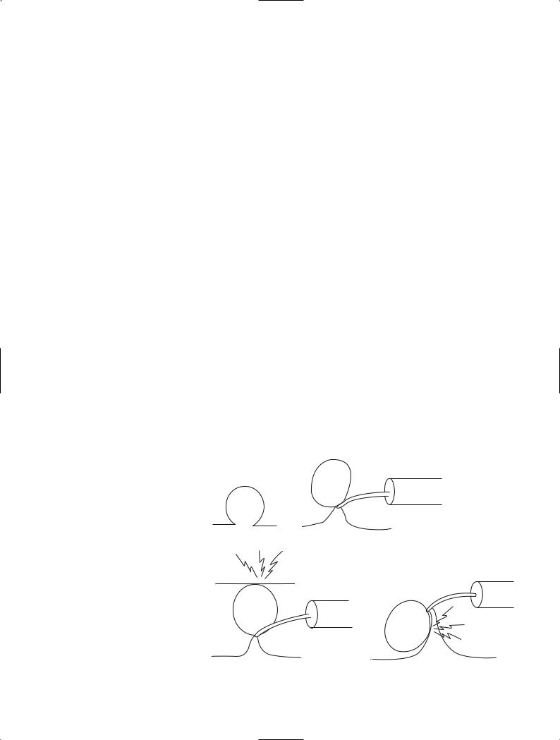
Snare resection is the procedure of choice for the endoscopic treatment of colorectal lesions diagnosed as cancer. For snare resection high-frequency current is used. The snare is used either alone or with other auxiliary techniques. The former procedure is conventionally referred to as polypectomy, but the term “simple snare resection” is more appropriate because it directly contrasts it with the latter procedure, termed combined snare resection [2] (Table 1). Combined snare resection can be done by two or more basic techniques. A lesion can be lifted with the grasping forceps and encircled with a snare [3] (Fig. 2). Alternatively, two snares can be employed. One snare grasps part of the lesion and the other serves a supplementary role (double-snare technique) [2]. A two-channel scope is required for these procedures. Two separate scopes may also be used. The grasping forceps are inserted via one scope, and the snare is introduced through the other (double-scope technique) [3]. The lesion may also be first grasped with an indwelling snare and ligated. A snare is then placed around the artificially produced constriction. Recently,
a
b
c
d
e
injected fluid
Fig. 2. Various combined snare techniques. a Grasping forceps and snare technique. b Double-snare technique. c Double-scope technique. d Detachable snare and snare technique. e Preinjection polypectomy (endoscopic mucosal resection, EMR)
some investigators have recommended that physiological saline solution is injected into the submucosa immediately below the lesion [4], thereby raising the lesion and facilitating grasping with a snare. This technique is referred to as preinjection polypectomy [5] or subcutaneous injection polypectomy [6]. Recently, this technique became well known as endoscopic mucosal resection (EMR). The original technique was termed endoscopic electroresection [7] and can be traced back to Rosenberg [8].
Both simple snare resection and combined snare resection can be used to resect lesions en bloc or in a piecemeal fashion. The resection procedure used should be clearly recorded.
In addition to physiological saline solution, epinephrine or dye can be added to the local injection solution.
3. Management of Colorectal Cancer by “Hot Biopsy” and Snare Resection |
203 |
In general, physiological saline solution is safest and least expensive. A hood can be attached to the scope tip and the lesion encircled with a snare loop. The lesion can then be aspirated into the hood and strangled.
3.1 Simple Snare Resection
Simple snare resection is undertaken with only highfrequency current and a snare. The more familiar term for this procedure is polypectomy. Cancer is frequently present in adenomas measuring more than 1 cm in diameter. Lesions with a relatively fine pedicle have a low risk of submucosal invasion by tumor. Even if such invasion occurs it is usually minimal. This similarly applies to mobile lesions with a distinct constriction. The likelihood of complete resection of cancer is increased by placing the snare at the level of the surrounding mucosa and strangling the lesion. To avoid burns caused by concentration of large amounts of high-frequency current on the muscularis propria, some distance must be physically created between the muscularis propria and the lesion. After strangulation, current should not be immediately applied. First, the lesion should be lifted into the center of the lumen to separate it from the muscularis propria (Fig. 3), or the scope should be pulled distally. Excessive pulling of the scope should be avoided because it can induce peristalsis, or the lesion may be hidden behind the distal semilunar folds. Lesions with a long pedicle that are lifted into the lumen may come in contact with the contralateral mucosa. This causes the high-frequency current to be halved, resulting in low generation of heat at the strangulation site. The time required for resection is prolonged, and the contralateral mucosa adjacent to the lumen may be
affected by heat and become purely white. If this occurs, the lesion and snare must be gently moved during the application of current to avoid continued contact with the same site of the contralateral mucosa. Some lesions with a large head cannot be lifted into the center of the lumen. In this case, it is also necessary to slightly change the point of contact with the lesion as described above.
Nonpedunculated hemispheric lesions and sessile lesions are difficult to completely strangle with the snare loop, even when caught by a snare without difficulty. The presence of large amounts of tissue in the loop prolongs the current application time and increases the risk of burning the muscularis propria. Such lesions may also be associated with submucosal invasion. As compared with pedunculated and semipedunculated lesions, determining whether nonpedunculated hemispheric lesions and sessile lesions can be successfully treated endoscopically is more challenging. Endoscopic therapy is not indicated for lesions with a deep central depression and often cannot be used to treat lesions that have narrowing at the border with the surrounding mucosa, large nodules, fold convergence, or surface deformity suggesting mucosal defects. If such findings are unclear, the lesion and surrounding mucosa should be observed at higher magnification or EUS should be performed.
An important goal of endoscopic diagnosis is to determine whether cancer is present. Careful observation of the lesion base can provide vital clues suggesting whether endoscopic therapy is indicated. After resection, all specimens should be retrieved. Evaluation of sections including the resection margin is essential.
To decrease the risk of hemorrhage, coagulation waves are used for the high-frequency current. Nonpedunculated lesions are frequently treated with a blend
Fig. 3. Simple resection technique and caution during the procedure to avoid excess burn
204 V. Endoscopic Treatment
of coagulation waves and cutting waves. Small amounts of remnant tumor have been shown to slough off due to the burning effect. However, persistence of even small numbers of tumor cells should be avoided.
Additional surgical resection is necessary when the resection margin is tumor positive, the tumor front shows undifferentiated or poorly differentiated carcinoma, or vascular permeation is present [9].
3.2 En Bloc Resection with Local Injection
Local injection into the submucosa has three main advantages: (1) the distance between the lesion and muscularis propria can be increased; (2) physiological saline solution acts as insulation; and (3) catching the lesion with a snare becomes easy. Even if insulation is minimal and lesions such as superficial type tumors cannot be ensnared, local injection markedly raises the lesion, facilitating encirclement by the snare loop. Increased distance between the lesion and muscularis propria decreases the concentration of heat on the latter.
In addition to superficial lesions, this technique is also effective for nonpedunculated lesions as described above. It is sometimes used to prevent hemorrhage when resecting large pedunculated lesions.
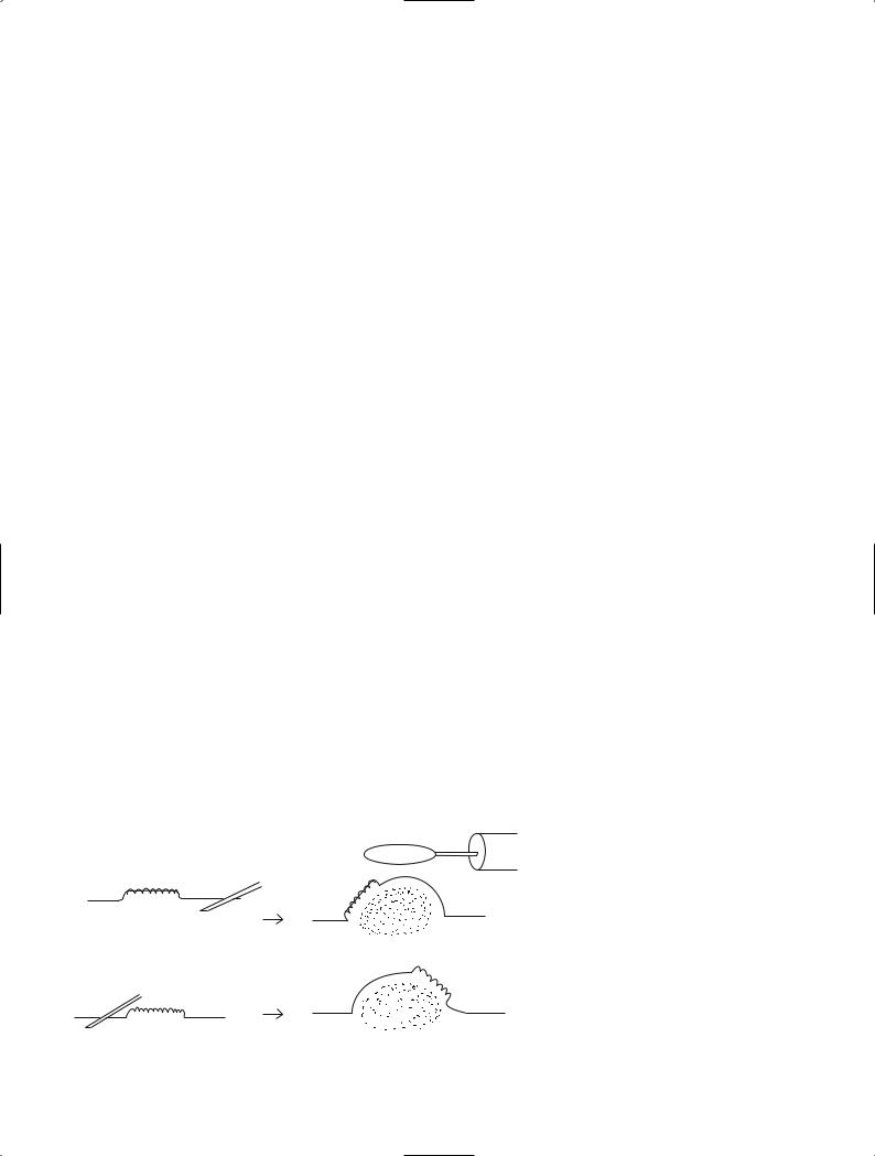
During local injection, the needle must correctly penetrate to the submucosa. The needle tip should therefore approach the lesion laterally and be inserted with a snap. The recommended angle from the mucosal surface is 30°to 45°. Strong insertion of the needle tip from steeper angles relative to the mucosal surface can result in penetration of the submucosa and muscularis mucosae, as well as penetration of the serosa. Insertion of the needle beyond the intestinal wall results in loss of physiological saline solution, with no change on the mucosal side. When physiological saline solution is injected into the subserosa, a protrusion slowly develops, elevating the lesion. However, the color of the mucosa surrounding the lesion remains unchanged.
Correct penetration of the needle into the submucosa and injection of physiological saline causes changes in structure and color tone: a round protrusion develops near the needle tip and a transparent grayish-blue tone appears. By changing the position of the needle during injection, the lesion can be guided to the top of the protrusion. Insertion of the injection needle into the submucosa at the anal side (the side near the scope) of large lesions can cause the lesion to move towards the oral side of the protrusion (Fig. 4). Therefore, the needle should be inserted and physiological saline injected into the submucosa at the oral side of lesions scheduled to be treated by en bloc resection. If the lesion starts to shift to an undesirable site during injection, the needle should be removed and reinserted into an appropriate site.
The snare is used in a conventional manner. It is placed around the lesion, including a small margin of normal mucosa, and the lesion is strangled. The highfrequency current should employ cutting waves for resection of superficial lesions and a combination consisting primarily of cutting waves and including coagulation waves for hemispherical lesions. Strangulation should be stronger than that used with coagulation waves alone, because superficial lesions are supplied by fine blood vessels.
Irrespective of the procedure, bleeding can be effectively managed by clipping. Clips should therefore be available during treatment.
All resected lesions should be retrieved. Superficial lesions should be spread out on cork or rubber plates, pinned in place, and fixed in 10% formalin. Lesions placed directly in formalin curl up, precluding preparation of complete specimens. This can lead to incorrect evaluation of the resection margin or the response to treatment.
3.3 Piecemeal Resection
In principle, lesions should be resected en bloc. To permit non-touch isolation, lesions must not be divided
NO
OK |
Fig. 4. Suitable site of injection for |
EMR
3. Management of Colorectal Cancer by “Hot Biopsy” and Snare Resection |
205 |
|
Fig. 5. Planned piecemeal resection and |
2nd |
|
suitable orders |
|
|
3rd
2nd
1st
1st
OK |
NO |
: suspicious of deeper invasion
unless absolutely necessary. Tissue retrieved after piecemeal resection performed in a single day usually cannot be treated similarly to specimens obtained by en bloc resection. Piecemeal resection carries an increased risk of error when evaluating tumor spread or properties of the tumor front. During resection of large lesions, the snare may not be able to be removed once the lesion is ensnared, even after the endoscopist realizes that complete en bloc resection is not feasible. These events may necessitate piecemeal resection. Some lesions considered resectable en bloc may be found to be larger than expected after application of current. Undetected parts of lesions can thus lead to remnant tumor. After retrieval, the lesion should be reconstructed as closely as possible. Pieces of the lesion should be pinned in place to maintain their configuration and then fixed in formalin.
Some large lesions undergo piecemeal resection by design. The procedure is similar to that described above. When using a snare, caution is required to avoid division of the lesion along the middle of the tumor (Fig. 5). Large nodules, flat regions differing from the surrounding tumor area, and central depressions are strongly associated with the tumor front. These findings should be included in a single specimen whenever possible.
Piecemeal resection involves the repeated application of high-frequency current to nearly the same site, and large amounts of tissue are present in the snare loop. Resection therefore requires large amounts of heat, even when only cutting current is very strongly concentrated on the target lesion. Consequently, local injection is essential, and the lesion must be drawn into the center of the lumen to adequately separate it from the muscularis propria.
Heat-induced perforation may occur 3–4 days after treatment. Patients must therefore rest after the procedure. Particular caution is required after piecemeal resection.
References
1.Williams CB (1973) Diathermy biopsy, a technique for endoscopic management of small polyps. Endoscopy 5: 215–218
2.Sakai Y (1992) Japanese classification of colorectal carcinoma and tentative classification by Japan Gastrointestinal Endoscopy Society (in Japanese). MB Gastro 9:95–100
3.Takekoshi T, Fujii A, Takagi K, et al (1988) Indication for endoscopic double snare polypectomy of gastric lesions (in Japanese with English abstract). Stomach Intest 23: 387–398
4.Watanabe S, Kishi H, Katagiri K, et al (1990) Endoscopic resection by double colonoscope method for giant colonic polyps (in Japanese with English abstract). Endosc Forum 6:274–278
5.Williams CB (1997) Personal communication
6.Waye JD (2000) Colonoscopic polypectomy. Diagn Ther Endosc 6:111–124
7.Deyhle P, Largiader F, Jenny S, et al (1973) A method for endoscopic electroresection of sessile colonic polyp. Endoscopy 5:38–40
8.Rosenberg N (1955) Submucosal saline wheal as safety factor in fulguration of rectal and sigmoidal polyp. Arch Surg 70:120–122
9.Japanese Society for Cancer of the Colon and Rectum (1997) Japanese classification of colorectal carcinoma. Kanehara, Tokyo