


5. New Trends in Endoscopic Ultrasonography
KENJIRO YASUDA
1. Introduction
Gastrointestinal carcinoma has been detected and diagnosed by barium meal X-ray study and endoscopic study with or without biopsy study. In particular, early and minute carcinoma can be detected only by endoscopic study. Due to the development and refinement of endoscopy, the diagnosis of early gastrointestinal (GI) carcinoma is now easily accomplished by endoscopic observation and biopsy study. Endoscopic detection of gastrointestinal lesions depends on the recognition of visible mucosal changes. However, the final diagnosis is performed by histopathological study of biopsy materials. Biopsy study is still very important to obtain the correct diagnosis of the lesion as carcinoma, dysplasia, adenoma, and hyperplasia, although it is sometimes possible to diagnose the lesion from the endoscopic investigation of mucosal surface details. In this chapter, endoscopic diagnosis of early carcinoma in the upper GI tract will be described.
2. How to Detect Early GI Carcinoma
Careful observation by endoscopy is important for detecting small lesions. When we focus on small lesions, detecting an abnormal area and performing a biopsy can be effectively carried out. This is easy to say theoretically, but in practice we need to learn the endoscopic observation skills and be familiar with small lesions.
For detection of early carcinoma of the upper GI tract, endoscopists have to learn to recognize early cancer lesions, which are observed with characteristic color changes and an irregular mucosal pattern.
It is useful to employ a dye-spraying method using iodine for esophageal carcinoma and indigocarmine for gastric and colorectal carcinoma. This is a valuable complementary technique but cannot directly detect gastric or colorectal lesions.
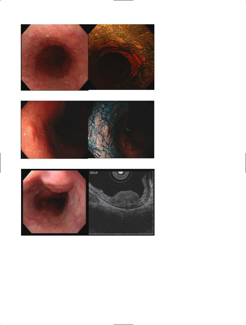
Figure 1 shows the early stage of esophageal carcinoma limited to the epithelium with and without iodine spray. The area of the lesion can be clearly detected after dye-spraying. Figure 2 shows a gastric carcinoma limited to the mucosa after spraying with
indigocarmine. The shape and irregularity of the lesion are clearly demonstrated.
3. Is Endoscopic Ultrasonography
(EUS) Effective?
Endoscopic ultrasonographic diagnosis of early GI tract malignancy is one of the recent clinical topics of importance. We can demonstrate the cross section of the GI wall ultrasonographically by using EUS. Nowadays two types of EUS instruments are available.
One is the conventional ultrasound endoscope with a radial scan transducer at the tip of the endoscope, while the other is an ultrasound catheter probe with a small radial scan transducer at the tip, which can be used through the working channel of the endoscope. The gastrointestinal wall can be delineated as five or more layered structures in the water-expanded GI lumen, which correspond well with histological layers. Higher frequency ultrasound scanners such as 20–30 MHz transducers can delineate a more precise picture of the GI wall.
The role of EUS is to evaluate the alteration of the GI wall by carcinoma based on the layered structure of GI wall, but we cannot detect the lesion by EUS except in the rare case of early stage of scirrhous carcinoma. The capability of EUS is to diagnose the depth of carcinoma invasion, which is an important factor in choosing the preferred treatment such as endoscopic resection (ER), laparoscopic surgery, or laparotomy. The diagnostic accuracy of depth of carcinoma invasion is around 80%, when we divide the lesions into mucosal
(m) carcinoma, submucosal (sm) carcinoma, carcinoma invading to the muscularis propria (mp), and deeper than the subserosal layer (ss) according to our criteria, which involve three hyperechoic layers of the GI wall.
The accuracy rate of diagnosis of a mucosal lesion, which is a good indication for endoscopic mucosectomy, is 90%. One of the most important diagnostic feats of EUS is to decide the indication for endoscopic treatment at the early stage of GI malignancy. Figure 3 shows EUS pictures of esophageal carcinoma limited to the submucosal layer obtained by an ultrasound probe with a 30 MHz transducer, and Figure 4 shows pictures of a gastric carcinoma limited to the mucosa demonstrated by a 20 MHz ultrasound probe.
181
182 IV. Detection of Early Cancer: Is Endoscopic Ultrasonography Effective
Fig. 1. Esophageal carcinoma limited to the epithelium with (A) and without (B) iodine spray. The extent of the lesion can be clearly detected after dye spraying (B)
A B
A
A
Recently, three-dimensional (3-D) reconstruction of EUS images obtained by a 3-D ultrasound probe has become possible. The significance of 3-D pictures is not only to make the image more easily understood but also to avoid overlooking the lesion. In addition, the therapeutic effect can be evaluated by measuring the mass volume using this method. Figure 5 shows the radial and linear images of gastric carcinoma obtained by 3-D probe and reconstruction of the lesion.
Fig. 2A,B. Gastric carcinoma limited to the mucosa with indigocarmine spraying (B). The shape and irregularity of the lesion are clearly
Bdemonstrated
Fig. 3A,B. Endoscopic ultrasonography (EUS) pictures of esophageal carcinoma limited to the submucosal layer obtained by ultrasound probe
Bwith a 30 MHz transducer (B)
4.Magnifying Endoscopy
Through advances of technology, high-resolution and high-magnification endoscopy with both fiberoptic and video-imaging systems has been developed and improved. There are reports of the diagnostic ability of high-resolution and high-magnification endoscopes. However, it has not been easy to manipulate these endoscopes in ordinary clinical examinations, especially
5. New Trends in Endoscopic Ultrasonography |
183 |
Fig. 4A–D. Early gastric carcinoma type IIa+IIc. A Endoscopic finding showing the redness. B Close-up view of the lesion. C Indigocarmine spraying. D Endoscopic ultrasonography pictures of gastric carcinoma limited to the mucosa demonstrated by a 20 MHz ultrasound probe, showing the normal submucosal layer
A B
C D
B
A
Fig. 5A–C. Three-dimensional (3-D) image of gastric carcinoma demon-
strated |
by ultrasound |
probe. |
|
A Radial and linear |
image (dual |
||
planes) |
obtained by |
3-D |
probe. |
B Endoscopic view. C Surface con- |
|||
struction of 3-D EUS image |
C |
||

184 IV. Detection of Early Cancer: Is Endoscopic Ultrasonography Effective
in the upper GI tract, because of the difficulty of focusing due to the movement by respiration and cardiac contractions and the darker imaging view in magnification.
By using new techniques of high-resolution and highmagnification electronic endoscopes, how can we diagnose the GI lesions? High-magnification endoscopes have a long history. The first models, which had a fiberoptic endoscope system, were developed in the late 1960s and attempted to reach a histological diagnosis of the lesions without biopsy. However, handling of endoscopes had some limitations because of the dark visual field and difficulty of focusing.
Progress in the field of electronic endoscopy gives us the hope that high-resolution and high-magnification endoscopes will be developed that are more easily manipulated. The most advanced video endoscope for the upper tract (GF-Q240Z), which can demonstrate magnified images up to 80¥, can be used in routine study although the focusing of this model is not so easy at maximum magnification. Nevertheless, this model can provide higher-resolution pictures, easier handling, and satisfactory brightness compared with previous ones.
We can observe the surface mucosal pattern (pit pattern) and capillary structure by using highmagnification endoscopy. Based on the analysis of the mucosal pit pattern obtained by magnification, histo-
logical changes of carcinoma, dysplasia, and adenoma can be assessed. However, it is not easy to diagnose the histological changes from the magnified images. In addition, we cannot observe the whole GI wall on the magnified image, even when we have some criteria on magnification image diagnosis. The role of high-magni- fication endoscopy is thus to magnify the target area when conventional endoscopy has detected some abnormality in ordinary pictures.
Figure 6 shows an example of high-magnification images of an advanced carcinoma in the prepyloric region. Gastric areas can be seen in the normal mucosa; on the other hand, irregular, wider areas and a nonstructured surface are observed in the carcinoma (Fig. 6C,D).
Figure 7 shows magnification images of a type IIc, depressed gastric mucosal carcinoma at the greater curvature of the gastric antrum. Magnification images delineate the mucosal lesion with irregularly sized and structured areas.
Figure 8 is a case of type IIa, slightly elevated gastric carcinoma limited to the mucosa at the posterior wall of the gastric body, showing the irregular pit pattern of the mucosal surface.
By gathering many cases and analyzing the images, it should be possible for magnifying endoscopy to help in the assessment of the histological diagnosis.
A B
|
Fig. 6A–D. Advanced gastric carci- |
|
noma at the posterior wall of the |
|
prepyloric region. A Ordinary endo- |
|
scopic picture of the lesion. B Dye- |
|
spraying image of the lesion. C,D |
C |
Magnification picture of the border of |
D benign and malignant areas |

5. New Trends in Endoscopic Ultrasonography |
185 |
Fig. 7A–D. Gastric mucosal carcinoma (type IIc) in the gastric antrum. A Conventional endoscopy picture of the lesion. B Close-up view at the site of the cancer showing the irregular areas with dye spraying. C,D Magnification images of the malignant depression with dye, showing the irregular mucosal pattern
A
C
A
Fig. 8A–D. A case of type IIa, |
|
slightly elevated type of gastric carci- |
|
noma limited to the mucosa. A Ordi- |
|
nary endoscopic view. B With dye |
|
spraying. C,D Magnification images |
|
of the lesion showing the irregular pit |
C |
pattern of the mucosal surface |
B
D
B
D

186 IV. Detection of Early Cancer: Is Endoscopic Ultrasonography Effective
5. Endoscopic Optical
Coherence Tomography
Optical coherence tomography (OCT) is a recently developed technique for demonstrating the crosssectional images with 10-times higher resolution than that of a 30 MHz ultrasound catheter probe. This system demonstrates the images by using broad-bandwidth illumination and recording the reflection of the illumination. We can observe the microscopic structure of tissues by this method, but the depth of imaging penetration is limited.
We have started to evaluate the clinical application of OCT using a prototype of the OCT probe made by Olympus (Japan) as of August 2000. This probe, which has the same aspect and same view angle of 360°as the ultrasound probe, can be used through the working channel of an ordinary endoscope, so we call this method endoscopic optical coherence tomography (EOCT). For EOCT scanning, water injection or balloon contact methods are not required, as the air does not obstruct the illumination beam.
Examined lesions were demonstrated with highresolution images, but with poor penetration. The depth of imaging penetration was 1.5–2.0 mm, but
A
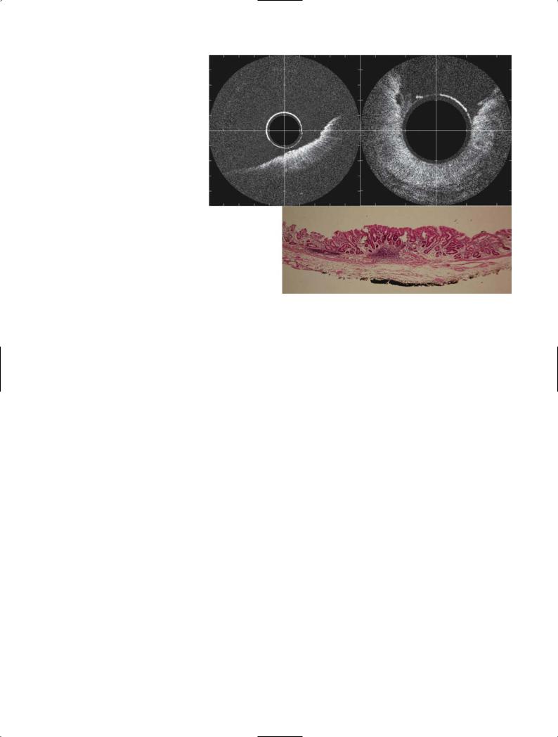
we were able to demonstrate the mucosal glandular structure, lamina propria, muscularis mucosae, and part of the submucosa individually. The esophageal wall can be demonstrated as a layered structure. From the surface, a low-reflective homogeneous layer and a high-reflective layer can be seen, behind which is a black layer that is thought to be mucosal muscle and an irregular low-reflective layer of the submucosa. Figure 9 shows an EOCT image of esophageal carcinoma, which demonstrates the thickening of the wall in good correspondence with resected findings. However, the differentiation of normal mucosa and cancerous mucosa was difficult.
The gastric wall is also observed as a layered structure, which is different from that of the esophagus. The surface layer shows the gland structure, behind which are three layers, high-, low-, and high-reflective layers, which are thought to be the lamina propria (high reflectivity), the mucosal muscle (low reflectivity), and the interface layer of the submucosal layer (high reflectivity). We expected OCT to demonstrate the gland structures by which we can differentiate between normal and malignant mucosa. Figure 10 shows the OCT images and mucosectomy specimen of a gastric carcinoma. However, histological diagnosis from OCT images seemed difficult, as we could not analyze the OCT
B
Fig. 9A–C. Endoscopic |
optical |
coherence tomography |
image of |
esophageal carcinoma limited to the mucosa. A Cross-sectional image of normal esophageal wall. B Cross-sec- tional image of the cancerous region showing the thickening of the wall, in good correspondence with resection findings. C Cross-sectional view of
Cresected specimen
5. New Trends in Endoscopic Ultrasonography |
187 |
Fig. 10A–C. Endoscopic optical coherence tomography (EOCT) image of gastric carcinoma limited to the mucosa. A,B EOCT findings of the gastric mucosal carcinoma showing the gland-like structure. C Endoscopic resection specimen showing the well-differentiated adenocarcinoma limited to the mucosa
A B
C
images of the cancerous region. Further investigation should be performed in this field.
Although the resolution was much higher than that of a 30 MHz ultrasound scanner, penetration of EOCT was too poor to use this method for assessing the depth of tumor invasion. However, by using this particular sophisticated instrument, we can expect to determine the histological nature of tissues in the near future. Endoscopic optical coherence tomography is expected to be a method of optical biopsy study in endoscopic examination in the future.
6. Diagnosis of Early GI Tract
Carcinoma in the Future
Presently, the diagnosis of early GI tract carcinoma is performed by endoscopic examination. Universal criteria cannot be established, because the ability to detect and diagnose is dependent on personal capability and experience. Other techniques such as blood tests or gastric juice tests, or gene analysis, might harbor the possibility to detect small GI tract carcinomas, but there are probably some difficulties involved in these diagnostic methods, as we will be unable to know the position of the lesions unless there is direct visualization of the GI lumen.
In the future, use of new EOCT instruments for detecting early and small lesions should become routine. The progress of information technology is so fast and beyond our expectation, so there is hope for detection and differentiation of GI lesions by autodiagnostic endoscopy. We are hopeful that therapeutic procedures for early GI tract carcinoma will be further improved for endoscopists.
References
1. Yasuda K, Nakajima M, Kawai K (1987) Fundamentals of endoscopic laser therapy (ELT) for GI tumors; new aspects with endoscopic ultrasonography (EUS). Endoscopy 19:s2–s6
2.Tio TL, Cohen P, Coene PP, et al (1989) Endosonography and computed tomography of esophageal carcinoma. Gastroenterology 96:1478–1486
3.Yasuda K, Nakajima M, Kawai K (1992) Endoscopic diagnosis and treatment of early gastric cancer using endoscopic ultrasonography (EUS). Gastrointest Endosc Clin North Am 2:495–507
4.Van Dam J (1994) Endosonography of the esophagus. Gastrointest Endosc Clin North Am 4(4):803–826
5.Rosch T (1995) Endosonographic staging of esophageal cancer: a review of literature results. Gastrointest Endosc Clin North Am 5:537–547

188 IV. Detection of Early Cancer: Is Endoscopic Ultrasonography Effective
6.Kida M, Tanabe M, Watanabe M, et al (1998) Staging of gastric cancer with endoscopic ultrasonography and endoscopic mucosal resection endoscopy. Dig Endosc 30(suppl):A64–A68
7.Yasuda K (2000) Gastrointestinal carcinoma. In: The handbook of endoscopic ultrasonography in digestive tract. Blackwell Science, Japan, pp 54–69
8.Penman ID, Shen EF (2002) EUS in advanced esophageal cancer. Gastrointest Endosc 56:s2–s6
9.Yasuda K (2002) EUS in the detection of early gastric cancer. Gastrointest Endosc 56:s68–s75
10.Yasuda K, Kamaguchi M, Morikawa J, et al (2005) Role of endoscopic ultrasonograhy (EUS) in the diagnosis of early esophageal carcinoma. Gastrointest Endosc Clin North Am 15:93–99