

Reference |
129 |
Differential Diagnosis: Since this does not appear like mass and is lobulated and widely distributed, bleeding is first on the differential.
Diagnosis: Aortic rupture.
Reference
1. Zylak CM, Standen JR, Barnes GR, Zylak CJ. Pneumomediastinum revisited. Radiographics. 2000;20(4):1043–57.
Chapter 7
Abnormal Bones, Soft Tissue,
and Other Findings
There are times when pathological processes occur outside the lungs and mediastinum, i.e., in the chest wall, to include bones and soft tissues. This is why we include these structures on the search pattern, in that one must think about those structures separately to deduce their potential involvement. I will also include a short synopsis of lines and tubes often seen in the ICU; they are easier to navigate than most think.
Mechanism: An abnormal process of the bones or soft tissues seen on the CXR, excluding lungs, mediastinum, and vascular structures already presented.
Case 7.1
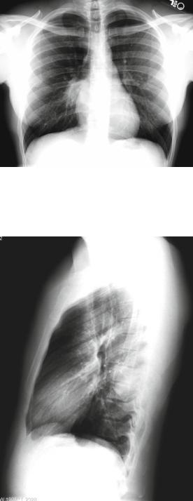
The example case Fig. 7.1a-f highlights the thought process to include those processes that occur outside the lungs. This is the case of a 24-year-old male US Marine with posterior chest wall/rib pain for 4–6 months, increased with deep inspiration. This case will also point out a few signs and fundamentals presented earlier in the book. This case is reprinted with permission from: Warnock et al. (2008) [1].
Findings: Poorly marginated opacity overlying right hila. On the lateral CXR, a partially marginated opacity is noted posteriorly.
Location/Pattern: The posterior location raises possibilities such as pleural based (since the obtuse angles posteriorly) or bone origin.
Differential Diagnosis: Because of the location and characteristics, it is best to approach this CXR by analyzing it based on broad possibilities: posterior lung mass (or focal consolidation), pleural-based mass or process, or bone process.
Posterior lung mass would include the lung mass pattern and posterior mediastinal differential.
L.R. Folio, Chest Imaging, DOI 10.1007/978-1-4614-1317-2_7, |
131 |
© Henry M. Jackson Foundation for the Advancement of Military Medicine, Inc. 2012 |
|
132 |
7 Abnormal Bones, Soft Tissue, and Other Findings |
Fig. 7.1a |
PA CXR |
demonstrating an ill-defined right hilar mass. Note the hilar overlay sign and boney destruction (see also Fig. 7.1c)
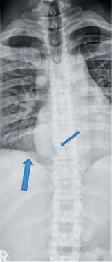
Fig. 7.1b Lateral CXR showing a positive spine sign/opacity posteriorly with obtuse angled margins superiorly and inferiorly
Case 7.1 |
133 |
Fig. 7.1c Thoracic spine plain radiograph better demonstrating bone destruction. Note the absence of medial right 9th rib (wide arrow) and moth-eaten T9 vertebral body on right (thin arrow)
Pleural based masses
•Solitary pleural density
•Loculated pleural effusion
•Mesothelioma
•Metastases
•Splenosis
Bone process
•Benign primary bone tumors include the following:
–Chondroblastoma
–Chondromyxoid fibroma
–Osteochondroma
–Giant cell
–Enchondroma
–Fibrodysplasia
•Malignant primary bone tumors include the following:
–Chondrosarcoma
–Osteoblastoma, aggressive variant
–Osteosarcoma
134 |
7 Abnormal Bones, Soft Tissue, and Other Findings |
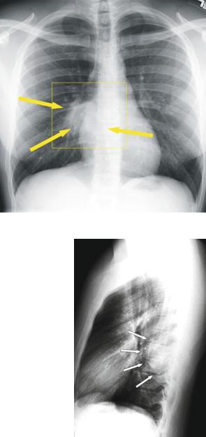
Fig. 7.1d PA CXR with arrows pointing to the mass
Fig. 7.1e Lateral CXR with arrows pointing to the mass
Diagnosis: This turned out to be a chondroblastoma with secondary aneurysmal bone cyst. Although this differential diagnosis is unusual, the point here is to use search pattern considerations of structures and regions other than lung and mediastinum.
Figure 7.1d–f show a large, ill-defined, lobulated mass located in the right paraspinal region in this patient.