

44 |
4 Abnormal Lung Patterns |
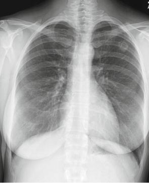
Fig. 4.13 CXR of infectious granulomatous disease
Infectious Granulomatous Disease
Infectious granulomatous diseases are frequently identified as nodules (or mass) on a CXR. This can happen with the following diseases.
•Tuberculosis
•Atypical mycobacterial diseases – especially Mycobacterium Avium-Intracellular (MAI)
•Fungal diseases
–Coccidioidomycosis
–Blastomycosis (North American and South American)
–Cryptococcosis
–Sporotrichosis
•Bacterial diseases, nocardiosis and/or actinomycosis
Case 4.4
The following case (Figs. 4.13 – 4.15) presents a 28-year-old female who had a CXR to monitor a preexisting lesion, though she was asymptomatic at the time of the CXR. She lived in central California.
Mass |
45 |
Fig. 4.14 Lateral CXR of infectious granulomatous disease
Findings: Opacity with central lucency in Left Upper Lobe posteriorly. Pattern: Mass, cavitary.
Differential Diagnosis
Since cavitary, consider the CAVITARY mnemonic differential. Wegener’s granulomatosis can appear just like this.
The standard mass differential:
•Malignancy
•Granulomatous
•Inflammation
•Benign neoplasm
•Congenital
Diagnosis: Coccidiomycosis.
In this case, history helped narrow the diagnosis further, as the patient grew up in the San Joaquin River valley: Coccidiomycosis is also known as San Joaquin Valley Fever.
Signs: Fever, cervical adenopathy, skin lesions, pleural effusion, friction rub, pulmonary rales.