

6 Ultrasound
Contents
Gallbladder and bile ducts 465
Liver 471
Hepatic doppler 477
Pancreas 483
Spleen 485
Kidneys 486
Scrotum and testicles 493
Vascular ultrasound 499
Thyroid and parathyroid 504
Uterus 507
Ovaries and adnexa 514
First trimester pregnancy 519
Second and third trimesters 531
464

Gallbladder and bile ducts
Gallstones and cholecystitis
Cholelithiasis
Single gallstone: Sagittal ultrasound of the gallbladder shows an echogenic gallstone (calipers) in the gallbladder neck, with posterior acoustic shadowing (arrows).
Multiple small gallstones: Sagittal ultrasound of the gallbladder in a different patient shows multiple small shadowing gallstones (arrows).
•Cholelithiasis is the presence of a gallbladder stone or stones, without associated inflammation.
•The classic clinical presentation of symptomatic cholelithiasis is colicky pain after eating a fatty meal, but it is common to see gallstones incidentally in asymptomatic patients.
•Risk factors for developing gallstones include female sex, obesity, pregnancy, middle age, and diabetes.
•The ultrasound diagnosis of gallstones is usually straightforward. Stones are echogenic with posterior acoustic shadowing and are often mobile. It is often helpful to reposition the patient (typically in the left lateral decubitus position) while scanning to assess whether the stones layer dependently to differentiate stones from polyps or other masses.
•A gallbladder completely filled with stones can be more challenging to identify. The wall-echo-shadow (WES) sign describes the appearance of a gallbladder full of multiple stones (or one giant stone).
Two parallel echogenic arcs represent the gallbladder wall and leading edge of the stone, with an intervening thin layer of hypoechoic bile. The gallstone typically casts a prominent shadow.
•The differential diagnosis of echogenic material within the gallbladder includes:
Gallstone(s) (mobile, shadowing). Gallbladder sludge (mobile, non-shadowing).
Gallbladder polyp (non-mobile, non-shadowing, often attached to the gallbladder wall via a stalk, may be vascular).
Hyperplastic cholecystoses (non-mobile, multiple polyps).
465

Acute calculous cholecystitis
Acute cholecystitis: Oblique sagittal ultrasound through the gallbladder demonstrates a thickened, echogenic gallbladder wall (arrows). The gallbladder contains numerous echogenic gallstones (red arrow).
•Acutecholecystitisisinflammationofthegallbladder,usuallyduetoagallstoneimpacting thecysticduct.Ultrasoundisthefirst-lineevaluationofsuspectedacutecholecystitis.
•Acute cholecystitis clinically presents with right upper quadrant (RUQ) pain and fever.
•Thereisno100%specificultrasoundfindingforacutecholecystitis.However,gallstonesare seen>90%ofthetimeandapositivesonographicMurphy’ssign(RUQpainwithpressure fromthetransducer)alsohasahighpositivepredictivevalue.Otherfindingsinclude:
Gallbladder wall thickening >3 mm.
Distended gallbladder >4 cm in diameter.
Pericholecystic fluid.
Color Doppler showing hyperemic gallbladder wall.
Hyperechoic fat in the gallbladder fossa (ultrasound correlate to CT finding of fat stranding).
•Complications of acute cholecystitis are rare but serious.
Emphysematouscholecystitisisgasinthegallbladderwallandhasahighriskofgallbladderperforation.
Gangrenous cholecystitis is necrosis of the gallbladder wall. Sonographic findings include layering echogenic material in the gallbladder lumen representing hemorrhage and sloughed membranes.
Gallbladder perforation appears as focal discontinuity of the gallbladder wall. Perihepatic ascites containing dirty echoes is often present.
•Surgical treatment of uncomplicated acute calculous cholecystitis is cholecystectomy. In patients who are not good surgical candidates, a temporizing percutaneous cholecystostomy tube can be placed prior to definitive surgical cholecystectomy.
Acalculous cholecystitis
•Acalculous cholecystitis is cholecystitis without gallstones, typically seen in very sick patients. Risk factors include sepsis, prolonged total parenteral nutrition, and trauma.
•The ultrasound appearance is similar to acute cholecystitis but without stones. Since many patients are ventilated or obtunded, it’s often not possible to evaluate for sonographic Murphy’s sign.
•Treatment of acalculous cholecystitis is typically interventional radiology percutaneous cholecystostomy. Unlike the treatment of calculous cholecystitis, cholecystostomy is often the definitive therapy.
Emphysematous cholecystitis
•Emphysematous cholecystitis is a rapidly progressive form of acute cholecystitis characterized by gas in the gallbladder wall. Emphysematous cholecystitis is associated with gallbladder ischemia causing bacterial translocation. Treatment is urgent surgery.
•On ultrasound, gas is usually present in both the gallbladder lumen and wall, which appears as echogenic lines and foci with posterior dirty shadowing.
466

Porcelain gallbladder
•A porcelain gallbladder is a calcified gallbladder wall due to either chronic irritation from supersaturated bile or repeated bouts of gallbladder obstruction.
•Porcelaingallbladderisassociatedwithanincreasedriskofgallbladdercancer,butthe incidenceiscontroversial.Ingeneral,prophylacticcholecystectomyisthestandardofcare.
•On ultrasound, the wall of the gallbladder is echogenic, and there are almost always associated gallstones.
•Thedifferentialdiagnosisofanechogenicgallbladderwallincludesaporcelain gallbladder,agallbladderpackedfullofstones(whichwillfeaturethewall-echo-shadow sign),oremphysematouscholecystitis(intramuralgaswillhavedirtyshadowing).
Courvoisier gallbladder
Courvoisier gallbladder: Sagittal ultrasound of the gallbladder (left image, marked with calipers) demonstrates a massively distended gallbladder. The common bile duct (right image, indicated by calipers) is also distended due to chronic malignant obstruction.
Case courtesy Julie Ritner, MD, Brigham and Women’s Hospital.
•TheCourvoisiergallbladderreferstoamarkedlydilatedgallbladder(originallydescribed asbeingsolargeastobedirectlypalpable)frommalignantobstructionofthecommon bileduct.
•A markedly distended gallbladder implies chronic obstruction of either the cystic duct (when seen in isolation) or the common bile duct (when seen in combination with dilation of the common bile duct and intrahepatic biliary dilation).
Hyperplastic cholecystoses
Overview of hyperplastic cholecystoses
•Thehyperplasticcholecystosesareaspectrumofnon-neoplasticproliferativedisorders causedbydepositionofcholesterol-ladenmacrophageswithinthewallofthegallbladder. Thecholecystosesrangefromabnormalitiesofthegallbladderwall(adenomyomatosis andstrawberrygallbladder)togallbladderpolypsextendingintothelumen.
Adenomyomatosis
•Adenomyomatosis is cholesterol deposition in mural Rokitansky–Aschoff sinuses.
It is important not to confuse with adenomyosis of the uterus: It may be helpful to remember that there are three L’s in gallbladder, and adenomyomatosis is a longer word than adenomyosis.
•Theultrasoundhallmarkofadenomyomatosisisthecomet-tail artifactduetoreflections offoftinycrystalsseeninafocallythickenedandechogenicgallbladderwall.
Strawberry gallbladder (cholesterolosis of the gallbladder)
•Strawberry gallbladder is a pathologic diagnosis that is not apparent by imaging. It is characterized by tiny mural cholesterol deposits likened to strawberry seeds.
467

Gallbladder polyps
•Most gallbladder polyps are benign cholesterol polyps that are part of the hyperplastic cholecystosis spectrum. Rarely (<5%), polyps may be premalignant adenomas.
•Clinically,gallbladderpolypsmaycauserightupperquadrant painorevencholecystitisifthecysticductisobstructed.
•The following characteristics, known as the six S’s, increase the risk for a polyp being malignant:
Size >10 mm or rapid growth. As a caveat, ultrasound has limited sensitivity and specificity in detecting small polyps (<10 mm), especially in the presence of gallstones.
Single: A solitary polyp is more suspicious for malignancy. In contrast, benign cholesterol polyps tend to be multiple.
Sessile (broad-based): Sessile morphology is suspicious. A polyp is more likely benign if pedunculated.
Stones: The presence of stones may induce chronic inflammation, which can predispose towards malignancy.
Primary Sclerosing cholangitis increases risk of malignancy. Sixty (age) or greater.
•In patients with several of these high-risk features, cholecystectomy should be considered in the presence of a polyp greater than 6 mm in size.
•The typical ultrasound appearance of a polyp is a nonmobile, non-shadowing polypoid lesion extending from the wall into the lumen of the gallbladder. There may be vascular flow in the stalk.
Gallbladder polyp:
Sagittal and transverse views of the gallbladder show
a small non-shadowing echogenic lesion (arrows). After repositioning the patient, this was shown to be nonmobile.
•Themaindifferentialconsiderationisadherentsludge,whichwillnothaveanyvascularflow.
Gallbladder cancer
Primary gallbladder carcinoma
•Gallbladder cancer is a rare malignancy with a poor prognosis. A typical clinical presentation may include right upper quadrant pain, weight loss, and jaundice.
•Risk factors for development of gallbladder cancer include:
Gallstones and chronic cholecystitis.
Porcelain gallbladder (somewhat controversial).
Primary sclerosing cholangitis.
Inflammatory bowel disease (ulcerative colitis more frequently than Crohn disease).
Adenomatous polyp >10 mm or >6 mm with multiple risk factors, as described above.
•Ultrasound shows a polypoid mass with increased vascularity in the gallbladder. There is often direct invasion into the liver.
Regional adenopathy occurs early.
Bile duct obstruction may be present.
Gallbladder metastases
•Metastases to the gallbladder are uncommon.
•Hepatocellular carcinoma can spread directly to the gallbladder through the bile ducts.
•Melanoma can spread hematogenously to the gallbladder mucosa.
468

Gallbladder: Common imaging patterns
Diffuse gallbladder wall thickening >3 mm (common causes in bold)
•Fluid-overload/edematous states:
Cirrhosis: Hypoalbuminemia leads to diffuse gallbladder wall thickening.
Congestive heart failure.
Protein-wasting nephropathy.
•Inflammatory/infectious:
Cholecystitis, usually with associated cholelithiasis.
Hepatitis.
Pancreatitis.
Diverticulitis.
•Infiltrative neoplastic disease:
Gallbladder carcinoma.
Metastases to gallbladder (rare).
•Post-prandial state.
Sagittal ultrasound of the gallbladder shows diffuse wall thickening to 8 mm (calipers). In this case, the wall thickening was due to cirrhosis and resultant hypoproteinemia.
Focal gallbladder wall thickening (common causes in bold)
•Hyperplastic cholecystoses: Adenomyomatosis and cholesterol polyp.
•Vascular: Varices.
Gallbladder varices due to portal hypertension: Sagittal grayscale ultrasound of the gallbladder (left image) demonstrates several hypoechoic, cystic-appearing structures within the gallbladder wall (arrows). Color Doppler (right image) confirms the vascular etiology.
Case courtesy Julie Ritner, MD, Brigham and Women’s Hospital.
•Neoplastic disease:
Adenomatous polyp.
Gallbladder carcinoma.
Adjacent hepatic tumor.
Non-shadowing “mass” in the gallbladder lumen
•Tumefactive sludge (mobile).
•Blood/pus (mobile).
•Gallbladder polyp (immobile).
•Gallbladder carcinoma (immobile).
Echogenic gallbladder wall
•Porcelain gallbladder.
•Gallbladder full of stones (signified by the wall-echo- shadow sign).
•Emphysematous cholecystitis.
469

Bile ducts
Bile duct anatomy
Sagittal view of the normal porta hepatis on CT and ultrasound:
ant post
|
sagittal CT |
|
|
|
common bile duct |
right |
common bile duct |
||
|
|
|
||
right |
|
|
hepatic |
|
|
|
|
||
|
|
artery |
|
|
hepatic |
|
|
|
|
artery |
|
|
|
|
|
|
|
|
|
|
|
|
|
|
(obscured) |
|
|
|
|
|
|
duodenum |
|
|
portal vein |
|
|
|
portal vein |
|
|
|
|
|
head |
|
feet |
head |
feet |
Choledocholithiasis
Choledocholithiasis: Sagittal ultrasound (left image) of the porta hepatis (in the same orientation as the reference anatomic image above) demonstrates common bile duct dilation (calipers) to 1.1 cm. Transverse scan (right image) through the region of the head of the pancreas shows an echogenic gallstone within the distal common bile duct (arrow).
Case courtesy Julie Ritner, MD, Brigham and Women’s Hospital
Choledocholithiasis is a stone in the common bile duct, generally treated with ERCP.
Mirizzi syndrome
Mirizzi syndrome is seen when a stone in the cystic duct causes inflammation and external compression of the adjacent common hepatic duct (CHD).
Essential for the surgeon to know about preoperatively because the CHD may be mistakenly ligated instead of the cystic duct. Additionally, inflammation can cause the gallstone to erode into the CHD and cause a cysto-choledochal fistula and biliary obstruction.
On ultrasound, a stone is typically impacted in the distal cystic duct, and the CHD is dilated. The cystic duct tends to run in parallel with the CHD
Pneumobilia
Pneumobilia is air in the biliary tree. It is commonly seen after biliary interventions, but may be due to cholecystoenteric fistula or rarely emphysematous cholecystitis.
On ultrasound, small echogenic gas bubbles are seen centrally in the liver with posterior dirty shadowing.
n contrast to pneumobilia, portal venous gas (which implies bowel ischemia until proven otherwise) is peripheral and causes a spiky appearance of the portal vein spectral Doppler waveform.
470

Cholangiocarcinoma
•Cholangiocarcinoma is cancer of the bile ducts. It classically presents with painless jaundice.Most cases of cholangiocarcinoma are sporadic, although key risk factors include chronic biliary disease (in the US) and liver fluke infection (in the Far East).
•The hilum is the most common location of cholangiocarcinoma. A hilar cholangiocarcinoma is known as a Klatskin tumor. Intrahepatic cholangiocarcinoma occurs uncommonly (10%).
•Ultrasoundplaysaroleintheinitialevaluationofadjacentadenopathyandvascular structures.Localnodesincludeportahepatisandhepatoduodenalligamentnodes.If moredistalnodaldiseaseispresent,thenthetumorisgenerallyconsideredunresectable.
Biliary ductal dilation
•A rule of thumb for assessing the common bile duct diameter (CBD) is to assume that the CBD ought to be 6 mm or less before age 60, but may still be normal if 1 mm larger per decade after that age. For example, an 8 mm duct in an 80-year-old patient may be considered normal.
Some sources, however, suggest very small differences with age (mean duct diameter of 3.6 mm for 60-year-old patients and 4.0 mm for 85-year-old patients).
For the hepatic ducts, >2 mm in size or >40% of the adjacent portal vein diameter is abnormal.
•Thecommonbileductisapproximately1.6mmwider(onaverage)inpatientswhohave undergonecholecystectomy,comparedtopatientswhohavenothadacholecystectomy.
•In general, malignancy causes more prominent ductal dilation than benign disease.
Liver
Diffuse metabolic parenchymal liver disease
Hepatic steatosis
Normal liver: Ultrasound of the liver and kidney shows the normal isoechoic appearance of liver relative to renal cortex.
Hepatic steatosis: Ultrasound in a different patient shows diffusely increased echogenicity of the liver when compared to the renal cortex.
•Hepaticsteatosisistheaccumulationofexcessfatwithinhepatocytesduetoametabolic derangement(obesityordiabetes),hepatotoxins(EtOH),orprolongedfasting.
•Ultrasound shows a diffuse increase in hepatic echogenicity. Normally, the liver and kidney should have the same echogenicity. With fatty infiltration, the liver appears more echogenic than the kidney. Hepatic steatosis also causes increased sound attenuation, leading to poor visualization of deeper structures.
•Focal fat sparing is a geographic area of hypoechogenicity in an otherwise fatty liver.
A characteristic location of focal fat sparing is the gallbladder fossa.
471

Cirrhosis
•Cirrhosis is the replacement of functioning hepatocytes with dysfunctional fibrotic tissue, due to long-standing repeated cycles of hepatocyte injury and repair.
•Micronodular cirrhosis causes cirrhotic nodules less than 3 mm in size and is most commonly associated with alcoholism.
•Macronodular cirrhosis features larger nodules (>3 mm) separated by wide scars and fibrous septae. Macronodular cirrhosis is caused by fulminant viral hepatitis which does not uniformly affect the liver.
•The typical ultrasound appearance of cirrhosis is a coarse, heterogeneously increased liver echotexture with a nodular external contour. In early cirrhosis, the superficial nodularity is best appreciated with a high-frequency linear probe. The caudate lobe is often spared and hypertrophies in response to increased demand (the caudate has direct venous drainage into the IVC and therefore can bypass the hypertensive portal system). End-stage cirrhosis is characterized by a shrunken, nodular liver.
•Signs of portal hypertension are often present, including an enlarged portal vein, splenomegaly, varices, portosystemic shunts, and a patent umbilical vein. Imaging of portal hypertension is discussed in detail in the liver Doppler section.
Liver infections
Viral hepatitis
Viral hepatitis: Sagittal image of the liver (left image) demonstrates increased echogenicity of the portal triads appearing as numerous echogenic dots (arrows) that produce a starry sky appearance. Sagittal view of the gallbladder in the same patient (right image) shows marked diffuse gallbladder wall thickening (calipers), which is commonly seen in acute hepatitis.
Case courtesy Julie Ritner, MD, Brigham and Women’s Hospital.
•Viral hepatitis is infection of the liver by a hepatotropic virus. Hepatitis B and C cause chronic disease.
•The most common ultrasound finding is a normal liver. Occasionally periportal edema produces the characteristic starry sky pattern of increased portal triad echogenicity.
•Acute hepatitis is often associated with diffuse severe gallbladder wall thickening.
Pyogenic abscess
•Pyogenic abscess is caused by pus-forming organisms and is usually due to spread from intestinal or biliary infection (most commonly E. coli).
•Infectionstartsasanill-definedareaofalteredechogenicity(phlegmonstage)that evolvesintoawell-definedhypoechoicstructurewithinternalechoes(matureabscess).
472
Amebic abscess
•Amebic abscess is caused by Entamoeba histolytica. A near-universal presenting symptom is pain, seen in 99% of patients. The most common location is near the dome of the right lobe.
•On ultrasound, an amebic abscess is indistinguishable from a pyogenic abscess and appears as a hypoechoic structure with low-level internal echoes.
•Antimicrobial therapy is usually sufficient treatment, and drainage is rarely necessary.
Echinococcal cyst (hydatid disease)
•Echinococcal cyst is caused by larvae of Echinococcus granulosus, most commonly found in endemic areas in the Middle East, Mediterranean, and South America.
•Thereisariskofanaphylaxiswithperitonealspillageofcystfluid,althoughtheseareoften biopsiedanddraineduneventfully.Medicaltreatmentisalbendazoleormebendazole.
•Classicultrasoundappearanceisalargelivercystwithnumerousperipheraldaughtercysts.
A highly suggestive finding is the change in position of daughter cysts as the patient is repositioned. The water-lily sign is an undulating membrane within the hydatid cyst.
Hydatid sand is a fine sediment caused by separation of the membranes from the endocyst.
Candidiasis
•Hepatic candidiasis is a rare infection in the immunocompromised due to Candida albicans or Candida glabrata.
•On imaging, there are multiple tiny targetoid lesions. The presence of concurrent similar-appearing lesions in the spleen is highly suggestive of hepatosplenic candidiasis.
Hepatic Pneumocystis jiroveci
•Hepatic Pneumocystis jiroveci is seen in disseminated disease in the severely immunocompromised. Hepatic infection is classically secondary to the use of inhaled pentamidine to treat Pneumocystis pneumonia, as pentamidine is not absorbed systemically and thus would not prevent hepatic infection.
•Ultrasoundshowsmultiplepunctateechogeniccalcificationsintheliverandoftenspleen.
Benign hepatic neoplasms (in order of frequency)
Cavernous hemangioma
Cavernous hemangioma: Transverse ultrasound through the right lobe of the liver demonstrates a circumscribed slightly heterogeneous echogenic mass
(calipers) with mild posterior acoustic enhancement.
Portal venous phase axial contrast-enhanced
CT demonstrates a hypoattenuating lesion with discontinuous peripheral nodular enhancement (arrows), typical of a hemangioma.
Case courtesy Julie Ritner, MD, Brigham and Women’s Hospital.
473
•Hepatic cavernous hemangioma is the most common benign hepatic neoplasm.
•The classic ultrasound appearance of hemangioma is a solitary, circumscribed, homogeneously echogenic mass with no flow on color Doppler. Posterior acoustic enhancement is nonspecific but may be present. When seen, posterior acoustic enhancement is thought to correlate with hypervascularity. A hypoechoic halo should never be seen – this finding suggests malignancy.
•Hepatic hemangioma can rarely have an atypical hypoechoic appearance when seen in a fatty liver.
•If a solitary, classic-appearing hemangioma is seen and the patient has an otherwise normal-appearing liver, normal LFTs, no known malignancy, and is asymptomatic, then no further workup is required.
•Any heterogeneity or atypical ultrasound findings should prompt consideration of an alternative diagnosis. The differential of a hyperechoic hepatic mass includes hyperechoic hepatocellular carcinoma or metastatic disease (even in the absence of a halo). In a patient with cirrhosis or any known primary malignancy, further workup (MRI or CT) is usually warranted if the mass is new, even if classic appearing.
Focal nodular hyperplasia (FNH)
•Focal nodular hyperplasia (FNH) is a benign hyperplastic hepatic mass with a central non-fibrotic stellate scar consisting of biliary ductules and venules.
•Ultrasound findings are nonspecific. The central scar is rarely seen on ultrasound, and even when it is, this finding can be seen in other lesions, including hepatocellular carcinoma, giant hemangioma, or adenoma.
•FNH is often difficult to detect on sonography. It may be nearly isoechoic to normal liver and manifest on imaging as a subtle displacement of the hepatic contour.
•Doppler findings of FNH include a spoke-wheel configuration of arterial vessels.
•MRI or Tc-99m sulfur colloid scintigraphy can confirm (classically, FNH has increased uptake of sulfur colloid). MRI is by far the more useful test.
Hepatic adenoma
•Hepatic adenoma is a benign liver tumor associated with oral contraceptives, anabolic steroids, and type I glycogen storage disease (von Gierke’s disease - in which case adenomas will be multiple).
•Due to high incidence of hemorrhage, adenomas are usually resected.
•Therearenospecificultrasoundfeaturestodistinguishanadenomafromotherhepatic masses.Anadenomamaybehyperechoic,isoechoic,orhypoechoicrelativetonormalliver.
•AdenomaisusuallyphotopeniconTc-99msulfurcolloidscintigraphy(incontrasttoFNH).
Hepatic lipoma
•Hepatic lipoma is a benign neoplasm composed of fat that appears as a welldefined hyperechoic mass. It may appear identical to hemangioma or hyperechoic hepatocellular carcinoma.
•When multiple, may be associated with tuberous sclerosis and renal angiomyolipomas.
Biliary cystadenoma
•Biliary cystadenoma is a benign cystic mass lined with biliary-type epithelium.
•Althoughbenign,mostaresurgicallyresectedsincemalignanttransformationmayoccur.
•Biliary cystadenoma appears as a multiseptated cystic mass on all imaging modalities. Mural nodules should be regarded with suspicion. The presence of mural nodularity suggests malignant transformation to cystadenocarcinoma.
474

Hepatic malignancy
Hepatic metastases
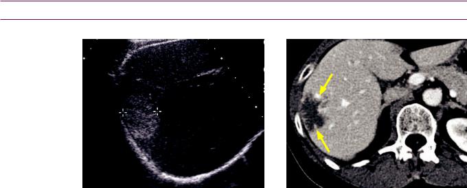
Innumerable liver metastases, initially difficult to see due to technique: Initial scanning with a lowfrequency vector probe (left image) demonstrates a coarsened hepatic echotexture without definite mass. This appearance may mimic cirrhosis. When a higher frequency curved probe is used (right image), innumerable target lesions (arrows) become apparent, consistent with innumerable hepatic metastases.
Case courtesy Julie Ritner, MD, Brigham and Women’s Hospital.
•Metastatic disease to the liver is far more common than primary hepatocellular carcinoma.
•Metastases can have a variable ultrasound appearance, although the classic finding is a hypoechoic rim producing a target sign.
•Hypoechoic hepatic metastases include:
Breast (can be either hypoechoic or hyperechoic).
Pancreas.
Lung.
Lymphoma.
•Hyperechoic hepatic metastases include:
Colon cancer is hyperechoic in greater than 50% of cases. A hyperechoic appearance may suggest a better prognosis.
Renal cell carcinoma.
Breast (can be either hyperechoic or hypoechoic). Carcinoid.
Choriocarcinoma.
•Calcified hepatic metastases (hyperechoic with acoustic shadowing) include:
Colon cancer (especially mucinous type).
Gastric adenocarcinoma.
Osteosarcoma (very rare).
•Cystic hepatic metastases include:
Ovarian cystadenocarcinoma.
Gastrointestinal sarcoma.
•Infiltrative metastases include:
Lung.
Breast. In particular, treated breast cancer may cause a pseudo-cirrhosis appearance.
Prostate.
475

Hepatocellular carcinoma (HCC)
•Hepatocellular carcinoma (HCC) is a hepatic malignancy arising in the setting of chronic inflammation.
•Patients with cirrhosis or chronic viral hepatitis are regularly screened for HCC with serum alpha-fetoprotein levels and ultrasound.
Ultrasound is not very sensitive to detect small HCC in end-stage cirrhotic livers.
•HCC has a variety of ultrasound appearances — therefore, a mass in a cirrhotic liver is considered HCC until proven otherwise.High Doppler flow may be present, especially at the periphery of the mass, due to arteriovenous shunting.
•HCC has a propensity for venous invasion. The portal veins should always be carefully evaluated in the presence of a hepatic mass. Internal Doppler flow within a venous clot suggests a tumor thrombus.
Fibrolamellar carcinoma
•Fibrolamellar carcinoma is a variant of HCC seen in young adults without cirrhosis and is not associated with elevated alpha-fetoprotein.
•Fibrolamellar carcinoma has a much better prognosis compared to typical HCC.
Hepatic lymphoma
•Primary hepatic lymphoma may present as a single mass or multiple masses.
•Lymphoma tends to be hypoechoic and may demonstate the target signtypicalof metastases.
Post-transplant lymphoproliferative disorder (PTLD)
•Post-transplant lymphoproliferative disorder (PTLD) is a type of lymphoma caused by Epstein–Barr virus that arises after solid organ or bone marrow transplant. Patients with renal transplants are at particular risk for development of PTLD. PTLD may occur anywhere, regardless of which organ was transplanted.
•Treatment is reduction/withdrawal of immunosuppression.
•PTLD appears as a mass with a variable and nonspecific ultrasound appearance. Therefore, it is important to mention PTLD if a liver mass is seen in a transplant patient.
Liver: common imaging patterns
Multicystic liver
•Multiple simple cysts.
•Caroli disease (saccular dilation of the intrahepatic bile ducts).
•Autosomal dominant polycystic kidney disease (ADPKD): Liver cysts seen in >50% of patients.
Liver cyst with internal echoes
•Simple cyst with internal hemorrhage.
•Liver abscess.
•Hematoma.
•Necrotic or cystic metastasis (ovarian cystadenocarcinoma or gastrointestinal sarcoma).
Multiple echogenic liver lesions
•Prior granulomatous disease exposure.
•Disseminated pneumocystis in AIDS. Classic history is treatment with inhaled pentamidine, which does not have systemic absorption.
476

Hepatic doppler
Portal veins
Anatomy
|
RHA = right hepatic artery |
|
LHA = left hepatic artery |
|
CHA = common hepatic artery |
RHA |
RPV = right portal vein |
LPV = left portal vein |
|
|
MPV = main portal vein |
|
|
splenic artery |
|
||
|
|
|
|
vein |
|
|
|
splenic |
pancreas |
||
|
portal triad |
|
|
|
|
|
|
|
|
|
|
|
superior |
inferior |
|
||
|
mesenteric |
|
|||
|
mesenteric |
|
|||
|
vein |
|
|||
|
vein |
|
|||
|
|
|
|
|
|
|
|
|
|
|
|
|
|
|
|
|
|
ortalhypertensionisincreasedpressureoftheportalvenoussystem.Itcanbeclassified inrelationtothehepaticcapillarybedaspre-sinusoidal,sinusoidal,orpost-sinusoidal:
Pre-sinusoidal: Insult is proximal to the hepatic parenchyma, such as portal vein thrombosis.
Sinusoidal: Insult is hepatic in origin, such as cirrhosis.
Post-sinusoidal:Insultisbeyondtheliver,suchasBudd–Chiari(hepaticveinthrombosis)orIVC thrombosis.
Normally, the portal veins and hepatic arteries flow in the same direction, toward the iver. This direction is called hepatopetal flow ( tal = toward). The normal portal venous waveform is above the baseline (hepatopetal) and gently undulating.
normal = hepatopetal ow (towards the liver)
hepatic arteries and portal veins ow in the same direction
|
RHA = right hepatic artery |
|
|
LHA = left hepatic artery |
|
RHA |
CHA = common hepatic artery |
|
RPV = right portal vein |
||
|
||
|
LPV = left portal vein |
|
|
MPV = main portal vein |
MPV
Apulsatileportalvenouswaveformisabnormal.Thedifferentialdiagnosisforapulsatile portalvenouswaveformincludestricuspidregurgitationandright-sidedCHF.Thedifferential diagnosisforhepaticveinpulsatilityissimilar,andisdiscussedinthefollowingsection.
477
ortal pressure is defined as a direct portal venous pressure of >5 mm Hg, although the portal venous pressure is not measured directly.
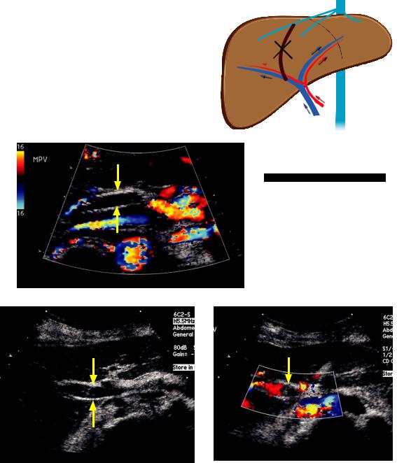
Ultimately, when portal venous pressure is higher than forward pressure, the portal venous flow will reverse, which is diagnostic for portal hypertension. Reversal of portal
venous flow is called hepatofugal |
flow (-fugal |
L tin root as fugitive). |
reversed = hepatofugal ow |
|
|
hepatic arteries and portal veins ow in opposite directions |
|
|
RHA
MPV
n addition to flow reversal, there are several secondary findings of portal hypertension:
Splenomegaly and splenic varices: Sagittal ultrasound |
Transverse Doppler ultrasound of the |
in the left upper quadrant shows an enlarged spleen |
eft lobe of the liver shows a recanalized |
(calipers) measuring 15 cm in craniocaudal dimension. umbilical vein, which is considered |
|
There are numerous tubular hypoechoic structures |
diagnostic of portal hypertension. |
(arrows) at the splenic hilum representing varices. |
|
Case courtesy Julie Ritner, MD, Brigham and Women’s Hospital
Low portal venous velocity (<16 cm/sec).
Dilated portal vein (13 mm is the maximal normal diameter in quiet respiration).
Splenomegaly.
Varices
Portosystemicshuntsareoftenpresent,mostcommonlygastro-esophageal,paraumbilical,or splenorenal.Notethatanisolatedportosystemicshuntmaynotbecausedbyportalhypertension.For instance,isolatedobstructionofthesplenicveinfrompancreatitisorneoplasmmayleadtoashunt.
A recanalized umbilical vein is a portosystemic shunt that is diagnostic of portal hypertension.
478

Transjugular intrahepatic portosystemic shunt (TIPS)
Portal hypertension (and reversal of portal flow) can be treated with a transjugular intrahepatic portosystemic shunt (TIPS), which connects a branch of the portal vein to a systemic hepatic vein.
Ultrasound is used for surveillance of TIPS patency, starting with a post-procedure baseline. Routine follow-up is performed according to the following schedule: In month, every 3 months for the first year, and then every 6 to 12 months.
Flow in a patent TIPS will be towards the hepatic veins, and flow in the portal veins will be towards the TIPS. Therefore, flow in the main portal vein will be hepatopetal and flow in the right and left portal veins will be hepatofugal (highlighted below with yellow circles).
Patent TIPS:
blood ows through TIPS to hepatic veins RHA RPV and LPV have reversed ow (toward TIPS)
MPV has hepatopetal ow (toward TIPS)
MPV
Patent TIPS: Color Doppler of the porta hepatis including the proximal TIPS shows flow within the TIPS (yellow arrow).
Case courtesy Julie Ritner, MD, Brigham and Women’s Hospital
US can evaluate for stenosis of the TIPS.
TIPS stenosis: Spectral Doppler of a TIPS shows a velocity of 45 cm/sec, indicative of stenosis due to slow flow.
Case courtesy Julie Ritner, MD, Brigham and Women’s Hospital
High intra-TIPS velocity >190 cm/sec or low intra-TIPS velocity of <90 cm/sec suggests stenosis.
ntra-TIPS velocity change of ±>50 cm/sec since the baseline study is also concerning for stenosis.
Low main portal vein velocity (<30 cm/sec) suggests TIPS stenosis.
479
f the TIPS becomes occluded, the right and left portal veins will “re-reverse” and become hepatopetal.
occluded TIPS: no blood ow through TIPS
RPV and LPV have “re-reversed”: RHA now have hepatopetal ow (away from TIPS) 
MPV has hepatopetal ow (towards occluded TIPS)
MPV
the porta hepatis including the proximal TIPS shows complete absence of flow within the TIPS (arrows).
Case courtesy Julie Ritner, MD,
Brigham and Women’s Hospital
Portal vein thrombosis
Grayscale transverse image of the porta hepatis shows echogenic debris within the main portal vein
(arrows). Color Doppler confirms partial portal vein thrombosis with lack of flow in the proximal portal vein (arrow).
Case courtesy Julie Ritner, MD, Brigham and Women’s Hospital
Thrombosis of the portal vein can be bland (simple thrombosis) or may be due to tumor invasion.
Bland portal vein thrombus can be caused by general hypercoagulable state or may be due to local inflammation from pancreatitis or hepatitis.
n infants, omphalitis or dehydration may also lead to portal vein thrombosis.
Tumor thrombus is most commonly caused by hepatocellular carcinoma.
480

•Ultrasound of portal vein thrombosis shows lack of portal venous flow, often with echogenic thrombus within the portal vein.
Expansion of the portal vein can be seen with either bland or tumor thrombus.
On color Doppler, flow within the thrombus suggests tumor thrombus.
•One potential pitfall to be aware of is slow (<16 cm/sec) or stagnant portal venous flow in the presence of portal hypertension which may mimic portal vein thrombosis.
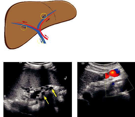
•Long-standing portal vein thrombosis leads to cavernous transformation of the portal vein, characterized by formation of multiple small periportal collaterals.
Cavernous transformation of the portal vein: Grayscale transverse image of the porta hepatis shows numerous tubular hypoechoic structures in the expected location of the portal vein (arrows). Color Doppler (right image) demonstrates flow within these collateral vessels, with no identifiable portal vein.
Case courtesy Julie Ritner, MD, Brigham and Women’s Hospital.
Portal venous gas
•Portal venous gas is due to abdominal catastrophe (ischemia and infarction) until proven otherwise. If the cause of the portal venous gas is unknown, CT should be performed emergently.
•Grayscale ultrasound shows peripheral patchy branching foci of hyperechogenicity that are often transient. Spectral Doppler of the portal vein features numerous characteristic spikes.
•In contrast to portal venous gas, pneumobilia tends to be more central.
Portal venous gas: Grayscale ultrasound through the liver shows numerous tiny echogenic foci (arrows) in a branching pattern throughout the liver, extending to the periphery.
Case courtesy Julie Ritner, MD, Brigham and Women’s Hospital.
481

Hepatic veins
Normal hepatic vein waveform
The hepatic veins feed into the IVC and the right side of the heart. The spectral
Doppler waveform of the hepatic veins is therefore affected by the cardiac cycle.
Thenormalhepaticvenouswaveformhasthreedistinctcomponents:TheA,S,andDwaves. Note that antegrade flowisdefinedasforwardflowinthenormalexpecteddirection.
A-wave atrial systole
retrograde (towards transducer)
anterograde (into heart;
away from transducer)
D-wave
ventricular S-wave diastole
ventricular systole
A-wave: Atrial systole, during which blood is forced retrograde (away from the heart) into the liver. S-wave: Ventricular systole, during which a large volume of blood returns to the right atrium.
D-wave: Ventricular diastole, during which a smaller volume of blood returns to the right atrium.
Increased hepatic vein pulsatility: Accentuated A-wave
ncreased hepatic vein pulsatility is caused by a right-sided cardiac abnormality, either right-sided heart failure or tricuspid regurgitation. Both conditions are characterized by accentuation of the A-wave due to increased retrograde flow during atrial systole.
tricuspid regurgitation |
right-sided CHF |
accentuated |
accentuated |
A-wave |
A-wave |
short |
D-wave |
preserved |
D-wave |
|
S-wave |
S-wave |
|||
|
|
Normally, the tricuspid valve closes at the beginning of ventricular systole (the beginning of the S-wave).
n tricuspid regurgitation, there is some degree of blood flow from the right ventricle into the right atrium during ventricular systole, allowing less blood to return to the right atrium from the hepatic veins and IVC during ventricular systole. This results in a decreased or even retrograde S-wave
n right-sided heart failure, the tall A-wave is due to increased right atrial pressure; however, in contrast to tricuspid regurgitation, the S-wave is normal since the tricuspid valve remains competent.
482

Decreased hepatic vein pulsatility
•Decreased hepatic vein pulsatility is seen in cirrhosis, Budd–Chiari (hepatic vein thrombosis), and hepatic veno-occlusive disease.
Pancreas
Pancreatitis
Acute pancreatitis
•Pancreatitis is inflammation of the pancreatic parenchyma, most often caused by alcohol or gallstones.
•Ultrasound is useful in the initial evaluation of pancreatitis to evaluate for gallstones or biliary obstruction.
•Usually, the pancreas appears normal in acute pancreatitis. The pancreas may be diffusely enlarged and relatively hypoechoic due to edema. More severe inflammation may cause the normally hypoechoic pancreas to become isoechoic to liver.
•Ultrasound has limited utility in evaluating complications of pancreatitis such as pancreatic necrosis or peripancreatic fluid collections.
Acute pancreatitis: Transverse ultrasound of the head and body of the pancreas shows a diffusely enlarged, heterogeneous pancreas (arrows) due to pancreatic edema.
Case courtesy Julie Ritner, MD, Brigham and Women’s Hospital.
•Complications of acute pancreatitis may be inflammatory, infectious, or vascular.
A pancreatic pseudocyst is usually detectable by ultrasound, although the full extent of large pseudocysts can be difficult to determine by ultrasound alone.
Infectious complications of acute pancreatitis include peripancreatic abscess, infected pseudocyst, and infected pancreatic necrosis.
The two most important vascular complications of pancreatitis are splenic vein thrombosis and splenic artery pseudoaneurysm, both of which can be characterized by Doppler ultrasound.
Chronic pancreatitis
•Chronic pancreatitis is caused by repeated bouts of acute pancreatitis (most commonly alcoholic).
•The classic ultrasound appearance of chronic pancreatitis is an atrophied gland, with diffuse calcifications and dilated and beaded distal pancreatic duct.
•Calculi within the pancreatic duct may also be seen.
483
Pancreatic neoplasms
Pancreatic adenocarcinoma
•Pancreatic adenocarcinoma is the most common pancreatic tumor, and is typically seen in older males.
•Small tumors are hypoechoic, while larger masses may be more heterogeneous. It can be difficult to identify the tumor extent on ultrasound because of infiltrative margins and invasion of the tumor into pancreatic parenchyma and adjacent structures.
•The most common location for a tumor to arise is the pancreatic head, where the mass often presents with ductal obstruction. The double duct sign represents dilation of both the pancreatic and common bile ducts caused by malignant obstruction.
Cystic pancreatic neoplasms
•Cystic pancreatic neoplasms are a diverse group of unrelated pancreatic tumors that may appear similar by ultrasound.
•Serous cystadenoma is a benign tumor seen in older females, consisting of multiple tiny cysts. A characteristic calcified scar is not often seen, but is very specific when present.
•Mucinous cystic neoplasm has malignant potential, and is usually seen in middle-aged females. Compared to serous cystadenoma, the cysts are larger in size. The mucin can generate numerous fine echoes.
•Intraductal papillary mucinous neoplasm (IPMN) is a neoplasm of variable and controversial natural history that communicates with either the main pancreatic duct or branch ducts. Demonstration of the ductal communication can be difficult by ultrasound.
Pancreatic endocrine tumors
•Tumors arising from the neuroendocrine cells of the pancreas may be either functioning or nonfunctioning.
•Functioning tumors are usually symptomatic, small at diagnosis, and identified through biochemical testing. In contrast, nonfunctioning tumors may be asymptomatic, and hence, large at the time of diagnosis.
•Intraoperative ultrasound continues to gain ground and is helpful in identifying small tumors at surgery.
•Insulinomas are the most common pancreatic endocrine tumors. Preoperative ultrasound detection is difficult and is successful less than 60% of the time. When seen, insulinomas are hypoechoic, encapsulated pancreatic nodules.
•Gastrinomas are the second most common pancreatic endocrine tumors. Liver metastases are present at the time of diagnosis in 60% of patients.
Pancreatic lymphoma
•B-cell lymphoma is the most common subtype of lymphoma to affect the pancreas, and is almost always associated with adenopathy and multi-organ involvement by the time the pancreas is involved.
•The typical ultrasound appearance of pancreatic lymphoma is a diffusely enlarged, hypoechoic gland.
484

Spleen
Patterns of disease
Splenic calcification
•Granulomatous disease: Calcifications may be scattered or diffuse.
•Splenic infarct.
•Hematoma.
•Calcified splenic artery aneurysm.
Cystic splenic lesion: Color Doppler should always be used to exclude a vascular etiology
•Splenic artery aneurysm or pseudoaneurysm.
•Hematoma.
•Abscess.
•Pancreatic pseudocyst.
Echogenic splenic lesion
•Hemangioma (can also be hypoechoic).
•Hamartoma.
•Lymphangioma.
Hypoechoic splenic lesion
•Laceration (in the setting of trauma).
•Abscess.
•Lymphoma.
•Sarcoidosis.
•Metastasis.
•Infarct (tends to be peripheral).
•Extramedullary hematopoiesis.
Splenomegaly (defined as >14 cm in sagittal plane)
•Mild to moderate splenomegaly:
Portal hypertension (most common).
Infection.
AIDS.
•Moderate to marked splenomegaly:
Leukemia/lymphoma.
Infectious mononucleosis.
•Massive splenomegaly:
Myelofibrosis.
485

Kidneys
Stones, obstruction, and hydronephrosis
Evaluation of kidney stones
•Ultrasound is an excellent modality for evaluation of nephrolithiasis, which may cause renal obstruction and resultant hydronephrosis.
•Anechogenicshadowingfocusinthekidney,ureter,orbladderissuspiciousforastone.
•After diagnosing a renal or ureteral calculus, one should always evaluate for the presence of hydronephrosis and perinephric fluid.
Approach to hydronephrosis
•The most common cause of hydronephrosis is an obstructing calculus.
Sagittal view of the left kidney shows marked hydroureteronephrosis (arrows show dilation of the proximal ureter).
Transverse view of the bladder in the same patient shows a 17 mm shadowing left UVJ calculus (calipers).
•Although hydronephrosis is usually due to ureteral obstruction, it is possible to have hydronephrosis without obstruction. For instance, vesicoureteral reflux or pregnancy may cause a dilated ureter without obstruction. Pregnancy preferentially affects the right side.
•Likewise, obstruction without hydronephrosis may also be seen in:
Very acute obstruction.
Obstructionwithdehydration,wherethereisinsufficienturineproductiontocreateapressurebackup.
Obstruction with ruptured fornix. Increased pressure from obstruction may cause a fornix to rupture, which would decompress the renal pelvis and spill fluid into the perinephric space.
Pitfalls in diagnosing hydronephrosis
•It can sometimes be difficult to distinguish between hydronephrosis and multiple renal sinus cysts. Renal sinus cysts are subsequently discussed and include peripelvic and parapelvic cysts. On imaging, renal sinus cysts will show a single or multiple discrete cystic lesions that do not communicate with each other.
•In true hydronephrosis, all the dilated fluid-filled spaces are contiguous.
Resistive index (RI) may be helpful in diagnosing obstruction
•The renal resistive index (RI) may be elevated in acute obstruction, thought to be due to cytokine-mediated renal artery vasoconstriction.
The resistive index is calculated with pulse-width Doppler of the renal segmental or arcuate arteries.
RI = (PSV – EDV)/PSV
PSV = peak systolic velocity EDV = end diastolic velocity
486

Higher resistive indices correlate with higher resistance.
With no diastolic flow, RI = PSV/PSV = 1
Reversal of diastolic flow technically causes RI >1, although in such cases RI is not measured.
•A RI of >0.7 on the affected side, or a difference of >0.1 between kidneys, suggests acute obstruction.
Bilateral elevated RIs (>0.7) are nonspecific and can be due to any number of medical renal processes.
•The resistive index is not used to diagnose chronic obstruction.
Ureteral jets may be helpful but are controversial
•A ureteral jet is flow of urine into the bladder as seen by color Doppler.
•Flow from the kidney to the bladder would be completely eliminated in complete obstruction, so theoretically the presence of a ureteral jet rules out a complete obstruction. However, ureteral jets are very commonly seen even with stones, and jets are often absent in normal patients.
Solid renal masses
Angiomyolipoma (AML)
•An angiomyolipoma is a benign hamartoma made up of blood vessels (angio), smooth muscle (myo), and fat (lipoma).
•Although benign, there is an increased risk of hemorrhage if >4 cm in size. The hemorrhage may be caused by microaneurysm rupture within the vascular elements of the AML.
•On ultrasound, AML is echogenic due to the fat component. There is considerable overlap between the ultrasound appearance of AML and renal cell carcinoma.
About one third of AML demonstrate shadowing, which is a specific finding for AML.
Multiple AML are seen in tuberous sclerosis.
Oncocytoma
Oncocytoma: Sagittal ultrasound through the right kidney (left image) demonstrates an exophytic solid renal mass (arrows) that is isoechoic to cortex. Color Doppler suggests a spoke-wheel pattern of vascularity.
Case courtesy Julie Ritner, MD, Brigham and Women’s Hospital.
•Oncocytoma is a benign renal tumor arising from tubular cells.
•On ultrasound, oncocytoma is indistinguishable from renal cell carcinoma (RCC). It may be hypoechoic, isoechoic, or hyperechoic. A spoke-wheel vascular pattern is sometimes seen on color Doppler.
•Due to imaging overlap with RCC, oncocytomas are treated surgically, even if the typical stellate or spoke-wheel vessels are seen.
487

Renal cell carcinoma (RCC)
Renal cell carcinoma: Sagittal ultrasound through the kidney shows a hypoechoic solid mass (arrows) with heterogeneous echotexture in the interpolar region. The mass demonstrates vascularity on color Doppler (right image).
Case courtesy Julie Ritner, MD, Brigham and Women’s Hospital.
•Renal cell carcinoma (RCC) is the most common solid renal mass.
•The staging of RCC uses the Robson system, which is discussed in the genitourinary section.
•RCC is most often isoechoic to renal cortex, but can occasionally be hypoechoic or even hyperechoic (mimicking AML). A hypoechoic rim and intratumoral cystic changes are typically seen only in RCC, which may help to distinguish it from AML.
•In the presence of a renal mass, the renal veins must be carefully evaluated as RCC has a propensity for venous invasion. Venous invasion is Robson stage IIIA, and the presence of venous invasion has important implications for surgical approach.
•Color and spectral Doppler are helpful in differentiating bland renal vein thrombus (which would not be stage IIIA) from tumor thrombus. Tumor thrombus will have color Doppler flow with an arterial waveform.
Renal lymphoma
•Renal lymphoma (most commonly high-grade B-cell) may disseminate hematogenously orspreaddirectlyfromtheretroperitoneumtothekidney.Primaryrenallymphomais veryrareandofuncertainoriginasthereisnonativelymphoidtissuewithinthekidney.
•Themostcommonimagingpresentationofrenallymphomaismultiplehypoechoicrenal masses.Retroperitonealadenopathyisusuallypresent.Asolitarymassisanuncommon presentation.Diffuselymphomatousinfiltrationproducingnephromegalyisrare.
Renal cysts and cystic masses
Potential pitfalls in diagnosing a cystic lesion
•Renal scanning should be performed with multiple angles of insonation to differentiate hydronephrosis from a renal sinus cyst (parapelvic or peripelvic cyst). In hydronephrosis, the dilated spaces will all connect.
•Color Doppler should always be utilized, as a renal artery aneurysm may mimic a cyst in grayscale.
Simple cortical cyst
•Asimplerenalcystshouldhavethesonographichallmarksofasimplecyst,featuringan imperceptiblythinwall,anechoicinternalcontents,andposteriorthroughtransmission.
•Harmonic imaging can be helpful in confirming the diagnosis of simple renal cyst by eliminating artifactual low-level internal echoes.
488

Renal sinus cyst
•Acystintherenalsinusmaybeaperipelvicorparapelviccyst.Peripelviccystsare secondarytolymphaticobstructionandareoftenmultiple.Incontrast,aparapelviccyst isarenalparenchymalcystthatherniatesintotherenalsinusandisusuallysolitary.
•When multiple renal sinus cysts are present (most commonly peripelvic cysts), the appearance may mimic hydronephrosis. In contrast to hydronephrosis, renal sinus cysts will not be contiguous with each other.
Renal abscess
•Renal infection, discussed below, may appear as a complex cystic renal mass.
Cystic renal cell carcinoma
•Although most cases of RCC present as a solid renal mass, a significant minority may present as a complex cystic lesion. Worrisome ultrasound findings of a complex cystic mass include thick septa, irregular wall thickening, and a mural nodule.
•The Bosniak classification of complex renal masses is based on CT appearance and depends on enhancement. The Bosniak classification is described in the genitourinary imaging section.
Renal infection
Acute diffuse pyelonephritis
•Pyelonephritis is infection of the renal parenchyma, usually by gram-negative urinary tract organisms that ascend from the lower genitourinary tract.
•The most common ultrasound appearance of pyelonephritis is a normal kidney. Occasionally generalized renal edema and engorgement can be seen.
Focal pyelonephritis
•Focal pyelonephritis is a focal or multifocal infection of the renal parenchyma.
•Theclassicultrasoundappearanceisahypoechoicmass(ormasses)withlow-amplitude echoesthatdisruptsthecorticomedullaryjunction.Adistinctwallislacking.
Renal abscess
•Arenalabscessisafocalnecroticparenchymalinfectionwithadefinedwall.Urinalysismay benegativeupto30%ofthetimeiftheinfectiondoesnotinvolvethecollectingsystem.
•Small abscesses (<3 cm) often undergo a trial of conservative medical therapy, while larger abscesses are typically drained.
•Ultrasoundshowsafluid-filledrenalmasswithadistinctwall,whichmaybemultiloculated.
Emphysematous pyelonephritis
•Emphysematous pyelonephritis is a complication of acute pyelonephritis characterized by replacement of renal parenchyma by gas. It is caused by gas-forming organisms, most commonly E. coli. Emphysematous pyelonephritis is almost exclusively seen in diabetic or immunocompromised individuals.
•Emphysematous pyelonephritis is a surgical emergency requiring broad-spectrum antibiotics and emergent nephrectomy. Mortality can reach 40%.
•Ultrasound shows high-amplitude echoes in the renal parenchyma representing gas locules with posterior dirty acoustic shadowing.
Tuberculous pyelonephritis
•Tuberculous pyelonephritis, caused by hematogenous spread of M. tuberculosis, is characterized by focal cavitary renal lesions with calcification.
•Putty kidney is an atrophic, calcified kidney seen in end-stage renal tubercolosis.
489

Xanthogranulomatous pyelonephritis
•Xanthogranulomatous pyelonephritis results from repeated cycles of chronic lowgrade infection caused by an obstructing calculus that leads to fibrofatty replacement of renal parenchyma.
•On ultrasound, the kidneys are enlarged with areas of mixed echogenicity. A central stone is nearly universally present, which may be staghorn in morphology.
Pyonephrosis
Pyonephrosis due to malpositioned nephroureteral stent: Initial ultrasound (left image) shows moderate hydronephrosis with a subtle echogenic dependent fluid–debris level (arrows). Low-level echoes are present within the collecting system. The nephroureteral stent is not visualized.
Subsequent ultrasound less than 12 hours later (right image) shows marked progression of hydronephrosis, a much larger fluid–debris level (arrows), and low level internal echoes within the dilated collecting system. Scanning of the distal ureter (not shown) revealed a malpositioned nephroureteral stent as the cause of obstruction.
Case courtesy Julie Ritner, MD, Brigham and Women’s Hospital.
•Pyonephrosis is infection of an obstructed collecting system and is a surgical emergency. Treatment is emergent relief of obstruction, either with percutaneous nephrostomy or ureteral stent.
•Ultrasoundfeaturesechoeswithinadilatedcollectingsystem.Afluidlevelmaybepresent.
HIV associated nephropathy
•The HIV virus may directly infect the renal parenchyma to produce HIV nephropathy, most commonly resulting in focal segmental glomerulosclerosis (FSGS). HIV nephropathy clinically presents with nephritic renal failure.
•The kidneys are characteristically echogenic. Enlarged echogenic kidneys are specific for HIV nephropathy, although the kidneys are enlarged only about 20% of the time.
Multicystic renal disease
Autosomal dominant polycystic kidney disease (ADPKD)
•Autosomal dominant polycystic kidney disease (ADPKD) is the most common cause of multiple renal cysts in adults. ADPKD is associated with cysts in the liver and other visceral organs.
•15% of patients have saccular cerebral aneurysms.
•The natural history of ADPKD is renal failure by middle age.
•ADPKD does not confer an increased risk of renal cell carcinoma; however, complex cysts with internal hemorrhage are difficult to distinguish from renal cell carcinoma.
•Imaging of ADPKD shows markedly enlarged kidneys with innumerable cysts of varying size and echogenicity.
490

Autosomal recessive polycystic kidney disease (ARPKD)
•Autosomal recessive polycystic kidney disease (ARPKD) is a diagnosis of infancy. Prognosis is poor. If the child survives infancy, hepatic fibrosis usually develops.
•ARPKD presents in utero as enlarged echogenic kidneys since the cysts are too small to be individually resolved by ultrasound.
Acquired renal cystic disease
•Patients on long-term dialysis often develop many small renal cysts superimposed upon atrophic kidneys. Acquired cystic disease does confer an increased risk of renal cell carcinoma, in contrast to ADPKD.
Imaging of renal transplant
Approach to renal transplant
•The goal of ultrasound evaluation after renal transplant is to determine whether there is a treatable surgical or vascular complication. Ultrasound is not useful for differentiating among the various kinds of parenchymal rejection.
•The transplanted kidney is implanted in the right or left iliac fossa (right more commonly), and is often very well imaged due to its superficial location.
•An elevated RI (>0.7) suggests renal dysfunction, but this finding is nonspecific.
Surgical complications following renal transplant
•Ureteral obstruction is apparent on ultrasound as hydronephrosis.
•Fluid collection (blood, pus, urine) is highly dependent on timing:
Immediately postoperative: Hematoma. |
3–4 weeks postoperative: Abscess. |
1–2 weeks postoperative: Urinoma. |
2nd month and beyond: Lymphocele. |
Vascular complications following renal transplant
•Renal vein thrombosis: The renal artery Doppler may show reversal of diastolic flow.
•Renal artery stenosis:Elevatedflowvelocitiesareseenatthesiteofstenosis,withaparvus et tardus waveformdistaltothestenosis.Usuallytakesseveralweekstomonthstodevelop.
•Pseudoaneurysm is usually due to renal biopsy.
Medical complications
•Medicalcomplicationsgenerallycannotbedifferentiatedonultrasound.Biopsyisnecessary fordiagnosis,althoughthetimeelapsedsincethetransplantmaybeahelpfulclue.
•Hyperacute rejection: Occurs in first few hours after transplant.
Hyperacute rejection is very rare, and is due to ABO blood type incompatibility.
•Acute tubular necrosis (ATN): Occurs in the immediate few postoperative days.
ATN is usually a sequela of pre-implantation ischemia.
•Acute rejection: Occurs within three months of transplant.
•Chronic rejection: Occurs after three months of transplant.
•Drug toxicity may caused by cyclosporine, which is nephrotoxic.
Post-transplant lymphoproliferative disorder (PTLD)
•Post-transplant lymphoproliferative disorder (PTLD) is a type of lymphoma that is thought to be due to immune suppression and Epstein–Barr virus proliferation.
•PTLD can arise anywhere in the body. Any new mass in any organ in a transplant patient should raise concern for potential PTLD.
•UltrasoundofrenalPTLDwillshowanamorphoushypoechoicmassthatmaysimulatea fluidcollectionongrayscaleimages.Unlikefluid,PTLDwilldemonstrateDopplerflow.
491

Renal: Specific imaging patterns
Medullary nephrocalcinosis
differential of medullary nephrocalcinosis
Medullary nephrocalcinosis: Sagittal ultrasound through the right kidney (left image) shows diffusely echogenic renal pyramids (arrows). Coronal CT MIP in bone windows (right image) in a different patient demonstrates symmetric cloud-like renal medullary calcification bilaterally.
Ultrasound case courtesy Julie Ritner, MD, Brigham and Women’s Hospital.
•Any cause of hypercalcemia and hypercalciuria can cause medullary calcification.
•Hyperparathyroidism is the most common cause of medullary nephrocalcinosis.
•Renal tubular acidosis (distal type).
•Medullary sponge kidney is caused by ectatic tubules in the medullary pyramids leading to stasis and
stone formation.
•Papillary necrosis.
•In a child, treatment with furosemide can lead to medullary nephrocalcinosis.
Cortical nephrocalcinosis
• Much more rare than medullary nephrocalcinosis, cortical nephrocalcinosis is due to
|
|
diffuse cortical injury. |
|
|
|
corticalofdx -nephro calcinosis |
• |
Acute cortical necrosis. |
|
||
|
• |
Hyperoxaluria (rare). |
|
• |
Alport syndrome. |
d |
• Autosomal recessive polycystic kidney disease. |
|
|
|
|
|
|
|
Echogenic kidneys
•Echogenic kidneys are most commonly due to medical renal disease, such as diabetic nephropathy, glomerulosclerosis, acute tubular necrosis, etc.
•HIV nephropathy causes enlarged and echogenic kidneys.
Echogenic renal mass
ifferentiald of echogenicrenal mass |
• Angiomyolipoma (AML). A shadowing echogenic renal mass is relatively specific for AML. |
|
• Malignant neoplasm (atypical appearance). |
||
• |
Sloughed papilla, secondary to papillary necrosis, may appear as an echogenic mass in the collecting |
|
|
• |
Renal calculus. |
|
• |
Intrarenal gas. |
• Milk of calcium, caused by crystals precipitating out of supersaturated solution.
system.
492

Scrotum and testicle
Scrotal anatomy
Epididymis: Function and anatomy
•The epididymis carries sperm away from the testicle to the vas deferens.
•Theepididymisiscomposedofhead,body,andtail.Theheadmaymeasureupto10mm.
•The epididymis is normally hypoechoic and has less blood flow compared to the testicle. Relatively increased epididymal blood flow can be seen in epididymitis.
Mediastinum testis: Function and anatomy
•Themediastinumtestisisfibroustissueinthehilumofthetesticle,fromwhichfibroussepta radiatetowardsthetesticularperiphery.Itprovidesstructuralsupporttotheretetestis.
Rete testis: Function and anatomy
•The rete testis is a network of tubules that carries sperm from the seminiferous tubules to the vas deferens. It functions to concentrate sperm.
Testicular masses
Approach to a testicular mass
•Intratesticular masses are usually malignant (90–95%). Conversely, most extratesticular masses are benign in an adult, although a pediatric mass in this location may be malignant.
•The retroperitoneum should always be evaluated if an intratesticular mass is seen. Likewise, if retroperitoneal adenopathy is seen in a reproductive-age male, the testicles should always be examined.
•Most scrotal masses are hypoechoic relative to normal testicular parenchyma.
•On Doppler, most masses will have increased vascularity with high diastolic flow, producing a low resistance waveform.
Malignant germ cell tumor (GCT): Seminoma
Seminoma: Grayscale (left image) and color Doppler show a heterogeneous hypoechoic vascular mass (yellow arrows) in the left testis. Note the presence of numerous tiny echogenic foci (red arrows) representing microlithiasis.
Case courtesy Julie Ritner, MD, Brigham and Women’s Hospital.
•Seminoma is the most common testicular malignancy. It has a favorable prognosis.
Seminoma typically occurs in middle-aged men. Uncommonly, hCG may be elevated.
The spermatocytic subtype of seminoma occurs in slightly older men (mid 50s) and has excellent prognosis with orchiectomy only. Tumor markers are not elevated.
493
Malignant germ cell tumors: Nonseminomatous germ cell tumors (NSGCT)
•Nonseminomatous germ cell tumors (NSGCT) include embryonal carcinoma, teratoma, yolk sac tumor, choriocarcinoma, and mixed subtypes.
Mixed germ cell tumor is the most common NSGCT, and is the second most common primary testicular malignancy after seminoma. The most common components of mixed NSGCT are embryonal carcinoma and teratoma.
Embryonal cell carcinoma in its pure form is rare and in adults is typically seen as a component of mixed germ cell tumors. The infantile form, called endodermal sinus tumor or yolk sac tumor, is the most common testicular tumor of infancy. AFP is elevated.
Teratoma is rare in its pure form in adults, but is seen in 50% of mixed NSGCT. Teratoma is classified as mature, immature, and malignant. In adults, teratomas are usually malignant. In children, teratomas are usually benign, with the mature subtype most commonly seen.
Choriocarcinoma is the most aggressive and rare NSGCT. Choriocarcinoma metastasizes early, especially to brain and lung. Metastases tend to be hemorrhagic. hCG is always elevated and gynecomastia may result from elevated chorionic gonadotropins.
•NSGCT generally occur in younger patients compared to seminomas, typically in young men in their 20s and 30s. NSGCT tend to be more aggressive than seminomas. Local invasion into the tunica albuginea and visceral metastases are common.
•A heterogeneous testicular mass that contains solid and cystic components and coarse calcification is a typical appearance for a NSGCT. It is not possible to distinguish the various subtypes of NSGCT on sonography.
Burnt-out germ cell tumor
•Burnt-out germ cell tumor is a primary testicular neoplasm that is no longer viable in the testicle even though there is often viable metastatic disease, especially retroperitoneal.
•In the testicle, focal calcification with shadowing is characteristic. A mass may or may not be present.
•Treatment is orchiectomy in addition to systemic chemotherapy.
Testicular microlithiasis
•Testicular microlithiasis is multiple punctate testicular calcifications.
•There is a controversial association between microlithiasis and testicular neoplasm. While the overall absolute risk for developing testicular cancer remains very small in the presence of microlithiasis, the relative risk may be increased.
•Currentguidelinesdonotsupportscreeningbyultrasoundortumormarkers,butpatients withmicrolithiasismayperformself-examinationsandbeseeninfollow-upasneeded.
•At least five microcalcifications must be present per image to be called microlithiasis. If there are fewer than five microcalcifications the term limited microlithiasis is used.
•Microlithiasis can produce a starry sky appearance if calcifications are numerous.
Intheliver,hepatitiscancauseastarry sky appearanceduetoincreasedechogenicityoftheportaltriads.
Testicular metastases
•The most common metastases to the testicles are leukemia and lymphoma, as the relevant chemotherapeutic agents do not cross the blood–testis barrier.
•Hematologic malignancies typically present in older patients, tend to be bilateral, and may be infiltrative with diffuse testicular enlargement.
Benign testicular tumors
•An epidermoid isakeratin-filledcystwithadistinctiveonion-ringappearanceofconcentric alternatingringsofhypo-andhyperechogenicity.Ifsuspected,localexcisionisperformed insteadofthestandardorchiectomytypicallyperformedforpresumedmalignantmasses.
494

•Sex-cord stromal tumors are 90% benign but are sonographically indistinguishable from malignant tumors. Orchiectomy is therefore the standard treatment.
Leydig cell tumor can present with gynecomastia due to estrogen secretion.
Sertoli cell tumor is associated with Peutz–Jeghers and Klinefelter syndromes.
Sarcoidosis
•Sarcoidosis may involve either the testis, the epididymis, or both. Scrotal involvement is rare, but presents clinically as painless scrotal enlargement.
•Theultrasoundappearanceoftesticularsarcoidisindistinguishablefromasolidmalignant mass.Ifsarcoidosisissuggestedbyclinicalhistory,thetesticularmassmustbebiopsiedto excludemalignancy.Withouttissuepathology,amasscannotbeassumedtobesarcoid.
Benign testicular tumor mimics
•Congenital adrenal rests are embryologic remnants of adrenal tissue trapped within the testis. These are typically seen in newborns with congenital adrenal hyperplasia.
Adrenal rests appear as bilateral hypoechoic masses and classically enlarge with ACTH exposure.
•Polyorchidism/supernumerary testis: An extra testicle has an identical imaging appearance to normal testicular parenchyma.
Extranumerary testes carry a slightly increased risk of torsion and testicular cancer.
Extra-testicular masses
•In contrast to intratesticular masses, extratesticular masses are usually benign. Up to 16% of extratesticular masses may be malignant, however, and ultrasound cannot reliably differentiate benign from malignant masses.
Benign extratesticular masses
•Spermatic cord lipoma is the most common extratesticular neoplasm overall.
•Benign adenomatoid tumor of the tunica albuginea is the most common epididymal neoplasm.
The “-celes” and cystic lesions
Hydrocele
•Ahydroceleisexcessfluidinthescrotumsurroundingthetesticle.Mostareasymptomatic.
•A hydrocele may be congenital (due to patent processus vaginalis in utero or infancy), idiopathic, or post-inflammatory. Regardless of etiology, there is never fluid at the bare area where the testicle is attached to the tunica vaginalis.
Hematocele
•A hematocele is blood in the scrotum due to trauma or torsion.
Varicocele
•A varicocele is a dilated venous pampiniform plexus in the scrotum. A primary varicocele is due to incompetent valves of the internal spermatic vein. A secondary varicocele is due to increased venous pressure caused by an obstructing lesion.
•Varicocele is a common cause of infertility, seen in up to 40% of males presenting to an infertility clinic.
•Varicoceles are much more common on the left as the left testicular vein drains into the left renal vein and the superior mesenteric artery can compress the left renal vein. In contrast, the right testicular vein drains directly into the infrarenal IVC.
•85% of varicoceles are left-sided and 15% are bilateral. An isolated right-sided varicocele should prompt a search for a right-sided retroperitoneal mass.
495

•On ultrasound, varicoceles appear as multiple tubular and serpentine anechoic structures >2 mm in diameter in the region of the upper pole of the testis and epididymal head. The varicoceles follow the spermatic cord into the inguinal canal and can be compressed by the transducer. Careful optimization of Doppler parameters shows the slow venous flow within the varicocele.
Epididymal cysts and spermatocele
•An epididymal cyst is an anechoic fluid-containing cyst that can occur anywhere in the epididymis.
•A spermatocele is cystic dilation of the epididymis filled with spermatozoa, usually occurring in the epididymal head. Classic ultrasound appearance is an epididymal cyst with internal low-level mobile echoes.
•A simple epididymal cyst and a spermatocele cannot always be reliably distinguished by ultrasound.
Simple testicular cyst
•Asimpletesticularcystmeetssonographiccriteriaforasimplecyst(smoothposterior wall,imperceptiblewallthickness,completelyanechoic,posteriorthroughtransmission).
Tubular ectasia of rete testis
Tubular ectasia of the rete testes:
Transverse color Doppler ultrasound of the right testicle (left image) shows cystic dilation at the mediastinum testes (arrow). There is no flow within the lesion. This appearance is highly suggestive of tubular ectasia, although an avascular mass may rarely have a similar appearance.
Sagittal ultrasound (right image) shows elongation of the cystic dilation (arrows) along the mediastinum testes, which is confirmatory for tubular ectasia.
Case courtesy Julie Ritner, MD, Brigham and Women’s Hospital.
•Tubular ectasia of the rete testis is nonpalpable, asymptomatic, cystic dilation of the tubules at the mediastinum testes caused by epididymal obstruction. Tubular ectasia is often accompanied by an epididymal cyst or spermatocele.
•Tubular ectasia of the rete testis is common in older patients and may be bilateral.
•Imaging shows numerous tiny dilated structures in the region of the mediastinum testis, often seen in conjunction with an epididymal cyst/spermatocele.
•Important to be aware of only as a tumor mimic. Tubular ectasia is benign and no treatment is necessary.
Tunical cyst
•The tunica albuginea is the capsule overlying the testis. A cyst of the tunica albuginea presents as a palpable superficial nodule that resembles a BB.
•Sonography shows a typically small, simple, extra-testicular cyst.
•No treatment is necessary.
496

Vascular disease of the testis
Testicular torsion
•Testicular torsion is twisting of the testicle around the spermatic cord and the vascular pedicle. Torsion presents with acute scrotal pain and is a surgical emergency.
•Torsion may lead to irreversible testicular infarction if not de-torsed within a few hours.
De-torsion within 6 hours has an excellent prognosis.
De-torsion after 24 hours has a poor prognosis for testicular salvage.
•The bell-clapper deformitypredisposestotorsionduetoasmalltesticularbarearea.The bareareaisthetesticularattachmentsiteandnormallypreventsthetesticlefromrotation.
•Ultrasound findings of torsion are dependent on the time elapsed since torsion:
Hyperacute (within a few hours): Ultrasound shows a hyperechoic and shadowing torsion knot of twisted epididymis and spermatic cord, with no blood flow in the affected testicle.
Acute (between a few hours and 24 hours): Affected testicle is enlarged and heterogeneous.
Missed torsion (>24 hours): Affected testicle is enlarged and mottled, with scrotal skin thickening and increased flow in the scrotal wall. A complex or septated hydrocele may be present.
Segmental infarction
•Segmental infarction is a focal testicular infarction that can be due to microvascular thrombosis from acute inflammation, vasculitis, or sickle cell disease.
•Patients are typically in their 30s and present with acute pain which may mimic epididymitis or torsion clinically.
•The typical appearance of infarction is a wedge-shaped hypoechoic area with no flow on Doppler.
•The primary differential consideration of infarction is a hypovascular tumor. Infarcted tissue may undergo necrosis, making differentiation from tumor even more difficult. MRI may be helpful to distinguish infarction from tumor in ambiguous cases to potentially spare the patient from orchiectomy.
Scrotal trauma
Hematoma
•Thesonographicappearanceofanacutescrotalhematomaisanechogenic,extratesticular masswithnoDopplerflow.Whenlarge,thehematomacancompressthetesticle.
•Whenthehematomaevolvesintoacomplex,multiseptatedmass-likelesion,the distinctionbetweentheextratesticularhematomaandthetesticlemaybecomedifficult. Properdistinctionisnecessarytoavoidmistakingthehematomaforatesticularmass.
Testicular contusion
•Testicular contusion produces a peripheral, hypoechoic lesion that may mimic tumor.
•Even with a history of trauma, a suspicious testicular lesion requires further evaluation to exclude malignancy, typically with a short-term follow-up.
Testicular rupture
•Testicularrupturecausescapsuledisruption,oftenwithprotrusionoftesticularparenchyma throughthedefect.Ruptureisoftenassociatedwithatesticularhematomaorcontusion.
•Prompt diagnosis is critical, as testicular viability is dependent upon timely repacking of the seminiferous tubules back inside the capsule.
•Testicular rupture results in disruption of the blood–testis barrier and may be associated with future infertility due to the formation of anti-spermatozoa antibodies.
497

Scrotal infection
Epididymitis
Epididymitis: Sagittal grayscale ultrasound (left image) of the testicle and epididymis shows a markedly enlarged epididymis measuring 1.7 cm (calipers). Incidental note is made of an epididymal cyst (arrow). The testicle has a normal sonographic appearance. Transverse color Doppler of the epididymis (right image) demonstrates markedly increased flow.
Case courtesy Julie Ritner, MD, Brigham and Women’s Hospital.
•Epididymitis is infection of the epididymis, almost always ascending from the urinary tract.
•The classic clinical presentation of epididymitis is acute unilateral scrotal pain.
•The main differential based on clinical presentation is testicular torsion. In contrast to torsion, epididymitis features normal testicular blood flow.
•A key ultrasound finding of epididymitis is an enlarged epididymis with increased Doppler flow relative to the testicle (normally, the epididymis has less Doppler flow than the testicle). An associated hydrocele may be present, which often contains lowlevel echoes.
Epididymo-orchitis
•Epididymo-orchitis is infection that has spread from the epididymis to the testicle.
•Epididymo-orchitis has a similar ultrasound appearance to epididymitis, but blood flow to the testicle will also be increased.
•Infection and secondary inflammation can cause venous hypertension, which is a risk factor for focal testicular ischemia.
Fournier gangrene
•Fournier gangrene is necrotizing fasciitis of the scrotum and perineum, a highly morbid and surgically emergent condition.
•Infection is usually polymicrobial.
•The key imaging finding is subcutaneous gas, which appears on ultrasound as multiple echogenic reflectors in the subcutaneous tissues with dirty shadowing.
498

Vascular ultrasound
General principles
Angle correction
The Doppler signal is proportional to cos(θ). There is no Doppler shift at 90°.
75° 90°
60°
45°
30°
15°
0°
amount of Doppler shift is proportional to cos(θ)
at 90° there is zero Doppler shift
All measurements of velocity should be made at a consistent angle (typically 60°).
Measurements should never be taken at an angle greater than 60°.
θ = 60°
θ 
Overview of peak systolic velocity (PSV)
eak systolic velocity (PSV) is usually the most accurate method to evaluate the degree of arterial stenosis. PSV is elevated proximal to and at the site of stenosis.
PSV may be decreased distal to a hemodynamically significant stenosis.
The differential diagnosis of increased PSV includes:
Downstream (distal) stenosis.
Compensatory flow, contralateral to an obstruction or severe stenosis.
Physiologic hyperdynamic state in a healthy young patient.
The differential diagnosis of decreased PSV includes:
Upstream (more proximal) stenosis.
Poor cardiac pump function.
Near-total occlusion.
499

Carotid artery
Color and spectral Doppler parameters
head |
feet |
color scale shows red is toward the probe
(above the baseline)
Doppler angle corrected to 60 degrees
waveform above the baseline
is toward the probe
Normalcarotidexamination:Duplexultrasoundoftherightinternalcarotidarteryshowsnormalspectral waveform.Thepeaksystolicvelocityis124cm/sec,withinthenormalrange.Thereisnocarotidplaque.
By convention, for images obtained in the sagittal plane, the patient’s head is on the
eft side of the image and the feet are on the right.
n general, ultrasound parameters are optimized so that arteries are red and normal arterial flow is above the baseline. Certain parameters need to be adjusted so that arteries above the heart (which are normally heading towards the head) appear similar to arteries below the heart (which normally are heading towards the feet):
The color scale can be changed: Colors above the baseline go towards the probe.
Spectral Doppler baseline inversion can be changed: Positive waveforms go towards the probe.
Evaluation of the carotid arteries
There are three components to the carotid artery exam: Evaluation of plaque morphology, hemodynamic evaluation, and waveform analysis.
Plaque morphology
Plaque morphology is evaluated on grayscale imaging (without Doppler) and is described in terms of absolute percent stenosis.
<50% cross-sectional area plaque would not be expected to be hemodynamically significant.
>50% luminal plaque is expected to show elevation in peak systolic velocity.
Hemodynamic evaluation of stenosis
Normal peak systolic velocity (PSV) in large arteries is 60–100 cm/sec.
PSV tends to be elevated at a site of significant stenosis. Per the Society of Radiologists
n trasound (SRU) criteria, established in 2003:
>125 cm/sec suggests >50% stenosis.
>230 cm/sec suggests >70% stenosis.
Potential pitfall: An occluded or nearly occluded artery may have no detectable flow.
An elevated ratio of internal carotid artery to common carotid artery (ICA/CCA) PSV is
a useful secondary sign of ICA stenosis.
<2 is normal.
>2 suggests >50% ICA stenosis
>4 suggests >70% ICA stenosis
500
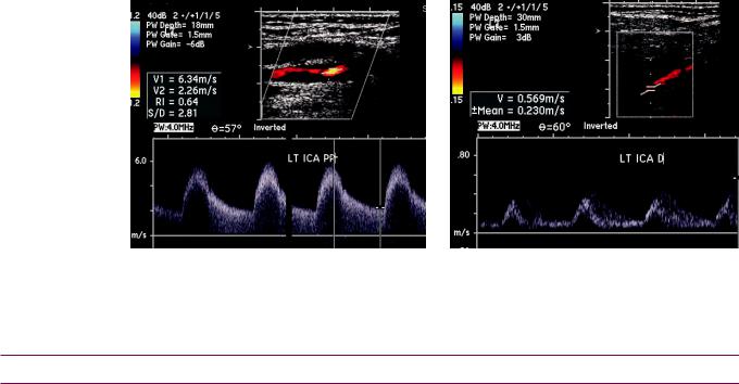
•End diastolic velocity of >100 cm/sec suggests >70% stenosis.
•In high and low flow states, the ICA/CCA ratio is more useful than the absolute PSV.
Waveform analysis
•Stenosis downstream (distal) to transducer (outflow lesion): Spectral waveform is high resistance and high velocity in morphology, characterized by decreased diastolic flow. The systolic upstroke is normal and rapid. Spectral broadening and aliasing may be present.
Spectral broadening describes the widened distribution of RBC velocities due to disruption of laminar flow.
Aliasing is an artifact where the highest velocities are shown to have a reversed flow.
•Stenosis upstream (proximal) to transducer (inflow lesion): Spectral waveform is low resistance and low velocity in morphology, with relatively increased diastolic flow. Systolic upstroke is slowed, producing the tardus et parvus waveform.
Carotid stenosis
Severe internal carotid artery stenosis: Spectral waveform of the proximal internal carotid artery (left image) shows spectral broadening and markedly elevated peak systolic velocity of 634 cm/sec,
consistent with severe stenosis. The grayscale images also show hypoechoic plaque. Evaluation distal to the stenosis (right image) shows a parvus et tardus waveform and decreased peak systolic velocity.
Case courtesy Julie Ritner, MD, Brigham and Women’s Hospital.
Renal artery stenosis
Renal artery stenosis: Criteria and protocol
•A peak systolic velocity of ≥180 cm/sec is consistent with renal artery stenosis.
Normal aortic and renal artery velocity is 60–100 cm/sec.
•Arenalarterytoaorticvelocityratioof>3.5isalsoconsistentwithrenalarterystenosis.
•Reduced or absent diastolic flow is suggestive of a stenosis distal to the area of interest.
•Aswiththecarotidartery,atardus et parvus waveformonspectralDopplerissuggestive ofastenosisproximal(upstream)tothetransducer,knownasaninflowlesion.
•An elevated renal resistive index (>0.7) is nonspecific, but may indicate renal artery stenosis. The resistive index is calculated as follows: RI = (PSV – EDV)/PSV, where PSV is peak systolic velocity and EDV is end-diastolic velocity.
RI is measured in the segmental arteries of the upper, mid, and lower poles.
Elevated resistive indices can also be seen in acute urinary obstruction or medical renal disease.
Atherosclerotic renal artery stenosis
•Atherosclerosis is by far the most common cause of renal artery stenosis, typically affecting the ostium of the renal artery.
501

Fibromuscular dysplasia (FMD)
Fibromuscular dysplasia (FMD) is a vasculitis that primarily affects the renal and carotid arteries in middle-aged females.
The mostcommonlocationofstenosisinFMDisthedistaltwothirdsoftherenalartery.
The classic angiographic appearance of FMD is a string of pearls caused by multifocal alternating stenoses and post-stenotic dilations.
Deep venous thrombosis (DVT)
Lower extremity venous system anatomy
The superficial venous system is composed of the great and small saphenous veins.
The great saphenous vein drains into the common femoral vein. Although the great saphenous vein is technically part of the superficial system, clots near the saphenofemoral junction are typically treated with anticoagulation because of their propensity to become dislodged.
The small saphenous vein drains into the popliteal vein (which continues proximally as the femoral
vein). Clots in the small saphenous vein are typically not treated.
Deep venous system anatomy mirrors arterial anatomy:
 common femoral vein
deep femoral vein
femoral vein
adductor hiatus
popliteal vein
anterior tibial vein peroneal vein
posterior tibial vein
lateral |
medial |
|
|
|
|
inguinal ligament. The CFV lies medial to the common femoral artery.
CFV tributaries include the deep femoral and femoral veins. The femoral vein was previously called the superficial femoral vein. The term superficial femoral vein should be avoided as it wrongly implies that this vein is part of the superficial venous system.
The three calf veins are the anterior tibial vein (lateral), peroneal vein (middle), and posterior tibial vein (medial), which join to form the popliteal vein (PV). The PV continues into the femoral vein.
Overview of the deep venous thrombosis (DVT) examination
Alowerextremityvenousultrasoundexamshouldincludevenouscompression,color
andspectralDoppler,andevaluationofvenousaugmentationandrespiratoryvariation.
Augmentation is the normal change in waveform when the calf is compressed. Lack of
augmentation suggests a distal venous obstruction between the calf and the transducer.
Respiratory variation is the normal change in waveform when the patient inspires. Lack of respiratory variation suggests a proximal venous obstruction.
502

•The popliteal, femoral, proximal deep femoral, and common femoral (including the saphenofemoral junction) veins should be imaged every 2–3 cm with and without compression.
Venous compression
•The hallmark sonographic finding of a DVT is a noncompressible vein with or without an intraluminal clot. A partially thrombosed vein may be partially compressible, while a completely thrombosed vein will not be compressible at all.
Color Doppler
•Color Doppler is almost always used to help localize the veins, but it is not necessary for diagnosing DVT.
•Normal color Doppler flow in a noncompressible vein is suspicious for nonobstructing thrombus.
Acute versus chronic deep venous thrombosis
•While the diagnosis of DVT is usually straightforward, distinguishing between acute and chronic thrombus can be difficult. Evaluation of the clot’s echogenicity is not a reliable way to determine the acuity of the clot as artifactual echoes within the vein lumen can overlap with the clot.
•Sonographic findings of chronic venous thrombus include clot retraction and poor visualization of the clot, only partial compressibility, irregularly echogenic and thickened vein walls, and prominent collateral veins.
Aortic disease
Abdominal aortic aneurysm (AAA)
Transverse grayscale ultrasound of the infrarenal abdominal aorta shows an aortic aneurysm measuring 5.6 cm in diameter (calipers), with extensive mural thrombus (arrows).
•Ultrasound is a principle screening modality for abdominal aortic aneurysm, with a proven mortality benefit in 65–79-year-old men who have ever smoked tobacco.
•If an aneurysm is present, the diameter is measured in three orthogonal planes.
•Aneurysms with an axial diameter of >5.5 cm should be considered for elective treatment.
•Aneurysms 3–5.5 cm are typically followed.
503
Aortic dissection
•In aortic dissection, a tear in the intima allows blood into the media. The characteristic intimal dissection flap is typically echogenic.
•Color Doppler may show flow in both true and false lumens, often with different flow rates.
Thyroid and parathyroid
Diffuse thyroid disease
Hashimoto thyroiditis (chronic lymphocytic thyroiditis)
•Hashimoto thyroiditis is an autoimmune disease that ultimately produces destruction of the thyroid gland parenchyma. It is the most common cause of hypothyroidism.
•Hashimoto thyroiditis can present with a variety of clinical findings, thyroid function test results, and imaging appearances, dependent on the duration and severity of the disease.
•Ultrasound may show either a diffusely nodular gland or a diffusely coarsened gland without a measurable nodule. The isthmus is characteristically thickened.
•Patients with Hashimoto thyroiditis are at increased risk of thyroid lymphoma. Any rapidly growing nodule should raise suspicion for lymphoma.
Graves disease
Graves disease: Sagittal grayscale ultrasound of the thyroid (left) demonstrates a diffusely enlarged gland with coarsened, heterogeneous echotexture. Color Doppler (right image) shows markedly increased Doppler flow representing the thyroid inferno sign.
Case courtesy Julie Ritner, MD, Brigham and Women’s Hospital.
•Graves disease causes autoimmune activation of the TSH receptor, stimulating thyroid hormone synthesis and secretion. Patients clinically present with thyrotoxicosis.
•The typical grayscale sonographic appearance of Graves disease is diffuse enlargement of the gland with a coarsened echotexture. The borders of the gland are often lobulated.
•The key color Doppler finding is the thyroid inferno sign, which represents marked hypervascularity caused by arteriovenous shunting and enlarged peripheral vessels.
504

Subacute thyroiditis (de Quervain thyroiditis)
Subacute (de Quervain) thyroiditis: Sagittal grayscale thyroid ultrasound (left image) demonstrates patchy areas of decreased echogenicity with no discrete nodule. Color Doppler (right image) does not demonstrate increased vascularity, in contrast to Graves disease.
Case courtesy Julie Ritner, MD, Brigham and Women’s Hospital.
•Subacute (de Quervain) thyroiditis is granulomatous inflammation of the thyroid, thought to be viral in origin. The gland is usually tender and adjacent cervical adenopathy is common.
•Ultrasound findings are non-specific and may feature a heterogeneous gland with patchy areas of decreased echogenicity.
•Subacute thyroiditis is treated with steroids. Follow-up ultrasound appearance can show a dramatic response to treatment.
Multinodular gland
•The term multinodular gland is preferred over multinodular goiter because goiter is a generic term for an enlarged gland, which can have numerous causes.
•On imaging, a multinodular gland will appear enlarged with innumerable mixed cystic and solid nodules.
Thyroid nodule and thyroid cancer
Approach to a thyroid nodule
•There are no definitive ultrasound features that distinguish benign from malignant nodules.
•Some institutions biopsy all solid nodules >1 cm by fine needle aspiration.
Thyroid cancer (malignant thyroid nodule)
Thyroidcarcinoma:Sagittal(leftimage)andtransversegrayscaleimagesthroughtheleftlobeofthethyroid demonstrateasolidnodule(calipers)withirregularborderscontainingpunctatecalcifications(arrows).
Case courtesy Julie Ritner, MD, Brigham and Women’s Hospital.
505

•A typical ultrasound appearance of a nodule suspicious for malignancy is a solid lesion with punctate calcifications and irregular margins.
A completely solid nodule is most suspicious. In general, there is decreasing likelihood of cancer with increasing cystic components.
In general, the likelihood of cancer is dependent on the pattern of calcification. Punctate calcifications are the most suspicious, followed by coarse or rim calcifications. Nodules without any calcification have the least risk of being malignant.
Taller-than-wide orientation is an ultrasound feature associated with thyroid cancer (analogous to the suspicious breast ultrasound finding of taller-than-wide orientation).
•Papillary cancer is by far the most common histologic subtype of thyroid cancer, and confers the best prognosis.
•Follicular and medullary subtypes are less common and more aggressive. The anaplastic subtype is very rare and has the worst prognosis.
•Thyroid lymphoma can be seen in patients with long-standing Hashimoto thyroiditis.
Malignant adenopathy
•Malignant lymph nodes often appear rounder in morphology than benign lymph nodes, with irregular margins and speckled or central calcifications.
•Metastatic adenopathy from papillary thyroid cancer has a tendency to undergo cystic degeneration. In some cases (especially in young women), a cystic lymph node may be the only presenting feature of thyroid cancer and the thyroid gland may be completely normal by ultrasound.
Parathyroid imaging
Normal parathyroid glands
•The parathyroid glands are normally not visible on ultrasound unless enlarged (due to parathyroid adenoma or hyperplasia).
•The inferior parathyroids are located posterior to the inferior tip of the thyroid.
•The superior parathyroids are located at the posterior aspect of the mid-thyroid.
Parathyroid hyperplasia
•In parathyroid hyperplasia, all four parathyroid glands are enlarged and usually visible on ultrasound.
Parathyroid adenoma
•A parathyroid adenoma represents a single overactive parathyroid.
•A nuclear medicine Tc-99m sestamibi scan localizes the parathyroid tissue if an adenoma cannot be seen by ultrasound.
506

Uterus
Pelvic anatomy
Space of Retzius
The space of Retzius is an extraperitoneal potential space between the pubic
symphysis and the bladder.
A mass in the space of Retzius (such as a hematoma) can displace the bladder
posteriorly.
n contrast, pelvic or abdominal masses will displace the bladder inferiorly or anteriorly.
fallopian tube
Cervix
The cervix is seen transvaginally in the sagittal plane as the most proximal portion of the uterus directly posterior to the angle of the bladder.
|
|
ovary |
rectus |
|
endometrium |
|
uterus |
|
abdominis |
||
|
|
space of |
|
|
Retzius |
The cervix is attached to the posterior edge
of the bladder by the parametrium.
The cervix and uterus normally form a
90-degree angle.
pubic bone
tum
Nabothian cysts are normal retention cysts due to occlusion of cervical glands.
True and false pelves
The linea terminalis is a bony landmark separating the true (inferior) pelvis from the false (superior) pelvis. The linea terminalis is a composite of the arcuate line of the ilium, the iliopectineal line, and the pubic crest.
Normally, the uterus and ovaries are in the true pelvis.
The dome of a full bladder extends into the false pelvis, pushing small bowel out of the true pelvis. The bladder acts as a sonographic window into the true pelvis.
Normal variant uterine positions
About 20 degrees of uterine anteflexion is normal. As the bladder fills, the degree of
anteflexion decreases.
Retroversion of the uterus may cause poor visualization of the fundus transabdominally.
Retroflexion of the uterus may cause even more severe sound attenuation of the uterine fundus.
507

Scanning orientation
normal anteflexed uterus
patient lying on her back
normal anteflexed uterus
transvaginal orientation flipped as if patient were standing on her head
The sagittal scan plane is rotated 90 degrees between transabdominal and endovaginal orientation. The patient typically empties her bladder prior to endovaginal scanning.
508

Congenital uterine malformations
Overview of uterine malformations
normal didelphys bicornuate septate
•Uterine malformations are due to abnormal development of the paired Müllerian ducts, which normally fuse during embryogenesis.
Complete failure of fusion → didelphys uterus.
Partial failure of fusion → bicornuate uterus.
Failure of resorption of inter-Müllerian septa → septate uterus (by far most common uterine anomaly).
•Congenital uterine abnormalities may be associated with urinary tract abnormalities such as renal ectopia or agenesis. The kidneys should be evaluated if a uterine malformation is seen.
•Uterine anomalies increase the risk of reproductive problems since the uterine cavity (or cavities) are abnormally small and/or abnormal in contour.
•TheAmericanFertilitySociety(nowknownastheAmericanSocietyofReproductive Medicine)classifiesMüllerianductanomalies.ClassIisuterineagenesis/hypoplasia,classII isaunicornuateuterus,andclassesIIIthroughVIIrepresenttheanomaliesdiscussedbelow.
Didelphys uterus (class III)
•Adidelphysuterusistwocompletelyseparateuteriandcervices,withcomplete endometrium,myometrium,andserosalsurfacesoneachside.75%haveavaginalseptum.
Bicornuate uterus (class IV)
•Abicornuateuterushastwouterinefundi,withasharedproximalloweruterinesegment.
•Abicornuateuterusmaybebicornisbicollis(twocervices)orbicornisunicollis(onecervix).
Septate uterus (class V)
•Aseptateuterusconsistsoftwouterinecavities,dividedbyafibrousormuscularseptum.
•Septate uterus is the most likely of all uterine anomalies to be implicated in pregnancy loss since the fibrous septal tissue or myometrium is relatively avascular.
Arcuate uterus (class VI)
•An arcuate uterus is a small inpouching or concave surface of the fundus, which is considered a normal variant rather than an anomaly.
Diethylstilbestrol (DES) uterus (class VII)
•In utero exposure to diethylstilbestrol (DES) causes the fetus to develop a hypoplastic uterus with a T-shaped endometrial contour and is associated with an increased risk of clear cell vaginal cancer. DES hasn’t been used since the 1970s.
Endometrium
Measuring the endometrium
•Thethickestportionoftheendometriumshouldbemeasuredtransvaginallyinthesagittal orientation.Ideally,theendometriumshouldbemeasuredinthemenstrual phase.
•Endometrial fluid is not included in the measurement: If endometrial fluid is present, the flanking endometrium is measured and the two components are summed.
509

Cyclical endometrial thickness
Days 1–4: Menstrual phase. Endometrial thickness <4 mm.
The endometrium is a thin, echogenic stripe in the menstrual phase.
MENSTRUAL PHASE
menstrual phase
thin echogenic line thickness <4 mm
< 4 mm
Days 5–9 Early proliferative phase. Endometrial thickness 4–8 mm.
Days 10–14: Late proliferative (periovulatory) phase. Endometrial thickness 6–10 mm.
Estrogen effects dominate in the proliferative phase, causing increased functional zone thickness.
The endometrium becomes trilaminar with a hypoechoic zone between the endometrial cavity and
the peripheral echogenic endometrium.
PROLIFERATIVE PHASE
proliferative phase
trilaminar endometrium thickness 4−10 mm
4-10 mm
Days 15–28: Secretory phase: Endometrial thickness 7–14 mm.
Progesterone effects dominate in the secretory phase, causing the functional layer to becomes even thicker, soft, and edematous as the spiral arteries become tortuous. The functional layer increases in echogenicity and becomes isoechoic relative to the basal layer.
The endometrium reaches its maximum thickness and echogenicity in the late secretory phase.
SECRETORY PHASE
secretory phase
homogeneously thickened thickness 7−14 mm
7-14 mm
510

Endometrial polyp
•An endometrial polyp can cause mucous discharge or irregular vaginal bleeding between cycles. Most endometrial polyps are benign, but larger polyps (>1.5 cm) or polyps occurring in postmenopausal patients may have malignant potential.
•Ultrasound shows a focal nodular area of endometrial thickening, often with a feeding vessel by Doppler. A polyp is more definitively diagnosed by sonohysterogram, where saline is instilled into the uterus prior to transvaginal ultrasound.
Tamoxifen effect
•Tamoxifen is an estrogen agonist/antagonist used in the treatment of breast cancer. It acts as an antagonist at the breast and an agonist at the endometrium.
•Tamoxifen can cause endometrial hyperplasia, metaplasia, and carcinoma.
•Ultrasound shows irregular, cystic endometrium, which may simulate endometrial cancer or endometrial cystic atrophy.
•Most women on tamoxifen are screened by ultrasound every 6 months for endometrial carcinoma.
Endometrial cancer and postmenopausal endometrial thickness
Endometrial cancer: Grayscale ultrasound of the uterus (left image) shows a mildly echogenic, irregular endometrial mass (arrows). Fluid in the endometrial canal has likely accumulated due to cervical stenosis. Color Doppler shows vascularity within the mass.
Case courtesy Julie Ritner, MD, Brigham and Women’s Hospital.
•Over 95% of endometrial carcinoma presents with postmenopausal bleeding. The main risk factor for endometrial cancer is prolonged estrogen exposure, which occurs with nulliparity, obesity, late menopause, and tamoxifen.
•If the patient is not bleeding, the postmenopausal endometrium should be <8 mm thick. Although an incidentally thickened endometrium may be a normal hyperplastic response to estrogen exposure, a thickened endometrium should always be regarded with suspicion for malignancy in a postmenopausal woman.
If the endometrium is thicker than 8 mm in a postmenopausal woman, the patient should be evaluated further, typically via endometrial biopsy with or without hysteroscopy.
Although uncommonly seen in the absence of bleeding, the finding most suggestive of endometrial carcinoma is the presence of ill-defined margins separating the endometrium and the myometrium.
•If the patient is bleeding and the endometrium is less than 5 mm, the bleeding is caused by endometrial atrophy. There is negligible risk of endometrial cancer if the thickness is less than 5 mm.
•Postmenopausal bleeding with an endometrium thicker than 5 mm may represent endometrial carcinoma and further workup is needed. Note that the average endometrial thickness with endometrial carcinoma is 21 mm.
511

Ectopic endometrium
Endometriosis
•Endometriosis is ectopic endometrial tissue outside of the endometrial cavity.
•An endometrioma is a hemorrhagic focus of ectopic endometrial tissue.
•The classic ultrasound appearance of an endometrioma is a well-defined complex cyst with homogeneous low-level internal echoes and increased through transmission. Small linear echogenic foci are often seen at the cyst periphery. This classic appearance isn’t always seen and occasionally an endometrioma may appear similar to a neoplasm.
•While the ovary is the most common site of involvement, endometriosis may affect the adnexa, pelvic viscera, or even organs outside of the pelvis, such as the brain.
Adenomyosis
•Adenomyosis is endometrial tissue within the myometrium. Adenomyosis typically presents with menorrhagia and pain.
•Ultrasound shows heterogeneous myometrium, typically more prominent in the posterior wall, associated with subendometrial cysts. The uterus may be globular and enlarged and there is often poor differentiation of the endometrial–myometrial border. Focal adenomyosis, known as an adenoma, may simulate a fibroid.
Myometrium
Fibroid (leiomyoma)
•Fibroids are extremely common benign tumors of smooth muscle seen in 25% of white women and 50% of black women over age 30.
•The typical ultrasound appearance of a fibroid is a slightly heterogeneous, hypoechoic uterine mass with linear bands of shadowing.
Calcification is often seen.
May undergo cystic degeneration and appear as an anechoic mass with posterior through transmission.
•Fibroid location:
Intramural: Location in the myometrium is the most common fibroid location.
Submucosal: A submucosal fibroid may bulge into the endometrial canal, producing pain and bleeding.
A submucosal fibroid can be resected hysteroscopically if >50% of the fibroid is intraluminal.
An intracavitary fibroid is a variant of a submucosal fibroid located nearly entirely within the uterine cavity.
Subserosal: A subserosal fibroid may simulate an adnexal mass if pedunculated, but Doppler will show blood supply coming from uterus.
Cervical: Rare, may simulate cervical cancer.
Fibroid: Ultrasound shows a hypoechoic myometrial mass (calipers) with linear bands of shadowing.
•A lipoleiomyoma is a variant of fibroid that contains fat and is echogenic.
Leiomyosarcoma
•Malignant transformation of a fibroid to leiomyosarcoma is extremely rare.
•A “funny looking fibroid” is much more likely to be a benign, inhomogeneous fibroid rather than a leiomyosarcoma.
•Tamoxifen increases the risk of leiomyosarcoma in addition to endometrial carcinoma.
512

Miscellaneous uterine disease
Endometrial fluid
•It is never normal to have more than a tiny amount of fluid in the endometrial canal.
•In a premenopausal woman, endometrial fluid can be due to bleeding from menses or spontaneous abortion.
•In a postmenopausal woman, endometrial fluid can be due to cervical stenosis, and a careful evaluation for cervical malignancy should be performed.
Uterine infections
•Endometritis is inflammation or infection of the endometrium, and is commonly seen postpartum, typically with no specific findings on ultrasound.Gas in the uterus may be normal up to 3 weeks postpartum (seen in 7% of normal cases), but gas in the uterus later than 3 weeks after delivery may represent endometritis.
•Pyometra (pus within the uterus) is very rare and usually due to outflow obstruction. An evaluation for cervical malignancy should be performed.
Intrauterine device (IUD)
•The ultrasound appearance of an intrauterine device (IUD) is dependent on the type of IUD:
Mirena IUD (delivers progesterone): Shadowing structure in endometrial canal.
Conventional IUD: Highly echogenic.
•Potential complications of an IUD are rare but serious:
Increased risk of infection with prolonged IUD use, especially actinomycosis.
When pregnancy occurs in the presence of an IUD, there is increased risk for ectopic pregnancy. Uterine perforation is very rare.
Uterine arteriovenous malformation (AVM)
•Uterine arteriovenous malformation may be congenital (very rare) or acquired iatrogenically (e.g., from a D&C).
•Grayscale and color Doppler appearance shows an enlarged, heterogeneous, and multicystic uterus. The appearance is similar to gestational trophoblastic disease (discussed in the first trimester of pregnancy section), but with negative β-hCG.
Post-Cesarean section complications
•Bladder-flap hematoma is a rare complication of a low-transverse Cesarean section, where a postsurgical hematoma forms in the vesicouterine space (posterior to the bladder, between the bladder and the uterus).
Ultrasound of a bladder-flap hematoma will show a complex mass posterior to the bladder.
•Subfascial hematoma is also a rare complication of Cesarean section due to extraperitoneal hemorrhage within the prevesical space (anterior to the bladder).
Ultrasound shows a complex mass anterior to the bladder.
•It is important to distinguish a subfascial hematoma from a bladder-flap hematoma as the surgical approach for repair is different.
513
Ovaries and adnexa
Anatomy and physiology
dual blood supply of the ovaries |
|
four segments of the fallopian tube |
|
ovarian artery |
interstitial/intramural |
||
(from aorta) |
|
isthmus ampulla |
|
and vein |
fundus |
||
|
|||
|
infundibulum |
||
|
|
||
|
|
ovary |
|
|
|
fimbria |
|
|
|
endometrium |
|
(fromuterineinternalartery iliac) |
|
cervix |
|
and vein |
vagina |
||
|
|||
•There is a dual blood supply to the ovary:
The ovarian artery comes directly off the aorta to supply the lateral aspect of the ovary.
A branch of the uterine artery arises from the internal iliac artery to supply the medial aspect of the ovary.
•The fallopian tube is divided into four segments, from proximal to distal:
Interstitial (intramural) is the narrowest segment.
Isthmus.
Ampulla.
Infundibulum.
Cyclical changes in the ovaries
•Day 5–7 of the menstrual cycle: Multiple follicles become apparent in the ovary.
•Day 8–13: One (or more) dominant follicles arise.
4–5 days before ovulation, the dominant follicle grows at the rate of approximately 2–3 mm/day. The maximal diameter of the dominant follicle is approximately 2 cm.
The day prior to ovulation, a hypoechoic ring forms around the dominant follicle, which represents the granulosa layer separating from the theca.
•Day 14: Ovulation. Physiologic bleeding occurs into the follicle at the time of ovulation, at which point the follicle is called the corpus hemorrhagicum. After ovulation, the corpus hemorrhagicum becomes the corpus luteum.
•Day 15–20: The corpus luteum retains fluid over the next 4–5 days to reach a maximal size of approximately 3 cm.
•Day 20–28. If pregnancy doesn’t occur, the corpus luteum involutes to become the corpus albicans, which cannot be seen by ultrasound.
•If pregnancy does occur, the corpus luteum develops into a gland secreting hCG. A prominent corpus luteum may be mistaken for an ectopic pregnancy due to its similar appearance. However, an ectopic pregnancy will only very rarely be in the ovary.
514

Ovarian cysts
Physiologic simple cyst (in a premenopausal patient)
SRU consensus (premenopausal)
•A simple ovarian cyst is a round or oval anechoic structure with smooth and imperceptibly thin walls, posterior acoustic enhancement, and lack of worrisome features such as solid components, septations, or internal flow on color Doppler.
•A simple ovarian cyst is a follicle that physiologically enlarges from estrogen stimulation as a normal part of the menstrual cycle.
•TheSocietyofRadiologistsinUltrasound(SRU)publishedaconsensusin2010regarding managementofasymptomaticovarianandadnexalcystsimagedatultrasound.
•Cysts≤3cmdonotneedtobedescribedinthereport,andthereisnoneedforfollow-up.
•Cysts >3 and ≤5 cm should be mentioned in the report and described as benign, with no follow-up
necessary.
•Cysts >5 and ≤7 cm are almost certainly benign but should be followed annually.
•Cysts >7 cm should be evaluated by MRI or surgery, as a full ultrasound assessment is difficult.
Postmenopausal simple ovarian cyst
consensusSRU |
(post menopausal) |
• Cysts ≤1 cm do not need to be reported or followed. |
|
• Cysts >1 cm and ≤7 cm are almost certainly benign, but should be described and followed annually |
|||
|
|
||
|
|
with ultrasound (similar to premenopausal cysts >5 and ≤7 cm). |
|
|
|
• Cysts >7 cm should be evaluated by MRI or surgery, as a full ultrasound assessment is difficult (similar |
|
|
|
to a premenopausal cyst of the same size). |
|
|
|
|
Functional cyst
•A functional cyst is the result of a follicular cycle that did not execute normally. Functional cysts include follicular cysts, corpus luteal cysts, and theca-lutein cysts.
•A follicular cyst is a simple cyst larger than 25 mm, representing a follicle that did not undergo ovulation.
•A corpus luteal cyst may grow to greater than 3 cm if it fails to involute normally.
A corpus luteal cyst can have variable appearances, but will often look like a complex ovarian cyst.
High diastolic flow is often present, which can also be seen in ovarian cancer.
•Theca-luteincystsareoftenmultipleandarisefromelevatedhCG.Theycanbeseeninmolar pregnancy,multiplegestations,orinfertilitypatientsongonadotropinsorclomiphene.
•A hemorrhagic cyst is the result of hemorrhage into a functional cyst, most commonly a corpus luteum. Ultrasound findings can be suggestive, although a complex cyst should be followed-up at least once to ensure resolution.
Hemorrhagic cyst: Transvaginal color Doppler of an ovary shows a large complex ovarian cystic mass containing weblike internal echoes, with no flow on color Doppler. Followup ultrasound confirmed resolution.
515

An acutely hemorrhagic cyst may be hyperechoic and potentially mimic a solid mass, but will usually show posterior enhancement. As the clot dissolves, the internal echo pattern becomes more complex to produce characteristic web-like internal echoes. Retractile mural clot features concave margins and absent Doppler flow. in contrast, a solid mural nodule features a convex margin and internal flow.
Ovarian hyperstimulation syndrome (OHSS)
Ovarian hyperstimulation syndrome: Sagittal grayscale ultrasound of the right upper quadrant (left image) shows a large amount of ascites. Right lower quadrant ultrasound (right image) shows a markedly enlarged ovary (calipers measure greater than 8 cm), with numerous enlarged follicles. The patient was receiving infertility treatment.
•Ovarian hyperstimulation syndrome (OHSS) is a complication of fertility treatment, thought to be due to VEGF dysregulation causing capillary leak.
•The criteria for diagnosis of OHSS include abdominal pain, enlargement of the ovary to greater than 5 cm, and presence of either ascites or hydrothorax. At least one additional laboratory or clinical symptom must be met, including elevated hematocrit (≥45%), elevated WBC (>15,000), elevated LFTs, acute renal failure, or dyspnea.
•OHSS increases the risk of ovarian torsion and ectopic pregnancy.
Polycystic ovarian syndrome (PCOS)
•Polycystic ovarian syndrome is a clinical syndrome of obesity, insulin resistance, anovulation, and hirsutism secondary to excess androgens.
•Ultrasoundcriteriainclude>12smallfollicles(mostoftenarrangedaroundtheperipheryof theovary),nonegreaterthan9mmindiameter,andanovarianvolume>10mL.Ovarian volumeiscalculatedbymultiplyingthediameterofthreeorthogonalplanesby0.52.
•The ovarian stroma is typically very vascular when evaluated by color Doppler.
•A differential consideration is normal ovaries under the influence of oral contraceptives, although contraceptives will not increase the vascularity of the ovary.
Adnexal cystic lesions
Paraovarian cyst
•A paraovarian cyst is a simple cyst separate from the ovary, thought to be developmental in origin.
•Paraovarian cysts are considered normal if <5 cm.
•The main differential is an ovarian cyst. Ovarian cysts should be reported (and followed) if they are greater than 3 cm, while paraovarian cysts do not need to be followed unless they are greater than 5 cm.
516
Peritoneal inclusion cyst
•A peritoneal inclusion cyst is a septated fluid collection formed by adhesions from prior surgery. The ovary is always closely associated with the peritoneal inclusion cyst, either trapped within or adjacent to it.
•It is important not to recommend surgery for treatment of a peritoneal inclusion cyst, as further surgery may create additional adhesions.
•The main differential of a peritoneal inclusion cyst is a cystadenoma, which has thick septations and tends to exert mass effect.
Dilated fallopian tube
•The fallopian tube may become distended due to infection, inflammation, or traction from pelvic adhesions.
•A hydrosalpinx is a fluid-filled fallopian tube lacking internal echoes. Ultrasound shows a dilated, anechoic, paraovarian tubular structure with incomplete septations. The incomplete septations represent infoldings of the tubular walls.
•Hematosalpinx is a blood-filled fallopian tube that can be seen in the setting of a ruptured ectopic pregnancy or endometriosis. Imaging will show internal echoes within the dilated tube.
•Pyosalpinx is a pus-filled fallopian tube resulting from pelvic inflammatory disease. As in hematosalpinx, imaging will show internal echoes within the dilated tube.
Vascular adnexal disease
Adnexal torsion
•Adnexal torsion results from twisting of the ovarian vascular pedicle. This results in pain and potential vascular compromise to the ovary.
•Acute pain is usually localized to the affected side. Pain may be episodic, especially if the torsion is intermittent. Torsion occurs mainly in reproductive-age women, and commonly occurs in pregnancy. Torsion occurs more commonly on the right side due to the position of the sigmoid colon, which inhibits free rotation of the left adnexa. Torsion may clinically mimic appendicitis.
Diagram demonstrates dual blood supply of the ovary, with the yellow curved arrow representing adnexal torsion.
•Theovarymaybepredisposedtotorsionbyalead-pointmass,mostcommonlyadermoid.
•Because of the dual blood supply to the ovary (lateral from the ovarian vessels off the aorta, and medial from the uterine vessels from the internal iliac), flow may still be detectable by color Doppler even with torsion.
•The classic ultrasound presentation of torsion in a patient with acute pelvic pain is an enlarged ovary with free fluid and abnormal ovarian Doppler. The vascular pedicle may be twisted, which is very specific when seen. However, in the real world, the imaging findings tend to be less specific and may include:
Enlarged ovary >4 cm in diameter.
Unusual position of the affected ovary, which may even be found on the contralateral side.
Follicles pushed to the periphery of the ovary.
Free fluid in the pelvis.
Variable Doppler findings: Complete lack of flow is concerning, although this is rarely seen. Other
Doppler findings include intermittent flow, venous flow on spectral imaging, and even normal flow.
517

Ovarian neoplasm
Dermoid cyst
Dermoid cyst: Grayscale ultrasound image of the right ovary (left image) shows a complex ovarian cyst with a densely echogenic, shadowing focus centrally representing the Rokitansky nodule (arrow). Color Doppler shows the dot-dash sign, echogenic shadowing, and no significant internal Doppler flow.
•Dermoidcyst,alsocalledamaturecysticteratoma,isthemostcommonovarianneoplasm. Technically, a teratoma containsallthreeprimitivegermcelllayers,whileadermoid cyst maycontainonlytwo.Ingeneraluse,however,thesetermsareinterchangeable.
•Dermoid cysts are benign. Malignant transformation is very rare and typically occurs in postmenopausal patients.
•A dermoid cyst can act as a lead point for adnexal torsion.
•The classic ultrasound appearance of a dermoid cyst is a complex ovarian cyst with an echogenic Rokitansky nodule, a mural nodule containing solid elements. The imaging appearance can be variable, however, and other common imaging features include:
The dot-dash patterndescribesinterruptedechogeniclinesthoughttobeproducedbykeratinfibers. The tipoftheicebergsigndescribesobscurationofthedeepercontentsduetohigh-attenuationmaterial.
•CT or MRI will confirm the presence of fat in ambiguous cases.
Ovarian cancer
• Ovarian cancer is the sixth most common cancer in females, but is the leading cause of death from gynecologic malignancy as it commonly presents at an advanced stage.
• Ultrasound findings suggestive of a malignant mass include:
Mural nodule. |
High flow on color Doppler. |
Thick or irregular walls or septae. |
Presence of ascites. |
Solid components. |
Papillary projections. |
|
|
• Thefourhistologictypesofovarianneoplasmare:Epithelialneoplasm(comprisestwo thirdsofallovarianneoplasms),germcelltumor,sexcord-stromaltumor,andmetastasis.
The three subtypes of epithelial neoplasm are serous, mucinous, and endometrioid. Endometrioid carcinoma may arise from an endometrioma.
Teratoma (dermoid cyst), discussed above, is a germ cell tumor. Struma ovarii is a subtype of teratoma that is composed of mature, functioning thyroid tissue.
Sex cord-stromal tumors include fibroma, thecoma, and fibrothecoma. Meigs syndrome is the triad of benign ovarian fibroma, ascites, and right pleural effusion. Tumors containing thecal cells produce estrogen and may cause endometrial carcinoma.
Metastasis to the ovary is usually from an extra-pelvic primary. Common extra-pelvic primary cancers that may metastasize to the ovary include gastric and breast cancer. A Krukenberg tumor is an ovarian metastasis of a mucin-producing tumor, typically gastric or colonic. Endometrial cancer may also metastasize to the ovaries.
518

First trimester pregnancy
Imaging of the early pregnancy
Gestational sac
•The gestational sac is the earliest imaging finding in early pregnancy.
•The intradecidual sign and the double decidual sac sign are two findings that may aid in the detection of very early pregnancy.
Theintradecidualsignrepresentsthegestationalsacwithinthethickeneddecidua,seenat≤5weeks.
The double decidual sac sign represents two echogenic rings encircling the gestational sac. It is most useful when seen, where it confirms the presence of an intrauterine pregnancy (IUP). The absence of a double decidual sign is considered indeterminate and may suggest either an IUP or the pseudogestational sac of an ectopic pregnancy.
A pseudogestational sac, in contrast, is an intrauterine fluid collection surrounded by a single decidual layer, seen in the context of ectopic pregnancy.
Practically, these signs are of limited clinical utility. With a positive pregnancy test and normal adnexae, any fluid collection in the uterus is overwhelmingly likely to represent a very early intrauterine pregnancy (IUP), regardless of the presence of the intradecidual, double decidual sac, or pseuodogestational sac signs.
•A gestational sac should be seen by transvaginal ultrasound if the β-hCG is greater than 1,500. The gestational sac is normally seen by 5 weeks.
•The mean sac diameter (MSD) is the average diameter of the gestational sac measured in three orthogonal planes. The MSD is not routinely measured.
If the MSD measures 8 mm, a yolk sac should be visible. If a yolk sac is not present, the pregnancy is unlikely to be successful.
If the MSD measures 16 mm, an embryo should be visible. If an embryo is not seen when the MSD is 16 mm or greater, the pregnancy is unlikely to be successful.
•A subchorionic hematoma is a potential complication of early pregnancy caused by bleeding of the chorionic attachment.
A small subchorionic hematoma surrounding the gestational sac is of no clinical significance. A large subchorionic hematoma will cause an approximately 40% chance of pregnancy failure.
Yolk sac
•Unlike in a chicken’s egg, the fetal yolk sac doesn’t contain any nutrients. It is a vestigial structure that functions in the early circulation before the development of the heart.
•The yolk sac is normally seen by 5.5 weeks.
•If the yolk sac is abnormally large (>6 mm), the pregnancy has a high chance of failure.
Heartbeat and heart rate
•A heartbeat is almost always detected when the embryo is large enough to be seen. It is unusual to see an embryo with a measurable crown rump length (CRL) without a heartbeat. If no heartbeat is seen in a visible embryo, the pregnancy has a high risk of failure.
•It has long been accepted that a heartbeat should always be seen with a CRL of 5 mm, and lack of a heartbeat was felt to be 100% diagnostic of a failed pregnancy.
Normalembryo(measuring3.7mm)andyolksac.
Thisembryohadnormalheartrateof112bpm.
519

•Recent literature, however, suggests that definitive diagnosis of pregnancy failure based on absent heartbeat be withheld until the embryo has reached a size of 7 mm. Based on this, it may be prudent to recommend follow-up if no heartbeat is seen in an embryo under 7 mm, although the chance of a successful pregnancy in such a case is low.
•Absence of a heartbeat by a gestational age of 6.5 weeks or greater is 100% diagnostic of a failed pregnancy. Note that it is only possible to be certain of pregnancy dating if the patient has had a previous ultrasound to establish early dating, or if the patient underwent IVF with a known transfer date. The date of the last menstrual period is not reliable enough.
•If the early heartbeat is less than 90 bpm, there is very little chance that the pregnancy will be successful. There is no such thing as a “too fast” heart rate. In fact, embryos with a faster heart rate have the highest chance of normal outcome.
If the CRL is ≤4 mm, ≤90 bpm is considered slow and ≥100 is normal.
If the CRL is 5–9 mm, ≤110 bpm is considered slow and ≥120 is normal.
•Heart rate is measured using M-mode Doppler.
Normal heart rate:
M-mode Doppler shows a calculated heart rate of 112 bpm, which is normal in this 6-week embryo with a 3 mm CRL.
Pregnancy dating
Dating convention
•Early human civilizations may have associated sex with pregnancy, but not until recently has ovulation (occurring approximately 14 days prior to the first day of menses) been associated with conception.
•The modern convention for dating pregnancy is a holdover from ancient times. Gestational age is calculated from the first day of the last menstrual cycle, not from conception. Therefore, a “6 week” pregnancy has really only been growing for 4 weeks. For IVF patients with a precisely known implantation date, 2 weeks are added to be consistent with the dating of spontaneous pregnancies.
Assigning gestational age
•Between 5 and 6 weeks gestation, gestational age is determined based on three typical appearances of the early pregnancy.
•Gestational sac only (with or without double sac sign): 5.0 weeks.
•Gestational sac with a yolk sac, but without an embryo: 5.5 weeks.
•Gestational sac with an embryo <3 mm and heartbeat: 6 weeks.
•For embryos ≥3 mm in length, the crown rump length is used to assign gestational age using established reference tables. The CRL can estimate gestational age up to 12 weeks. After 12 weeks, dating is estimated using multiple fetal measurements.
520

Early pregnancy prognosis
guarded pregnancy prognosis: follow-up ultrasound recommended
MSD ≥8 mm no YS
mean sac diameter ≥8 mm with no yolk sac
MSD ≥16 mm
YS but no embryo
mean sac diameter ≥16 mm
|
with a yolk sac but no embryo |
|
HR <90
After 6 weeks
YS ≥6 mm |
embryo ≤7 mm |
regardless of embryo |
with no heartbeat |
A yolk sac ≥6 mm portends a |
Any visible embryo should have |
poor prognosis even if the |
a heartbeat. If the heartbeat is not seen, |
embryo has a normal heart rate. |
there is very little chance of successful |
|
pregnancy. |
definite pregnancy failure:
known gestational age ≥6.5 |
|
with no heartbeat. |
|
The only ways to reliably know a precise gestational age are if a previous ultrasound was performed to establish dating, or the patient underwent in vitro fertilization with a known embryo transfer date.
embryo >7 mm with no heartbeat
521

Ectopic pregnancy
“rule-out ectopic” patient
(newly positive pregnancy test and pain or bleeding)
15% chance of ectopic before any imaging performed
|
|
normal IUP |
|
normal |
abnormal |
|
||
|
adnexa |
|
|
adnexa |
|
|
|
|
|
|
|
100% normal IUP |
consider |
|
no ectopic |
heterotopic |
|
|
|
pregnancy |
|
|
(very rare) |
|
|
|
no IUP |
|
|
|
|
|
|
|
|
|
extrauterine |
|
tubal ring or |
|
normal |
|
embryo or |
|
adnexal mass |
|
|
|
|
|
adnexa |
||
|
mass with yolk sac |
|
(no embryo or YS) |
|
|
|
|
|
|
||
|
|
|
|
|
|
100% ectopic |
tubal ring: |
5−33% risk of ectopic |
|||
|
|
95% risk of ectopic |
if patient stable, |
||
|
|
adnexal mass: |
follow-up US performed |
||
|
|
92% risk of ectopic |
|
|
|
Overview of ectopic pregnancy
•Ectopic pregnancy is defined as a pregnancy outside of the endometrial cavity. Hemorrhage and resultant hypovolemic shock may be life-threatening to the mother.
•The classic clinical presentation of ectopic pregnancy is a positive pregnancy test, vaginal bleeding, pelvic pain, and tender adnexal mass. This presentation is seen in less than 50% of patients.
Risk stratification
•Any woman with a newly positive pregnancy test and either pain or bleeding is classified as a rule-out ectopic or clinically suspected ectopic patient and has about a 15% chance of having an ectopic pregnancy before any imaging is performed.
•A rule-out ectopic patient may have an intrauterine pregnancy (IUP), ectopic pregnancy, or spontaneous abortion. The IUP may be normal, abnormal, or too early to detect.
•The role of imaging is to determine if an IUP is present and to evaluate the common locations (especially the adnexae) where an ectopic may potentially be found.
Definite diagnoses are the exception
•More often than not, it is not possible to definitively diagnose ectopic pregnancy, except in the following situations:
An extrauterine gestational sac that contains either an embryo or a yolk sac is 100% diagnostic of an ectopic pregnancy.
A normal IUP, seen in conjunction with normal adnexae, is 100% diagnostic of a normal IUP.
•As previously discussed, the pseudogestational sac sign has been reported in the setting of ectopic pregnancy. However, identifying a structure that may be a
pseudogestational sac can be a pitfall, as this may represent a very early gestational sac instead. One needs to be very careful before concluding that an ectopic pregnancy is present based solely on the presence of a presumed pseudogestational sac, as incorrect diagnosis may result in administration of cytotoxic therapy that can have disastrous consequences for an intrauterine pregnancy. Careful clinical management and close imaging follow-up should be used if an ectopic is suspected and a pseudogestational sac sign is an isolated imaging abnormality.
522

Ectopic location
abdominal |
interstitial, |
tubal: ~95% |
|
(rare) |
including cornual |
||
|
|||
|
2−3% |
|
ovary <1% 
 Cesarean section scar (rare)
 cervical (rare)
Most (~95%) ectopic pregnancies occur in the fallopian tube, with the ampullary segment being the most common site.
t is rare for an ectopic pregnancy to be in the ovary (<1% of all ectopics).
An especially dangerous location for an ectopic pregnancy is the interstitial portion of the fallopian tube. An interstitial ectopic carries an especially high risk of catastrophic hemorrhage due to its propensity for delayed rupture and proximity to the ovarian vessels.
Ultrasound of an interstitial ectopic shows absent myometrium along the lateral edge. The interstitial line sign represents a thin, echogenic line extending from the endometrial canal to the center of the interstitial ectopic mass. This echogenic line is thought to represent the nondistended, empty endometrial canal.
A heterotopic pregnancy is a simultaneous IUP and ectopic pregnancy. Patients undergoing assistive reproductive techniques are at increased risk for heterotopic pregnancy. Prior to the popularity of these fertilization procedures, heterotopic pregnancies were extremely rare. Given the increased prevalence of assistive reproductive techniques, however, the adnexae must be carefully evaluated even in the presence of an IUP.
Heterotopic pregnancy: Transverse grayscale ultrasound of the uterus (left image) demonstrates a normal intrauterine pregnancy. Transverse scanning of the left adnexa shows a rounded hypoechoic adnexal mass (AD) distinct from the left ovary (LO).
Case courtesy Julie Ritner, MD, Brigham and Women’s Hospital
523
•Ectopic pregnancy may also occur (rarely) in a prior Cesarean section scar, in the cervix, or in the abdomen. When in the abdomen, the ectopic pregnancy can become large before causing symptoms.
Imaging findings of ectopic pregnancy
ovary
Adnexal ectopic: Sagittal grayscale endovaginal ultrasound of the uterus (left image) shows a normal uterus, with no evidence of intrauterine pregnancy. Ultrasound of the adnexa (right image) shows an adnexal ring (yellow arrows), discrete from the normal ovary. In a patient with a suspected ectopic, the presence of an adnexal ring and no intrauterine pregnancy has a 95% positive predictive value of being an ectopic pregnancy.
Case courtesy Julie Ritner, MD, Brigham and Women’s Hospital.
•In the absence of an IUP, an adnexal mass has a 92% positive predictive value of being an ectopic.
•In the absence of an IUP, an adnexal ring has a 95% positive predictive value of being an ectopic.
•An extrauterine embryo (with or without heartbeat) or mass with yolk sac has a 100% predictive value of being an ectopic.
•The nonspecific ring of fire sign describes increased peripheral color Doppler flow surrounding an adnexal mass. This sign is rarely helpful as it can be seen in both ectopic pregnancy and corpus luteum.
•In a rule-out ectopic patient, an ovarian mass is overwhelmingly more likely to be a corpus luteum than an ectopic pregnancy unless a definite embryo or yolk sac is identified. In contrast, an extra-ovarian mass would be concerning for ectopic. In
ambiguous cases, it may be helpful to gently apply pressure with the ultrasound probe and watch for movement of the mass with respect to the ovary to identify if a mass is ovarian or adnexal in origin.
Following serial hCG
intrauterine pregnancy: |
ectopic pregnancy: |
spontaneous abortion: |
β-hCG rises exponentially |
β-hCG plateaus |
β-hCG falls |
•The pattern of hCG levels over time may be a helpful adjunct to imaging in followingup the rule-out ectopic patient.
524
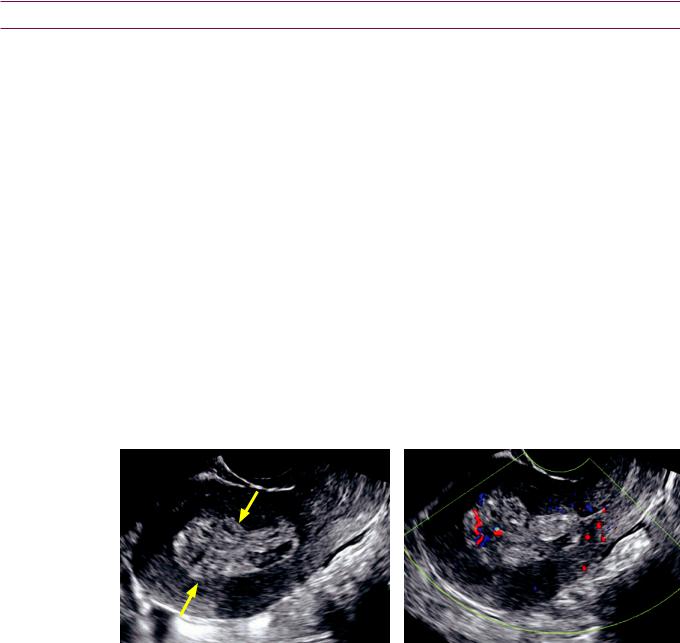
Miscellaneous first trimester disorders
Gestational trophoblastic disease (hydatidiform molar pregnancy)
•Gestational trophoblastic disease, also called hydatidiform molar pregnancy (or molar pregnancy for short), is invasive neoplastic overgrowth of the trophoblast into the myometrium or beyond. The trophoblast normally develops into the placenta.
•Theclassicclinicalpresentationofmolarpregnancyishyperemesis,markedlyelevated hCG,andanenlargeduterus.Thepatientmayalsopresentwithpainlessvaginalbleeding.
•Complete hydatidiform mole does not contain any fetal parts. It is caused by loss of the egg’s DNA prior to fertilization by the sperm and has a diploid karyotype of 46,XX (most commonly) or 46,XY. A complete mole may progress to metastatic choriocarcinoma.
Chorioadenoma destruens is a complete mole that invades the myometrium.
Molar pregnancy is associated with theca lutein cysts, which arise in response to elevated hCG.
•Partial hydatidiform mole is associated with some fetal development. It is usually caused by two sperm fertilizing the same egg and has a triploid karyotype of 69,XXX, 69,XXY, or 69,XYY. Partial mole is less likely to progress to choriocarcinoma.
•On ultrasound, molar disease causes uterine enlargement with a classic heterogeneous and multicystic snowstorm appearance. Visualization of fetal parts suggests a partial mole.
•Treatment of a molar pregnancy is endometrial suction curettage and close follow-up of serum hCG levels.
Retained products of conception (RPOC)
Retained products of conception: Sagittal grayscale ultrasound through the uterus demonstrates a markedly thickened and heterogeneous endometrium (arrows). Color Doppler shows vascularity within the endometrium, especially toward the uterine fundus. These imaging findings are suggestive of, but not diagnostic of, RPOC. Retained products of conception were proven at curettage.
Case courtesy Julie Ritner, MD, Brigham and Women’s Hospital.
•Retained products of conception (RPOC) are placental or fetal tissues that remain in the uterus after delivery, miscarriage, or termination.
•Untreated RPOC can lead to continued maternal bleeding and endometritis.
•The sonographic findings of an endometrial blood clot and RPOC overlap and it is often not possible to differentiate between these two entities. An endometrial mass, with or without Doppler flow, in the appropriate clinical context has only about a 50% positive predictive value. The presence of Doppler flow is not diagnostic of RPOC, as endometrial color Doppler flow can be detected in the presence or absence of RPOC. Color Doppler flow is seen more commonly with RPOC, however.
•A normal-appearing uterus, with an endometrial thickness less than 10 mm, is highly unlikely to contain RPOC.
525

Multiple gestations and placentation
•The placentation type (chorionicity and amnionicity) substantially affects the risk for pregnancy complications and influences how closely the pregnancy should be followed. The placentation should always be stated when first describing a multiple gestation.
•The zygosity (number of fertilized eggs) cannot always be determined by ultrasound. Monozygotic twins can have any placentation type, depending on when the developing zygote splits. Dizygotic twins, however, are always diamniotic/dichorionic, and only dizygotic twins can be different sexes.
Monozygotic (“identical”) twins arise from a single egg fertilized with a single sperm.
Dizygotic (“fraternal”) twins arise from two individually fertilized eggs.
•The chorionicity is the number of placentas.
Monochorionic twins share a single placenta.
Dichorionic twins each have a separate placenta.
•Amnionicity is the number of amnions.
Monoamniotic twins share a single amniotic sac.
Diamniotic twins each have a separate amniotic sac.
•By convention, chorionicity is stated before the amnionicity when stating the placentation. For instance, the abbreviation mono/di refers to monochorionic/ diamniotic twins.
Overview of complications by placentation type
•The more that the twins share, the greater the risk of complications.
•Di/di twins have an increased risk of premature delivery and low birth weight compared to singleton gestations.
•Mono/di twins have an increased risk of complications related to a shared placenta, including twin–twin transfusion, acardiac twin syndrome, and twin embolization.
•In addition to being at risk for the same complications as mono/di twins, mono/mono twins are also at risk for cord entanglement and being conjoined.
Early counting of multiple gestations
•If two separate gestational sacs are identified, the placentation is di/di. The zygosity is indeterminate.
Although dizygotic twins are always di/di, early splitting of a single fertilized egg can also lead to di/ di monozygotic twins.
•If a single gestational sac contains two yolk sacs, the placentation is mono/di, and the twins are monozygotic.
Dizygotic (“fraternal”) twins |
dizygotic twins |
•Dizygotic twins result when two
eggs are each fertilized by a different sperm.
• Dizygotic twins are always dichorionic/diamniotic.
dichorionic, diamniotic (100%)
526

Monozygotic (“identical”) twins
monozygotic twins
(single egg fertilized with a single sperm)
split 0−4 days
(33% of all monozygotic twins) embryo still in fallopian tube up to blastomere stage
split 4−8 days
(66% of all monozygotic twins) after implantation in uterus
after di erentiation of cytotrophoblast and syncytiotrophoblast (future placenta)
dichorionic diamniotic
(dual placentas, dual amnions)
monochorionic
diamniotic
(shared placenta, dual amnions)
|
split >8 days |
|
(1% of all monozygotic twins) |
embryo |
after development of |
|
chorion and amnion |
embryo
embryo
monochorionic monoamniotic
(shared placenta, shared amnion)
Monozygotic twins are the result of splitting of the blastocyst or embryo, formed by fertilization of a single egg by a single sperm. Although “identical” monozygotic twins are always the same sex, they may not be identical phenotypically due to local differences in the uterine and placental environment.
33% split early (0–4 days), before formation of either the placenta or amnion, leading to dichorionic/diamniotic twins
66% split intermediate (4–8 days), after formation of the placenta but before the amnion has developed, leading to monochorionic/diamniotic twins
1% split late (>8 days), after formation of the chorion and amnion, leading to monochorionic/monoamniotic twins. Very late splitting may result in conjoined twins, which are always monochorionic/monoamniotic.
527

Di/di (dichorionic/diamniotic) twins
•Di/di twins each have a separate placenta and amniotic sac.
•On ultrasound, two placentas can usually be separately identified.
•The inter-twin membrane will be relatively thick as there are two layers of chorion and two layers of amnion separating each twin.
•In the second and third trimesters, the thickness of the inter-twin membrane is less reliable to determine chorionicity because the membrane becomes thinner as gestation progresses.
•The twin peak sign (also called lambda sign)representsatriangle-shapedplacentalinfolding attheinterfaceoftheplacentaandthethickinter-twinmembranethatisseenindi/di twins.Thetwin peak signismostusefulwhenitisdifficulttodistinguishtwoplacentas.
•If the twins are different sexes, they must be dizygotic twins, which are always di/di.
Mono/di (monochorionic/diamniotic) twins
•Mono/di twins share a placenta, but have separate amniotic sacs.
•The shared placenta is usually apparent on ultrasound.
•The inter-twin membrane is thin, as it is composed of only two layers of amnion.
Mono/mono (monochorionic/monoamniotic) twins
•Mono/mono twins share both a placenta and a single amniotic sac.
•Mono/mono twins have a shared placenta with no intervening membrane between the twins. Intertwined cords are diagnostic of mono/mono placentation when seen.
•Isolated lack of visualization of an inter-twin membrane is not sufficient to diagnose mono/mono twins. If no inter-twin membrane (or intertwining cord) is seen, then the amnionicity cannot be determined. Because mono/mono twins are so rare, it is more common to have mono/di twins with non-visualization of the inter-twin membrane.
Conjoined twins
•Conjoined twins are caused by late (>13 days) incomplete division of the embryo.
Complications of monochorionic twins
Twin–twin transfusion syndrome (TTTS)
•Twin–twintransfusionsyndrome(TTTS)isacomplicationofmonochorionictwins(either mono-ordi-amniotic)causedbydisproportionatebloodflowbetweenthefetuses.
•The donor twin transfers excess blood flow to the recipient twin. The donor twin is small and has oligohydramnios. The recipient twin is larger and has polyhydramnios.
•There are three criteria to diagnose TTTS by ultrasound:
1)Disproportionate fetal sizes, with at least 25% discrepancy.
2)Disproportionate amniotic fluid, with the small twin having oligohydramnios and the large twin having polyhydramnios.
3)Single shared placenta (monochorionic).
•There is a spectrum of severities of TTTS. In the earliest stages, the donor twin’s bladder is still visible and the direction of umbilical artery Doppler flow is normal. Later stages are marked by fetal hydrops or death.
•A stuck twin describes severe oligohydramnios in the donor (small) twin. A stuck twin has so little amniotic fluid that the amnion is wrapped around the twin like shrink wrap.
•Treatment options of TTTS include laser ablation of placental arteriovenous fistulas, therapeutic amniocentesis from the recipient (large, poly) twin, or selective coagulation of the umbilical cord of the less viable twin.
528
Acardiac twins
•Acardiac twinning, also called twin reversed arterial perfusion (TRAP) sequence, is a severe variant of twin–twin perfusion syndrome. Similar to TTTS, acardiac twinning is a complication of monochorionic twins (either monoor di-amniotic).
•In acardiac twins, the donor fetus supplies circulation to itself and an acardiac twin, enabled by placental fistulous connections. The acardiac twin has rudimentary or no development of structures above the thorax.
•Doppler of the umbilical arteries and vein shows reversed flow in the acardiac twin.
Normally, the umbilical arteries carry deoxygenated blood out of the fetus, pumped by the fetal heart. In the acardiac twin, the umbilical arteries carry nutrient-depleted, poorly oxygenated blood into the fetus, pumped by the donor twin’s heart. Doppler of the acardiac twin’s umbilical arteries show an arterial waveform going into the fetus.
Normally, the umbilical vein carries oxygenated blood into the fetus, from the placenta. In the acardiac twin, the umbilical vein carries deoxygenated blood out of the fetus. Doppler of the acardiac twin’s umbilical vein shows a venous waveform going out of the fetus.
•Treatment is coagulation of the acardiac twin’s umbilical cord.
Twin embolization syndrome
•When one monochorionic twin dies in utero, the surviving twin is at risk for twin embolization syndrome, which can cause CNS, gastrointestinal, or renal infarcts.
•In general, prognosis for a surviving monochorionic twin is very poor when one twin dies in utero. In contrast, prognosis is generally good for a surviving dichorionic twin when one twin dies in utero.
529

Evaluation of the first trimester embryo
The first trimester embryo is too small for a complete fetal survey; however, a few key anatomic structures can be identified and evaluated.
Crown–rump length (CRL)
The crown–rump length (CRL) is used to assign gestational age from 6–12 weeks.
Measuring the CRL is straightforward in the first trimester as the fetus cannot flex or extend the neck.
Prosencephalon and rhombencephalon
By 8 weeks, the forebrain (prosencephalon) can be distinguished from the hindbrain (rhombencephalon). Both prosencephalon and rhombencephalon are hypoechoic, although the rhombencephalon is much more prominent. Absence of these structures may be the earliest finding of anencephaly.
Ventral abdominal wall
The midgut normally herniates through the ventral abdominal wall in the first trimester. During this herniation, the midgut rotates 270 degrees around the axis of the superior mesenteric artery (SMA).
Physiologic midgut herniation is usually complete by 12–13 weeks. Therefore, a pathologic ventral wall defect, such as omphalocele or gastroschisis, is generally not diagnosed before 13 weeks.
t is common to see some fullness at the base of the umbilical cord before 13 weeks, which usually represents physiologic midgut herniation. If the fullness is especially prominent then it may be prudent to bring the patient back for a follow-up at 13 weeks to evaluate for a true ventral wall defect.
Nuchal translucency
|
anterior |
|
abdominal wall |
midgut rotates |
midgut |
270˚ counterclockwise |
|
around SMA |
|
|
aorta |
physiologic |
SMA |
midgut |
|
herniation |
|

Second and third trimesters
Second and third trimester measurements
Head measurements
•The biparietal diameter (BPD) is measured from the outer edge of the skull closest to the transducer to the inner edge of the skull farthest from the transducer.
The plane of measurement is at the level of the thalami and cavum septum pellucidum. The skull should be completely visualized all the way around.
The corrected BPD incorporates the occipital frontal diameter (OFD) and a correction factor.
•The occipital frontal diameter
(OFD) is measured from the middle of the frontal skull to the middle of the occipital skull.
The measurement plane is the same as that used to measure the BPD, at the level of the thalami and cavum septum pellucidum.
The scanning plane for head measurements is at the level of the thalami (yellow arrows) and cavum septum pellucidum (red arrows).
The BPD (calipers marked 1) is measured from outer edge to inner edge of calvarium.
The OFD (calipers marked 2) is measured from middle to middle edge of calvarium.
Case courtesy Carol Benson, MD, Brigham and Women’s Hospital.
Abdomen measurements
•The abdominal diameter is measured from outer skin-to-skin in AP and transverse at the level of the intrahepatic umbilical vein, portal vein, and fetal stomach.
•Ideally, the abdomen should be round, with less than 1 cm difference between the AP and transverse measurements.
•The entire circumference of the skin should be well visualized.
•The best measurements are often obtained if the anterior–posterior axis of the fetal abdomen is angled approximately 45 degrees so that the artifacts from the spine are minimized.
The scanning plane for abdominal diameter is at the junction of the umbilical vein and portal vein (yellow arrow). The stomach (red arrow) should be visualized. Note how the anterior–posterior axis of the abdomen is angled approximately 45 degrees to minimize artifacts from the spine.
Case courtesy Carol Benson, MD, Brigham and Women’s Hospital.
Femur length
•The femur length is most accurately measured when the femur is closest to the transducer, perpendicular to the sound beam.
531
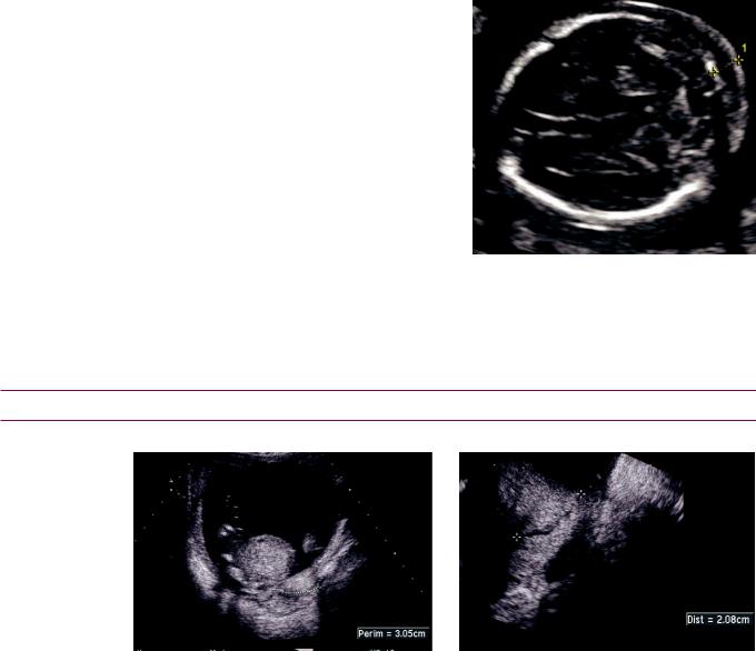
Amniotic fluid index (AFI)
•Toquantifytheamnioticfluidindex(AFI)between16and42weeks,thelargestvertical pocketoffluidismeasured(incm)ineachofthefourquadrantsandsummed.AFIvaries withgestationalage.Inborderlinecases,thesubjectiveassessmentshouldtakeprecedent. SomereferencesstatethatanAFIbetween7and25isnormal,butthesecutoffsvary.
Oligohydramnios: AFI ≤6.3 cm is ≤2.5th percentile. Peaks at 24 weeks: 9.0 cm = 2.5th percentile.
Polyhydramnios: AFI ≥19.2 cm is ≥97.5th percentile. Peaks at 36 weeks: 27.9 cm = 97.5th percentile.
•The amount of amniotic fluid should always be subjectively assessed.
Nuchal fold (second trimester only)
•A thickened nuchal fold is the most sensitive and specific ultrasound finding to suggest Down syndrome.
•Compared to nuchal lucency, measurement of nuchal fold is performed later in pregnancy. In contrast to the nuchal lucency, the nuchal fold is measured in the axial plane at the level of the posterior fossa.
•The nuchal fold is only measured from 16–20 weeks.
<5 mm is normal. 5–5.9 mm is borderline.
≥6 mm is a major marker for trisomy 21.
•A very thick nuchal fold may represent a cystic hygroma, which is associated with Turner syndrome (45,X).
Ultrasound of a 20-week fetus at the level of the brain and posterior fossa demonstrates a thickened nuchal fold >6 mm (calipers).
Case courtesy Beryl Benacerraf, MD,
Diagnostic Ultrasound Associates, Boston.
Evaluation of the cervix in second and third trimesters
Cervical shortening
Shortened cervix: Transabdominal grayscale ultrasound (left image) shows a borderline shortened cervix, measuring 3.05 cm. A transvaginal ultrasound more accurately demonstrates the length of the cervix, which is shortened, measuring 2.08 cm in length.
•Shortening of the cervix is a risk factor for pre-term delivery. A cervical length <3 cm is abnormal. The presence of cervical funneling (change in shape) is an ancillary finding.
The mnemonic trust your vaginal ultrasound canbeusedtorememberthesequenceofcervical funneling.AT-shapedcervixisnormal.Asfunnelingprogresses,thecervixresemblesY, V, and U shapes.
•Priortoviability(24weeks),treatmentiscervicalcerclage.After24weeks,treatmenttends tobeconservative(bedrest)duetoconcernformembranerupturewithanyprocedure.
532

Placenta
Placental embryology and physiology
•The placenta is formed by fetal chorion and maternal endometrium.
•The mature placental circulation allows exchange of oxygen and nutrients between maternal and fetal vessels through a membrane, although the blood does not admix.
Single umbilical artery
Two vessel cord (single umbilical artery):
Color Doppler through the fetal bladder demonstrates a single umbilical artery
(arrow).
Case courtesy Beryl Benacerraf, MD,
Diagnostic Ultrasound Associates, Boston.
•The normal umbilical cord has two umbilical arteries and a single umbilical vein. A single umbilical artery is associated with fetal anomalies (most commonly cardiovascular) in up to 50% of fetuses.
•There is an increased incidence of a single umbilical artery in trisomies 13 and 18.
Abnormalities of placental thickness
•Placental thickness is not routinely measured, but in fetal hydrops the placenta may become thickened.
•In polyhydramnios, the placenta usually becomes stretched and thinned. In the presence of polyhydramnios, if the placenta looks normal in thickness, or especially if the placenta looks thickened, concern should be raised for fetal hydrops.
Vasa previa
Grayscale ultrasound (left image) in the region of the fetal head and cervix demonstrates the position of the cervix (yellow arrows). Although the grayscale image appeared normal, color Doppler shows a placental vessel (red arrow) traversing the internal cervical os. Spectral Doppler (not shown) confirmed presence of a fetal arterial vessel.
Case courtesy Beryl Benacerraf, MD, Diagnostic Ultrasound Associates, Boston.
•Vasa previa is the traversing of fetal placental vessels across the internal cervical os, which can be caused by velamentous insertion or a placental succenturiate lobe.
Velamentous insertion is the insertion of the umbilical cord outside the margin of the placenta.
A succenturiate lobe is an island of placental tissue separate from the main placenta, connected to the main placenta by blood vessels.
533
Placenta previa and other abnormalities of placental position
•Obstetrical terminology regarding abnormalities of placental position in relation to the internal cervical os is confusing. The terms partial previa, complete previa, incomplete previa, marginal placenta, and low-lying placenta are inconsistent among references.
•The edge of the placenta should be >3 cm from the internal cervical os.
•If the placenta is <3 cm from the internal cervical os, it is best to simply be descriptive and describe how far the leading edge of the placenta is from the internal cervical os.
•Intrueplacentaprevia,theplacentacoverstheinternalcervicalos.Placentapreviaisseen inapproximately0.5%to1%ofdeliveriesandrequiresacesareansectionforsafedelivery.
A potential pitfall is over-diagnosis in the first and second trimesters due to contractions or overfilling of the maternal blader. To definitively diagnose previa, the patient should have an empty bladder and realtime scanning should be performed to confirm the lack of active contractions.
Placental abruption
•Placentalabruptionisprematureseparationoftheplacentafromitsuterineattachment.
•Thereisanincreasedincidenceofabruptioninmaternalhypertension,drugabuse,trauma, orrapiddecompressionofadistendeduterus(e.g.,fromalarge-volumeamniocentesis).
•A subchorionic hematoma is a variant seen early in pregnancy, where a hypoechoic crescent is present around the gestational sac.
•Placental abruption can have variable ultrasound findings and may even appear normal. Because a normal ultrasound is seen in 20% of cases of abruption, a negative ultrasound cannot exclude abruption. Typically, ultrasound of abruption shows a placental hematoma, which can be subchorionic (most commonly), retroplacental, or pre-placental. On clinical exam, blood may be present in the vaginal canal.
•Acute abruption may be especially challenging to diagnose on imaging as the hematoma is isoechoic to placenta.
placenta
hematoma
Acute retroplacental abruption: Ultrasound of the placenta shows a subtle, minimally hyperechoic retroplacental hematoma (arrows at placental/hematoma interface), nearly isoechoic to placenta. Acute
retroplacental hematoma can be very difficult to identify. Realtime scanning or review of cine loops may be helpful.
Case courtesy Julie Ritner, MD, Brigham and Women’s Hospital.
•Chronic abruption features hypoechoic blood products within the placenta.
Chronic abruption: Ultrasound of the placenta shows a heterogeneous, primarily hypoechoic preplacental hematoma (arrows), which lacks Doppler flow.
Case courtesy Julie Ritner, MD, Brigham and Women’s Hospital.
534

Placenta accreta
•Placenta accreta is a spectrum of abnormally tenacious or deep attachment of the placenta into the myometrium, carrying a risk of hemorrhage at the time of placental separation.
•Accreta is thought to be caused by scarring (which may be secondary to prior Caesarean section, D&C, endometritis, or adenomyosis) and resultant endometrial deficiency. It is especially important to consider accreta if an anterior placenta is present with a history of prior Caesarean section. Placenta previa also increases the risk of accreta.
•Ultrasound findings of accreta include loss of the normal retroplacental clear space, abnormalities at the bladder/placental interface, and prominent vascular lacunar spaces. The presence of a moth-eaten placenta with vascular lacunar spaces near the bladder is highly specific for accreta.
•Placenta accreta is an umbrella term that describes three degrees of placental attachment/invasion. Confusingly, the term “placenta accreta” also describes one of the specific three degrees.
•In placenta accreta, the placenta attaches deeply into the myometrium but does not invade.
Ultrasound shows thinning or absence of the normal hypoechoic subplacental zone.
•In placenta increta, the placenta invades into the myometrium.
•In placenta percreta, placenta penetrates through the myometrium and into or through the serosa.
Placenta percreta:
Sagittal ultrasound of the cervix show a large cystic space (yellow arrows) within the inferior placenta at the placental/bladder interface, consistent with a large vascular lake. The placenta has nearly completely invaded through the serosa into the bladder wall (red arrow).
Case courtesy Julie Ritner, MD, Brigham and Women’s Hospital.
535

Fetal hydrops
Overview of hydrops
Fetal hydrops:
Transverse ultrasound of a fetal head shows diffuse skin thickening of the scalp (arrows).
Polyhydramnios is also present (although incompletely seen on this single image).
Case courtesy Beryl Benacerraf, MD,
Diagnostic Ultrasound Associates, Boston.
•Hydrops is a fluid-overload state characterized by at least two of the following: Ascites, pleural or pericardial effusion, skin thickening, polyhydramnios, and placental enlargement.
•Hydrops may be classified as immune or non-immune. Prognosis is variable but tends to be poor for non-immune hydrops.
Immune hydrops
•Immune-mediated hydrops is fetal hemolytic anemia caused by prior maternal exposure to fetal antigens, by far most commonly the Rh antigen.
•Prognosis is good if treated with intrauterine or peripartum fetal blood transfusions.
Non-immune hydrops
•Non-immune hydrops can be due to a diverse array of causes, most of which lead to a common pathway of extracellular fluid overload. Prognosis of non-immune hydrops tends to be poor, as the primary cause is often not effectively treatable.
•Etiologies include (more common causes in bold):
Primary cardiac abnormalities, including structural abnormalities and arrhythmias.
Extra-cardiac shunt, including vein of Galen malformation, hepatic hemangioendothelioma, and twin–twin transfusion syndrome, all of which may lead to high-output cardiac failure.
Infectious, especially parvovirus B19 and TORCH infections.
Decreased oncotic pressure, due to hepatitis and fetal nephrotic syndrome. Increased capillary permeability, which can be due to anoxic injury. Venous obstruction, seen in Turner (45,X) syndrome.
Fetal ascites
•Whenseenwithotherabnormalities,ascitesisoneofthecriteriafordiagnosisofhydrops.
•Isolated fetal ascites may be due to urinary obstruction and resultant calyceal or bladder rupture or meconium peritonitis.
Fetal pleural effusion
•Fetal pleural effusion is a criterion for diagnosis of hydrops.
•When seen in isolation (not a component of hydrops or due to any other abnormality), fetal pleural effusion is mostly commonly caused by congenital chylothorax. Fetal chylothorax is thought to be due to thoracic duct or lymphatic malformation. At birth the pleural fluid is simple, but after the baby begins to drink milk it becomes chylous.
•Fetal pleural effusions can also be seen in Turner or Down syndromes.
536

Amniotic fluid
Overview of fetal amniotic fluid
•Amniotic fluid surrounds the fetus and is required for normal development of multiple organ systems, including the lungs.
•The amount of amniotic fluid should routinely be assessed subjectively. It is not necessary to routinely measure the amount of amniotic fluid; however, if a
measurement is needed, the amniotic fluid index (AFI) may be used for quantification.
•Amniotic fluid is produced primarily by the fetal genitourinary tract and is excreted by the fetus as urine. A small amount of amniotic fluid is also produced by the fetal lungs and nasopharyngeal cavities.
•Disorders of the fetal genitourinary tract may cause oligohydramnios due to insufficient secretion of fluid.
•Amniotic fluid is absorbed primarily by fetal swallowing.
•Disorders of the gastrointestinal tract or central nervous system may cause polyhydramnios due to impairment of swallowing or absorption of fluid.
Oligohydramnios
•Oligohydramnios is too little amniotic fluid.
•Oligohydramnios may lead to Potter sequence, which describes the typical malformations induced by confinement from oligohydramnios including facial dysmorphism, pulmonary hypoplasia, club feet, and musculoskeletal contractures.
•Most commonly, oligohydramnios is associated with intrauterine growth restriction (IUGR) without a fetal structural anomaly.
•Although malformations are relatively uncommon, the genitourinary system must be carefully evaluated in the setting of oligohydramnios. Several genitourinary anomalies may lead to oligohydramnios, including:
Renal agenesis – fatal if bilateral.
Congenital bladder outlet obstruction, including posterior urethral valves.
Bilateral ureteropelvic junction obstructions.
Renal dysplasias, including autosomal recessive polycystic kidney disease (ARPKD).
Polyhydramnios
•Polyhydramnios is too much amniotic fluid.
•Greater than half of cases of polyhydramnios are idiopathic, with a normal fetus. The remainder may be associated with chromosomal abnormalities, diabetes, or structural defects (primarily of the gastrointestinal tract), including:
Primary upper GI obstruction or atresia, such as laryngeal, esophageal or duodenal atresia. Secondary obstruction, due to diaphragmatic hernia, gastroschisis, or omphalocele. Severe CNS anomalies (which often cause disorders in swallowing).
Monochorionic twin syndromes, such as twin–twin transfusion syndrome.
Placental abnormalities, such as chorioangioma. Chorioangioma is a benign placental hemangioma that may cause polyhydramnios when highly vascular.
537

Fetal brain and spine
Ventriculomegaly
Ventriculomegaly:
Transverse ultrasound of the fetal head shows moderate symmetrical dilation of the lateral ventricles, with the calipers measuring 14 mm. Regardless of the gestational age, the lateral ventricles should always measure <10 mm.
Case courtesy Beryl Benacerraf, MD,
Diagnostic Ultrasound Associates, Boston.
•Ventriculomegaly is enlargement of the cerebral ventricles. The term hydrocephalus is usually avoided because that implies ventriculomegaly due to obstruction.
•Throughoutgestation,thelateralventriclesshouldeachmeasurelessthan10mmwhen measuredattheatrium.Theatriumistheconfluenceofthelateralventricle,temporal horn,andoccipitalhorn.Thenormalchoroidplexushasaroundedborderinthislocation.
Mild ventriculomegaly: 10–12 mm; moderate: 12–15 mm; marked: >15 mm
•Normally, the choroid plexus fills the lateral ventricle. The dangling choroid sign represents the dependent drooping of choroid plexus seen in ventriculomegaly.
Ventriculomegaly may be present even in a ventricle measuring <10 mm if there is >3 mm of fluid between the medial margin of the ventricle and the choroid.
•Ventriculomegalyisasignthatsomethingelseiswrong,withadiversearrayofetiologies:
Primary CNS structural (aqueductal stenosis, Dandy Walker, Chiari II, holoprosencephaly, agenesis of the corpus callosum).
Genetic (trisomies 13 and 18).
Destructive (due to infection, hemorrhage, or infarct).
Idiopathic.
Anencephaly
•Anencephaly is a lethal anomaly with complete lack of development of fetal cerebral cortex and calvarium above the orbits.
•AFP is elevated in anencephaly, as there is direct exposure of neural tissue to the amniotic fluid.
•Anencephaly may cause polyhydramnios due to impairment in swallowing.
•Angiomatous stroma is residual neural-type tissue that may be tethered above the head and may be confused with an encephalocele.
•The differential of anencephaly is amniotic band syndrome, which is almost always asymmetric.
Sagittal paramedian 17-week fetal ultrasound through the head and thorax shows lack of calvarium above the level of the orbits. Echogenic foci superior to the anencephalic head likely represent angiomatous stroma
(arrow).
Case courtesy Beryl Benacerraf, MD, Diagnostic
Ultrasound Associates, Boston.
538

Cephalocele
•A cephalocele is a midline neural tube defect characterized by protrusion of intracranial structures outside of the calvarium. The occipital skull is the most common location.
•A meningocele contains only meninges. An encephalocele also contains neural tissue.
•In addition to cephalocele, the primary differential consideration of a mass posterior to the occipital skull is a cystic hygroma, which is a congenital lymphatic malformation and the most common fetal neck mass.
Dandy Walker malformation
•Dandy Walker is a diverse spectrum of diseases characterized by hypogenesis of the cerebellar vermis and resultant fourth ventricular dilation.
•Dandy Walker is associated with agenesis of the corpus callosum.
Chiari II/Myelomeningocele
•Chiari II is the combination of a small posterior fossa and a neural tube defect. By far the most common associated neural tube defect is a lumbar myelomeningocele. A myelomeningocele contains both neural elements and meninges.
•The banana sign describes the characteristic flattened cerebellar hemispheres in the small posterior fossa. The banana sign is very specific for Chiari II. In fact, if the banana sign is seen, then a myelomeningocele is presumed to be present even if not identified on ultrasound.
•The lemon sign describes flattening of the frontal bones, causing the calvarium to have the morphology of a lemon when seen axially. Unlike the banana sign, the lemon sign is not specific for Chiari II.
Fetal ultrasound through the brain and posterior fossa demonstrates the banana and lemon signs. There is flattening of the frontal bones (lemon sign, yellow arrows) and flattening of the cerebellar hemispheres (banana sign, red arrows).
Although the lemon sign is nonspecific, the banana sign is very specific for Chiari II.
Sagittal ultrasound through the thoracolumbar spine in the same fetus shows a lumbar open neural tube defect (arrow) representing either a meningocele or myelomeningocele.
Case courtesy Beryl Benacerraf, MD, Diagnostic Ultrasound Associates, Boston.
539

Holoprosencephaly
Holoprosencephaly:
Oblique axial/coronal ultrasound through the posterior brain shows fused thalami across the midline (arrows). A large dorsal cyst is present, largely replacing the visualized supratentorial brain. No falx is seen.
Case courtesy Beryl Benacerraf,
MD, Diagnostic Ultrasound
Associates, Boston.
•Holoprosencephaly is failure of midline cleavage of the primitive prosencephalon in early embryologic development. The most severe form, alobar holoprosencephaly, leads to fused thalami and a single monoventricle that may communicate with a large dorsal cyst. Brain tissue surrounds the monoventricle, forming a characteristic boomerang shape.
•Holoprosencephaly is associated with trisomy 13, facial hypoplasias, and midline facial anomalies including clefts.
Agenesis of the corpus callosum
Agenesis of the corpus callosum:
Transverse ultrasound through the fetal head shows ventriculomegaly and a colpocephalic configuration of the lateral ventricle with a dilated teardrop-shaped occipital horn
(calipers). There is a concave medial border (arrows) of the lateral ventricle representing protrusion of Probst bundles. The ventricle is oriented parallel to the falx.
Case courtesy Beryl Benacerraf,
MD, Diagnostic Ultrasound
Associates, Boston.
•Absence of the corpus callosum can be a difficult diagnosis to make. Because the normal corpus callosum is not always visualized on ultrasound, it is often necessary to rely on secondary abnormal morphology of the ventricular system to diagnose absence of the corpus callosum.
Absence of the cavum septum pellucidum (CSP) isassociatedwithagenesisofthecorpuscallosum.
Abnormal teardrop morphology of the lateral ventricles with dilated occipital horns, known as colpocephaly, is associated with absence of the corpus callosum. Colpocephaly is often seen together with ventriculomegaly.
Widely separated ventricular frontal horns and parallel configuration of the lateral ventricles both suggest agenesis of the corpus callosum.
The medialbordersofthelateralventriclesmaybeconcave,duetoprotrusionofProbstbundlesand thecingulategyrus.Probstbundlesareaxonsthatnormallyconstitutethecorpuscallosum.Inagenesis ofthecorpuscallosum,theProbstbundlesdonotcrossthemidline.Instead,theypursueanaberrant courseparalleltotheinterhemisphericfissure,indentingthemedialmarginofthelateralventricles.
A midlineinterhemisphericcystmaybepresent,representingsuperiorherniationofthethirdventricle.
•A minority of babies with agenesis of the corpus callosum have trisomy 8, 13, or 18.
540

Absence of the cavum septum pellucidum (CSP)
•The cavum septum pellucidum (CSP) should always be identified in a normal fetus.
•If the CSP is not seen, the primary consideration is agenesis of the corpus callosum, as the cavum septum pellucidum and corpus callosum are formed simultaneously.
•Uncommonly, the CSP may be absent in the presence of a normal corpus callosum. This may represent septo-optic dysplasia and fetal MRI should be recommended.
Hydranencephaly
•Hydranencephaly is complete cortical destruction due to infarct or infection. The brain parenchyma is obliterated and replaced by fluid.
•ThemostcommoncauseofhydranencephalyisinuterocompleteocclusionoftheMCA.
•In contrast to severe hydrocephalus, a cortical mantle is absent in hydranencephaly.
•In contrast to holoprosencephaly, a falx is typically visualized in hydranencephaly.
Choroid plexus cyst
Choroid plexus cyst:
Transverse ultrasound of a fetal head shows a hypoechoic cyst (arrow) located within the echogenic, otherwise normal-appearing choroid.
•Cysts within the choroid plexus are common (seen in 1–5% of fetal surveys). The vast majority of choroid plexus cysts are present in normal fetuses and resolve on followup scans. However, up to 50% of trisomy 18 fetuses will have choroid plexus cysts.
•Choroid plexus cysts can be considered an incidental finding in the absence of any other sonographic abnormality and with a normal maternal serum screen.
•A potential mimicker of a choroid plexus cyst is the spongy choroid, which describes a heterogeneous echogenic choroid that is a normal variant.
Vein of Galen malformation
Vein of Galen malformation:
Color Doppler ultrasound of the fetal head shows an enlarged midline vascular structure representing a vein of Galen malformation.
Case courtesy Beryl Benacerraf, MD,
Diagnostic Ultrasound Associates, Boston.
•Vein of Galen malformation is dilation of the vein of Galen (in the pineal region) caused by an arteriovenous fistula.
•Vein of Galen aneurysm is a shunt lesion that may cause high-output cardiac failure, leading to hydrops.
541

Fetal thorax
Evaluation of the fetal thorax
•Abnormal position of the heart in the thorax is an important clue to the possible presence of a thoracic anomaly. Abnormal cardiac position or axis may be secondary to a thoracic mass lesion or pulmonary hypoplasia.
Congenital diaphragmatic hernia (CDH)
•Congenital diaphragmatic hernia (CDH) is herniation of abdominal organs (most commonly bowel) into the thorax through a diaphragmatic defect. CDH is the most common fetal intrathoracic mass lesion.
•Most cases are isolated anomalies; however, a prominent minority of fetuses have other anomalies, most commonly congenital heart disease.
•Byfar,themostcommonlocationforCDHistheleftposteriorthorax,calledaBochdalek hernia(mnemonic:“backtotheleft”).ABochdalekherniamaydisplacetheheart.
•When a CDH occurs on the right it is called a Morgagni hernia. The diaphragmatic defect of a Morgagni hernia tends to be anterior, with the liver the most commonly herniated organ.
•ThetwoclassicfetalultrasoundfindingsofCDHareacysticintra-thoracicmassrepresenting thestomachand/orbowel,andabsenceofthestomachbubblebelowthediaphragm.
•Complications of CHD include pulmonary hypoplasia on the affected side, bowel obstruction with resultant polyhydramnios, and obstruction of venous return due to IVC compression, which may lead to ascites.
Bronchopulmonary foregut malformation
•Bronchopulmonary foregut malformations are a spectrum of congenital abnormalities of the fetal lungs and upper GI tract.
•Congenital pulmonary airway malformation (CPAM) is a hamartomatous proliferation of small airways that communicates with the bronchial tree. Blood supply is from the pulmonary circulation. CPAM was previously called congenital cystic adenomatoid malformation (CCAM).
CPAMcanbeclassifiedintothreetypesbasedonthesizeofcysts(TypeI–largecysts;TypeII–small cysts;TypeIII–tinycyststoosmalltoseeonultrasound).Todaythisclassificationisnotusedmuch becauseprognosisisdependentonsizeoftheentirelesionratherthanthesizeoftheindividualcysts.
CPAM is not associated with other anomalies (unlike CDH).
CPAM may appear to spontaneously regress on ultrasound, but will remain apparent on CT or MR.
•Sequestration is aberrant lung tissue with a systemic blood supply, usually from the aorta. The most characteristic location of sequestration is the left lower lobe.
The classic ultrasound appearance of sequestration is an echogenic mass at the left lung base. The sequestration may occasionally be subdiaphragmatic and simulate an adrenal mass.
SystemicbloodsupplyshouldbeconfirmedwithcolorDoppler.IntheabsenceoftheDopplerfindings, sequestrationmaybedifficulttodifferentiatefromCPAM.IncontrasttothefindingsofCPAM,cysts arelesscommon,thereislessmasseffect,andlocationisalmostalwaysinthelowerlobes.
•Bronchogenic, gastrointestinal duplication, and neurenteric cysts almost always appear as solitary, simple cysts on ultrasound.
Pulmonary hypoplasia
•Pulmonaryhypoplasiaisinadequatelungdevelopment.Hypoplasiacanbeduetoathoracic masslesion(suchasCDH),oligohydramnios,oraskeletaldysplasiaaffectingtheribs.
•It is important to evaluate the size of the fetal thorax in relation to the abdomen on coronal images. A small, bell-shaped fetal thorax suggests pulmonary hypoplasia.
542

Laryngeal or tracheal atresia
•Atresia of the upper airway, otherwise known as congenital high airway obstruction syndrome (CHAOS), is lethal and may cause bilateral enlarged echogenic lungs.
Fetal abdomen
•Anomalies of the fetal gastrointestinal tract may cause polyhydramnios due to disruption of swallowing or impaired absorption of swallowed amniotic fluid.
Esophageal atresia
•Esophageal atresia is a blind-ending esophagus, due to incomplete division of the foregut in early embryologic development.
•Esophageal atresia is usually associated with a tracheoesophageal fistula.
•The classic ultrasound findings of esophageal atresia are polyhydramnios and an absent stomach bubble.
Duodenal atresia (DA)
st
duo
Duodenal atresia: Two transverse images through the fetal abdomen (left image) show the dilated stomach (st) and dilated proximal duodenum (duo) representing the double bubble sign. The image on the right confirms that these two dilated structures connect (arrow).
Case courtesy Julie Ritner, MD, Brigham and Women’s Hospital.
•Duodenal atresia (DA) causes duodenal obstruction from lack of recanalization of the duodenal lumen. Duodenal atresia is the most common cause of fetal duodenal obstruction.
•Approximately one third of babies with DA will have Down syndrome. If a double bubble sign is seen, a careful screen for additional findings of Down syndrome should be performed (e.g., thorough heart exam, nuchal fold if 16–20 weeks, etc.).
•The classic appearance of DA is the double bubble sign, representing a dilated stomach and dilated proximal duodenum. The differential diagnosis of the double bubble sign includes duodenal web, stenosis, and annular pancreas.
Distal fetal bowel obstruction
•A distal fetal bowel obstruction may be structural or functional.
•Structural causes of distal bowel obstruction include jejunal atresia, ileal atresia, and anorectal malformation.
Anorectal malformation is commonly associated with additional abnormalities, including the
VACTERL association (vertebral, anorectal, cardiac, tracheoesophageal, renal, and limb anomalies).
•Functional bowel obstruction may be due to Hirschsprung disease or meconium ileus.
Hirschsprung disease results in a functional obstruction due to absent distal enteric ganglion cells.
Meconiumileuscausesobstructionfromimpactionofmeconiumintheileum.Nearlyallinfantswith meconiumileushavecysticfibrosis;however,meconiumileusisrarelyidentifiedbeforethe3rd trimester.
543
Fetal manifestations of meconium
•Meconium ileus is bowel obstruction caused by impacted meconium in a fetus with cystic fibrosis.
•Meconium peritonitis is peritoneal inflammation secondary to in utero bowel perforation and resultant spillage of meconium into the peritoneal cavity. This leads to peritoneal adhesions and, ultimately, dystrophic calcifications.
•Meconiumpseudocyst isacysticabdominalstructure,oftenwithperipheralcalcification, representingawalled-offbowelperforation.Itisasequelaofmeconiumperitonitis.
Hyperechoic small bowel
•Hyperechoic or echogenic bowel is a nonspecific finding that is associated with Down syndrome. Other causes of hyperechoic small bowel include TORCH infection, cystic fibrosis, and swallowing of intra-amniotic blood. It may also be associated with intrauterine growth restriction.
•If the bowel is only mildly echogenic (less echogenic than bone) and no mass effect is present, this appearance may represent a normal variant.
Omphalocele
•An omphalocele is a midline anterior abdominal wall defect with resultant herniation of intra-abdominal contents covered by a peritoneal membrane. Omphalocele is the most common anterior abdominal wall defect.
•The key to differentiate omphalocele from gastroschisis (discussed below) is the position of the umbilical cord insertion. In omphalocele, the umbilical cord inserts centrally at the base of the herniated sac.
•When small, omphaloceles often contain just bowel. Larger omphaloceles may also contain liver and carry a worse prognosis.
•Omphalocele is associated with other anomalies in 50–75% of cases, including cardiac anomalies, trisomies, and Beckwith–Wiedemann syndrome (a congenital overgrowth syndrome characterized by omphalocele, macroglossia, hemihypertrophy, and visceromegaly).
Gastroschisis
•Gastroschisis is a paraumbilical (usually right-sided) anterior abdominal wall defect, through which bowel herniates without a peritoneal covering. Gastroschisis is the second most common abdominal wall defect.
•Unlike omphalocele, gastroschisis is usually seen as an isolated anomaly.
•Gastroschisis is seen more commonly in very young (teenage) mothers.
•Prognosis is similar to a simple omphalocele (with no other
associated abnormalities).
Pentalogy of Cantrell
Gastroschisis: Transverse ultrasound through the fetal abdomen shows loops of bowel herniating outside of the anterior abdominal wall (yellow arrows), lateral to the normal midline cord insertion (red arrows). The bowel loops have a lobulated contour because there is no peritoneal covering.
Case courtesy Beryl Benacerraf, MD, Diagnostic
Ultrasound Associates, Boston.
•PentalogyofCantrellisararedisorderconsistingofectopiacordis(extra-thoracicheart), omphalocele,diaphragmaticdefect,pericardialdefect,anddisruptionofthesternum.
544
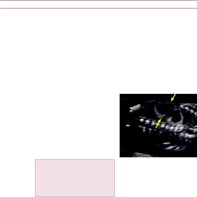
Fetal genitourinary tract
•Although oligohydramnios may be due to many causes, the complete genitourinary
(GU) tract (kidneys, ureters, bladder, and urethra) should be carefully evaluated in every fetus with oligohydramnios.
•Oligohydramnios due to a fetal GU malformation can be divided into three categories:
Fetal hydronephrosis (obstructive uropathy).
Cystic renal disease.
Bilateral renal agenesis.
•If the fetal bladder is not visualized (a bladder not filled with fluid will not be visible on ultrasound) the cause of oligohydramnios is likely a renal anomaly, such as:
Bilateral multicystic dysplastic kidneys.
Bilateral renal agenesis.
Autosomal recessive polycystic kidney disease (ARPKD).
•Normal fetal kidneys grow approximately 1 mm per week of gestation. For instance, a 20 week fetus should have kidneys approximately 2 cm in length.
Fetal hydronephrosis overview
•Screeningforfetalhydronephrosisisroutinely performedtoevaluateforpotentially treatablecauses,suchasobstructionor reflux,whichmayleadtoprogressive childhoodrenalfailureifundiagnosed.
•The axial diameter of the renal pelvis is measured to assess for hydronephrosis. The accepted range of normal depends on gestational age. Less than 4 mm is always normal and ≥10 mm is always abnormal.
Normal: <5 mm greater than 20 weeks; <4 mm at 16–20 weeks.
Equivocal: 5–9 mm greater than 20 weeks; 4–6 mm at 16–20 weeks.
Abnormal: ≥10 mm at 30 weeks;
≥8 mm at 20–30 weeks; ≥6 mm at 16–20 weeks.
Fetal hydronephrosis: Coronal fetal ultrasound shows bilateral hydronephrosis, severe on the right (arrows). This fetus has trisomy 18, which is associated with hydronephrosis.
Case courtesy Beryl Benacerraf, MD, Diagnostic
Ultrasound Associates, Boston.
•Hydronephrosis, hydroureter, and a normal bladder may be due to distal ureteral obstruction or reflux.
Distal ureteral obstruction from ectopic insertion of the ureter into the bladder is often associated with the upper pole moiety of a duplicated collecting system. The most common ectopic insertion of an upper pole moiety is inferior and medial to the normally inserting lower pole ureter.
A ureterocele is dilation of the intramural portion of the ureter which balloons out into the bladder and causes a functional obstruction at the ureterovesicular junction. Ureterocele is also associated with ectopic insertion of the upper pole moiety of a duplicated system.
Although not a physical obstruction, severe vesicoureteral reflux may cause hydronephrosis and hydroureter, but the bladder remains normal.
•Bladder outlet obstruction leads to hydronephrosis, hydroureter, and a dilated bladder.
Posterior urethral valves is by far the most common cause of bladder outlet obstruction.
Urethral atresia is a rare cause of bladder outlet obstruction.
•Hydronephrosis can lead to cystic renal dysplasia, a sign of irreversible renal damage.
545

Posterior urethral valves
•Posterior urethral valves is congenital obstruction of the posterior urethra due to a membranous flap in the proximal urethra.
•Ultrasound shows severe dilation of the posterior urethra resulting in the keyhole sign. The bladder is typically enlarged and thickened. Hydroureteronephrosis and oligohydramnios are usually present.
Autosomal recessive polycystic kidney disease (ARPKD)
Autosomal recessive polycystic kidney disease:
Transverseultrasoundthroughthefetalabdomen atthelevelofthekidneysshowsmassively enlargedandechogenickidneys(arrows).
There is complete absence of surrounding amniotic fluid, consistent with severe oligohydramnios.
Case courtesy Beryl Benacerraf, MD, Diagnostic
Ultrasound Associates, Boston.
•Autosomal recessive polycystic kidney disease (ARPKD) is a congenital disorder of diffuse collecting tubule dilation, leading to innumerable tiny renal cysts that are too small to be resolved by sonography.
•Ultrasound findings include very large and echogenic kidneys and severe oligohydramnios, caused by markedly reduced renal function.
•Prognosis is poor. ARPKD is associated with hepatic fibrosis if the baby survives infancy.
Multicystic dysplastic kidney (MCDK)
•Multicystic dysplastic kidney (MCDK) is thought to be the end result of fetal obstructive uropathy.
•If MCDK is unilateral and there are no associated abnormalities, the prognosis is excellent. MCDK may be fatal if bilateral, however.
•MCDK may affect only the upper pole of an obstructed duplicated system.
•On imaging, multiple non-communicating cysts are interspersed with dysplastic renal parenchyma. In contrast to hydronephrosis, the cysts of MCDK do not connect to the collecting system.
•The natural history of MCDK is gradual involution.
546

Fetal musculoskeletal imaging
Osteogenesis imperfecta (OI)
Osteogenesis imperfecta (nonfatal type): Third-trimester fetal survey shows an abnormal kinking (arrow) of the femur from prior in utero fracture.
Case courtesy Julie Ritner, MD, Brigham and Women’s Hospital.
•Osteogenesis imperfecta (OI) is a spectrum of congenital bone anomalies characterized by multiple fractures due to abnormal type I collagen. There are several types of OI, with type 2 being lethal. Type 2 can be diagnosed in the second trimester, but types 1, 3, and 4 are not typically diagnosed until the third trimester.
•OI causes severe shortening of the long bones (>3 SD below the mean). The long bones and ribs appear “wrinkled” due to multiple fractures. The thorax is usually small due to broken and structurally soft ribs.
•Unlike thanatophoric dysplasia, bony mineralization is decreased. Decreased calvarial mineralization causes the entire brain (including the nearfield) to be visualized well.
Thanatophoric dysplasia
•Thanatophoric dysplasia is a lethal skeletal dysplasia with characteristic telephone receiver femurs, severe limb shortening and bowing, and rib shortening.
•Platyspondyly is characteristic, which is flattening of the ossified portions of the vertebral bodies.
•The classic cloverleaf skull is caused by protrusion of the frontal and temporal lobes.
•Bony mineralization is normal.
Sacrococcygeal teratoma
Sacrococcygeal teratoma:
Parasagittal ultrasound of the fetal torso and spine shows a large, heterogeneous, solid and cystic mass (arrows) arising from the sacrum/coccyx.
In contrast to myelomeningocele, the lumbosacral spine demonstrates normal tapering and the overlying skin is intact.
Case courtesy Beryl Benacerraf, MD,
Diagnostic Ultrasound Associates,
Boston.
•Sacrococcygeal teratoma is a germ cell tumor of the sacrum. It often presents prenatally as a mixed solid and cystic complex mass.
•Sacrococcygeal teratoma may be associated with high output cardiac failure.
•Theotherprimarydifferentialconsiderationforadistalfetalspinalmassisa myelomeningocele,whichisassociatedwithspineabnormalitiesandadorsalskindefect.
547

Trisomies and syndromes
•Fetal anomalies often occur in associations. For instance, omphalocele may be a sentinel finding that signifies the presence of trisomy 13, 18, or Beckwith–Wiedemann syndrome. If a sentinel finding is seen, a careful search should be performed for associated anomalies seen in trisomies and syndromes.
Trisomy 13 from head to toe
|
• Holoprosencephaly and midline facial anomalies. |
||
13 |
• |
Encephalocele. |
|
• |
Congenital heart disease. |
||
trisomy |
|||
• |
Omphalocele. |
||
• |
Horseshoe kidney. |
||
• |
Polycystic kidneys. |
||
|
|||
|
• |
Polydactyly. |
|
Trisomy 18 from head to toe
|
|
Transverse image of the fetal skull shows a large |
3D ultrasound of the fetal face shows a cleft lip |
|
|
choroid plexus cyst (arrow) nearly replacing the |
(red arrow). |
|
|
entire choroid. |
|
|
|
Trisomy 18: Case courtesy Beryl Benacerraf, MD, Diagnostic Ultrasound Associates, Boston. |
|
|
|
||
|
• Strawberry sign: Inward bowing of the frontal bones creates a strawberry shape to the calvarium, with |
||
|
|
the tip of the strawberry projected anteriorly. |
|
|
• |
Choroid plexus cysts. |
|
|
• |
Facial cleft. |
|
18 |
• |
Micrognathia. |
|
|
|
|
|
trisomy |
• |
Cardiac anomalies. |
|
• |
Omphalocele. |
|
|
• |
Congenital diaphragmatic hernia. |
|
|
|
• |
Horseshoe kidney. |
|
|
• |
Hydronephrosis. |
|
|
• Clenched hand that never opens, with overlapping fingers. |
||
|
• |
Rocker bottom feet. |
|
548

Trisomy 21 (Down syndrome) from head to toe
• Increase in nuchal fold (>6 mm), measured between weeks 15 and 21, is the single most sensitive and
|
|
specific ultrasound finding for trisomy 21. |
|
|
• In contrast, nuchal translucency is measured earlier in the pregnancy (11–14) weeks, and is less |
||
|
|
specific for trisomy 21. |
|
21 |
• Absent ossification of nasal bone. |
||
• Cystic hygroma (although more common in Turner syndrome). |
|||
trisomy |
|||
• Congenital heart disease: VSD and endocardial cushion defect in particular. |
|||
|
|||
|
• |
Echogenic bowel. |
|
|
• |
Duodenal atresia. |
|
|
• |
Echogenic intracardiac focus. |
|
|
• Shortened femur and humerus. |
||
|
• Hypoplasia of the middle phalanx of the little finger. |
||
|
• |
Sandal gap toes. |
|
Beckwith–Wiedemann syndrome (BWS)
Wiedemann
Beckwith–
•Beckwith–Wiedemann syndrome (BWS) is a syndrome of overgrowth that carries an increased risk of childhood cancers. BWS is mostly sporadic, but 10–15% of cases follow an autosomal dominant inheritance.
•BWS increases the risk of developing Wilms tumor (the most common tumor in BWS),
hepatoblastoma, and other childhood tumors. The standard of care is screening with abdominal ultrasound every 3 months until age 8.
•Hemihypertrophy, organomegaly.
•Macroglossia.
•Omphalocele.
•Perinatal hypoglycemia.
Meckel–Gruber
Grubrer
Meckel–
•Meckel–Gruber is an autosomal recessive multi-organ syndrome.
•Encephalocele.
•Renal dysplasia causing multiple tiny renal cysts, which appear as echogenic kidneys, analogous to
ARPKD.
•Polydactyly.
Neonatal brain imaging
Germinal matrix hemorrhage
•The germinal matrix is the site of neuronal precursor cells located in the caudothalamic groove.
•Grade I: Hemorrhage confined to the germinal matrix.
•Grade II: Hemorrhage extends into the ventricles without ventriculomegaly.
•Grade III: Hemorrhage extends into the ventricles with ventriculomegaly.
•Grade IV: Hemorrhage extends out of the ventricles into the parenchyma.
549

References, resources, and further reading
General References:
Middleton, W.D., Kurtz, A.B. & Hertzberg, B.S. Ultrasound: The Requisites (2nd ed.). Mosby. (2003).
Rumack, C.M., Wilson, S.R., Charboneau, J.W. & Levine, D. Diagnostic Ultrasound (4th ed.). Mosby. (2011).
Hepatobiliary and Right Upper Quadrant:
Berk, R., Van der Vegt, J. & Lichtenstein, J. The hyperplastic cholecystoses: cholesterolosis and adenomyomatosis. Radiology, 146(3), 593(1983).
Breda Vriesman, A.C. van. et al. Diffuse gallbladder wall thickening: differential diagnosis. AJR. American journal of roentgenology, 188, 495-501(2007).
Chawla, S., Trick, W.E., Gilkey, S. & Attar, B.M. Does Cholecystectomy Status Influence the Common Bile Duct Diameter? A Matched-Pair Analysis. Digestive Diseases and Sciences, 55, 1155-60(2010).
Coss, A. & Enns, R. The investigation of unexplained biliary dilatation. Current gastroenterology reports, 11(2), 155-9.
Gallahan, W.C. & Conway, J.D. (2010). Diagnosis and management of gallbladder polyps. Gastroenterology clinics of North America, 39(2), 359-67, x.
Goyal, N. et al. (2009). Non-invasive evaluation of liver cirrhosis using ultrasound. Clinical radiology, 64(11), 1056-66(2009).
Hanbidge, A.E. et al. From the RSNA refresher courses: imaging evaluation for acute pain in the right upper quadrant. Radiographics, 24, 1117-35(2004).
McNaughton, D.A. & Abu-Yousef, M.M. Doppler US of the liver made simple. Radiographics, 31, 161-88(2011).
Rosenthal, S.J., Cox, G.G., Wetzel, L.H. & Batnitzky, S. Pitfalls and differential diagnosis in biliary sonography. Radiographics, 10(2), 285-311(1990).
Rybicki, F. The WES Sign. Radiology, 214, 881-2(2000).
Spence, S.C., Teichgraeber, D. & Chandrasekhar, C. Emergent right upper quadrant sonography. Journal of ultrasound in medicine, 28, 479-96(2009).
Yarmenitis, S.D. Ultrasound of the gallbladder and the biliary tree. European radiology, 12(2), 270-82(2002).
Pancreas:
Bruno, C., Minniti, S. & Schenal, G. The Role of Ultrasound in Acute Pancreatitis. In E.J. Balthazar, A.J. Megibow & R.P. Mucelli (Eds.), Imaging of the Pancreas: Acute and Chronic Pancreatitis (pp. 33-47). Springer. (2009).
DʼOnofrio, Mirko. The Role of Ultrasound (in Chronic Pancreatitis). In E. Balthazar, A. Megibow & Roberto Pozzi Mucelli (Eds.), Imaging of the Pancreas: Acute and Chronic Pancreatitis (pp. 139-48). Springer. (2009).
Martínez-Noguera, a. & DʼOnofrio, M. Ultrasonography of the pancreas. Conventional imaging. Abdominal imaging, 32(2), 136-49(2007).
Spleen:
Peddu, P., Shah, M. & Sidhu, P.S. Splenic abnormalities: a comparative review of ultrasound, microbubble-enhanced ultrasound and computed tomography. Clinical radiology, 59(9), 777-92(2004).
Renal:
Mostbeck, G.H., Zontsich, T. & Turetschek, K. Ultrasound of the kidney: obstruction and medical diseases. European radiology, 11(10), 1878-89(2001).
Paspulati, R.M. & Bhatt, S. Sonography in benign and malignant renal masses. Radiologic clinics of North America, 44(6), 787-803(2006).
Tublin, M.E., Bude, R.O. & Platt, J.F. The resistive index in renal Doppler sonography: where do we stand? American Journal of Roentgenology, 180(4), 885(2003).
Vourganti, S., Agarwal, P.K., Bodner, D.R. & Dogra, V.S. Ultrasonographic evaluation of renal infections. Radiologic clinics of North America, 44(6), 763-75(2006).
550

Scrotum and testicle:
Cokkinos, D.D., Antypa, E., Tserotas, P., Kratimenou, E., Kyratzi, E., Deligiannis, I. et al. Emergency ultrasound of the scrotum: a review of the commonest pathologic conditions. Current Problems In Diagnostic Radiology, 40(1), 1-14(2011).
Dogra, V., Gottlieb, R., Oka, M. & Rubens, D. Sonography of the scrotum. Radiology, 227, 18-36(2003).
Frates, C., Benson, C.B., Laing, F.C., Disalvo, N. & Doubilet, M. Solic Extratesticular Masses Evaluated with Sonography: pathologic correlation. Radiology, 204, 43-6(1997).
Pearl, M. & Hill, M. Ultrasound of the Scrotum. Seminars in Ultrasound, CT, and MRI, 28(4), 225-48(2007).
Vascular:
Bounameaux, H., Perrier, A. & Righini, M. Diagnosis of venous thromboembolism: an update. Vascular medicine (London, England), 15(5), 399-406(2010).
Grant, E.G. et al. Carotid artery stenosis: gray-scale and Doppler US diagnosis – Society of Radiologists in Ultrasound Consensus Conference. Radiology, 229(2), 340-6(2003).
Hamper, U.M., DeJong, M.R. & Scoutt, L.M. Ultrasound evaluation of the lower extremity veins. Radiologic clinics of North America, 45(3), 525-47, ix(2007).
Sidhu, R. & Lockhart, M.E. Imaging of Renovascular Disease. Seminars in Ultrasound, CT, and MRI, 30(4), 271-88(2009).
Spyridopoulos, T.N. et al. Ultrasound as a first line screening tool for the detection of renal artery stenosis: a comprehensive review. Medical ultrasonography, 12(3), 228(2010).
Thyroid:
Frates, M.C. et al. Management of thyroid nodules detected at US: Society of Radiologists in Ultrasound consensus conference statement. Radiology, 237(3), 794-800(2005).
Female pelvis:
Goldstein, S.R. Abnormal uterine bleeding: the role of ultrasound. Radiologic clinics of North America, 44(6), 901-10(2006).
Levine, D. et al. Management of Asymptomatic Ovarian and Other Adnexal Cysts Imaged at US: Society of Radiologists in Ultrasound Consensus Implications for Patient Care. Ultrasound Quarterly, 26, 121-31(2010).
Patel, M.D. Practical approach to the adnexal mass. Radiologic clinics of North America, 44(6), 879-99(2006).
Potter, A.W. & Chandrasekhar, C. a. US and CT evaluation of acute pelvic pain of gynecologic origin in nonpregnant premenopausal patients. Radiographics, 28(6), 1645-59(2008).
Ectopic and early pregnancy:
Bhatt, S., Ghazale, H. & Dogra, V.S. Sonographic evaluation of ectopic pregnancy. Radiologic clinics of North America, 45(3), 549-60, ix(2007).
Doubilet, P.M. & Benson, C.B. Emergency obstetrical ultrasonography. Seminars in roentgenology, 33(4), 339-50(1998). Doubilet, P.M. & Benson, C.B. First, do no harm.. to early pregnancies. Journal of Ultrasound in Medicine, 29(5), 685(2010).
Durfee, S.M., Frates, M.C., Luong, A. & Benson, C.B. The sonographic and color Doppler features of retained products of conception. Journal of ultrasound in medicine, 24(9), 1181-6; quiz 1188-9(2005).
Eyvazzadeh, A.D. & Levine, D. Imaging of pelvic pain in the first trimester of pregnancy. Radiologic clinics of North America, 44(6), 863-77(2006).
Lin, E.P., Bhatt, S. & Dogra, V.S. Diagnostic clues to ectopic pregnancy. Radiographics, 28(6), 1661-71(2008).
551

Multiple gestations:
Lewi, L. et al. Monochorionic diamniotic twins: complications and management options. Current opinion in obstetrics & gynecology, 15(2), 177-94(2003).
Shetty, a. & Smith, a.P.M. The sonographic diagnosis of chorionicity. Prenatal diagnosis, 25(9), 735-9(2005).
Placenta:
Baughman, W.C., Corteville, J.E. & Shah, R.R. Placenta Accreta: Spectrum of US and MR Imaging Findings. Radiographics, 28(7), 1905(2008).
Elsayes, K.M. Imaging of the Placenta: A Multimodality Pictorial Review. Radiographics, 29(5), 1371-91(2009).
Fetal screening and anomalies:
Bellini, C. et al. Etiology of nonimmune hydrops fetalis: a systematic review. American Journal of Medical Genetics. Part A, 149A(5), 844-51(2009).
Bethune, M. Time to reconsider our approach to echogenic intracardiac focus and choroid plexus cysts. The Australian & New Zealand Journal of Obstetrics & Gynaecology, 48(2), 137-41(2008).
Biyyam, D., Chapman, T. & Ferguson, M. Congenital Lung Abnormalities: Embryologic Features, Prenatal Diagnosis, and Postnatal Radiologic-Pathologic Correlation1. Radiographics, 98195, 1721-39(2010).
Chen, C.-P. Pathophysiology of increased fetal nuchal translucency thickness. Taiwanese journal of obstetrics & gynecology, 49(2), 133-8(2010).
Chow, J.S., Benson, C.B. & Doubilet, P.M. Frequency and nature of structural anomalies in fetuses with single umbilical arteries. Journal of ultrasound in medicine, 17(12), 765-8(1998).
Doubilet, P.M. et al. Choroid plexus cyst and echogenic intracardiac focus in women at low risk for chromosomal anomalies: the obligation to inform the mother. Journal of ultrasound in medicine, 23(7), 883-5(2004).
Eik-Nes, S.H. The 18-week fetal examination and detection of anomalies. Prenatal diagnosis, 30(7), 624-30(2010).
Fong, K.W. et al. Detection of fetal structural abnormalities with US during early pregnancy. Radiographics, 24(1), 157-74(2004).
Goldstein, R.B., & Filly, R.a. Prenatal diagnosis of anencephaly: spectrum of sonographic appearances and distinction from the amniotic band syndrome. AJR. American journal of roentgenology, 151(3), 547-50(1988).
Nyberg, D.a., Hyett, J., Johnson, J.-A. & Souter, V. First-trimester screening. Radiologic clinics of North America, 44(6), 837-61(2006).
Souter, V.L. & Nyberg, D.a. Sonographic screening for fetal aneuploidy: first trimester. Journal of ultrasound in medicine, 20(7), 775-90(2001).
552