

Книги по МРТ КТ на английском языке / Neurovascular anatomy in interventional neuroradiology Krings et al 2015
.pdfThe Dural Ring and the Carotid Cave
6 The Dural Ring and the Carotid Cave
6.1 Case Description
6.1.1 Clinical Presentation
In a 33-year-old woman with a long-standing history of migraines, an incidental aneurysm was discovered during routine MRI.
6.1.2 Radiologic Studies
See Fig. 6.1.
6.1.3 Diagnosis
Unruptured aneurysms of the superior hypophyseal artery above the distal dural ring.
6.2 Anatomy
The anatomy surrounding the area of the anterior clinoid process (ACP) and the so-called paraclinoid internal carotid artery (ICA) region is complex. The ACP connects medially to the planum sphenoidale via the roof of the optic canal and to the sphenoid body via the optic strut. The optic strut separates the optic canal (medially) from the superior orbital fissure (laterally). There are three dural folds in this region: the falciform ligament, the distal dural ring, and the proximal dural “ring,” also called the carotid-oculomotor membrane (COM).
The falciform ligament is the most cranial of the three dural folds. It lies above the optic nerve just proximal to the optic canal entry point, where the nerves enter into the optic canal. This ligament blends medially with the dura that covers the
Fig. 6.1 Computed tomographic angiography three-dimensional reconstruction (a) shows a medially oriented aneurysm (double arrows) in the clinoid segment of the ICA, arising below the origin of the ophthalmic artery (arrow). MRI coronal high-resolution T2-weighted images (b,c) show the location of the distal dural ring (DDR on b). The neck of the aneurysm is above the DDR, and the dome is therefore exposed to the subarachnoid space (arrowheads). Left ICA angiogram in oblique view before treatment (d) shows the clinoid segment medially oriented aneurysm before treatment. Plain radiography in oblique view (e) after deployment of two telescoping flow diverter stents demonstrates overlapping of the stents at the neck of the aneurysm. The left ICA angiogram after treatment in arterial (f) and venous (g) phases shows contrast stasis within the aneurysm sac, reflecting a satisfactory flow effect.
27

The Dural Ring and the Carotid Cave
planum sphenoidale. The falciform ligament is an important landmark at the time of microneurosurgical approach to aneurysms in this region.
The proximal dural “ring” or COM marks the transition of the cavernous to the clinoid segment of the ICA. The COM lines the lower margin of the ACP. It separates the ACP from the oculomotor nerve, extending from the oculomotor nerve medially to surround the ICA, forming a distinct ring only on the anterior and lateral margins of the artery. The ring is incomplete on the medial side of the ICA.
The distal dural ring (DDR) is formed by dura extending medially from the upper surface of the ACP, posteriorly from the upper surface of the optic strut, laterally from the distal end of the carotid sulcus, and anteriorly from the upper surface of the diaphragma sellae and posterior clinoid process. The carotid collar is a thin dural layer between the COM and DDR that surrounds the clinoid ICA. The carotid collar is loosely attached to the ICA except at the DDR, where it blends into a continuous dural layer. The DDR and COM join together to form a single layer at the posterior tip of the ACP, which will blend with the diaphragma sellae.
The clinoid segment of the ICA is the portion of the artery located between the proximal and distal dural ring. The DDR is the anatomical landmark that separates the extradural from the intradural ICA, or the clinoid segment from the ophthalmic segment of the ICA.
The DDR is firmly attached to the lateral wall of the ICA, but medially, the dura turns downward before attaching to the ICA wall and creating a focal recess. This recess between the DDR and medial ICA wall was called the “carotid cave” by Kobayashi and colleagues in 1989. The importance of this recess lies in the fact that the arachnoid can push through to create a subarachnoid space below the level of the DDR. These recesses can be present in ~75% of individuals, according to anatomic studies in cadavers.
There are two large ICA branches arising in the region of the carotid cave: the ophthalmic artery (OA) and the superior hypophyseal artery complex.
The OA originates more frequently from the intradural ICA, distal to the DDR, below the optic nerve. However, there are cases when the origin of the artery is in the clinoid segment (i.e., below the DDR), having a short extradural component before it becomes intradural—the so-called interdural origin (~7%). In addition, the OA may originate from the cavernous segment, as the dorsal OA, or less frequently, from the A1 segment, the so-called primitive ventral OA (see Case 7).
The superior hypophyseal artery complex is a group of one to five small branches that arise from the medial wall of the ICA in the vicinity of the OA (“paraclinoid” ICA). They can be located above or below the DDR and are considered the vessels involved in the development of carotid cave aneurysms.
located below the DDR are, in general, considered extradural, thus representing lesions with virtually no risk of subarachnoid hemorrhage. Extradural lesions will present clinically with symptoms related to mass e ect, and asymptomatic small aneurysms are, in our practice, managed conservatively. In contrast, lesions distal to the DDR are intradural and, therefore, have a risk for subarachnoid hemorrhage and may warrant treatment.
Carotid cave aneurysms, considered by some to be the most proximal intradural ICA aneurysms, present a unique challenge. These lesions can be located below the level of the DDR but still have their dome exposed to the subarachnoid space, as the carotid cave may contain an arachnoid membrane recess. This is more probable if the aneurysm is anteromedially oriented, where the carotid cave is deeper and can even go down to the level of the COM.
Direct visualization of the DDR and the carotid cave can be challenging. Surrogate bone landmarks have been suggested on unsubtracted angiography, including the base of the ACP, the superior surface of the ACP, and the tuberculum sellae. These markers may not su ce, as the anatomy of the DDR is more variable than that of the bony structures.
The origin of the OA has also been suggested as a marker for the location of the DDR. Aneurysms proximal to the OA would be considered extradural, and those at or distal to the OA would be considered intradural. This approach has two main flaws. First, up to 15% of patients will have an “atypical” origin of the OA, as discussed previously. Second, the approach does not account for aneurysms that arise from the superior hypophyseal arteries located in the carotid cave.
On computed tomography, the optic strut (posterior root of the ACP), which forms the floor of the optic canal, has been suggested as a surrogate marker for the DDR. As discussed before, the DDR may turn downward before attaching to the medial wall of the ICA and, therefore, may be located below the level of the optic strut.
Attempts to directly visualize the DDR have been performed with high-field MR studies using high-resolution coronal T2weighted fast spin-echo (T2W-FSE) as well as fusion MR angiography with 3D steady-state T2-weighting. These are promising techniques and will likely help in the understanding of the complex anatomy of the region, but large samples with surgical correlation to fully validate these methods are still lacking,
Carotid cave aneurysms carry the potential risk for a subarachnoid hemorrhage, but no cohort has separated this subtype of aneurysm from other paraclinoid aneurysms, so their natural history remains poorly understood. Treatment decisions regarding carotid cave aneurysms should be done on a case-by- case basis and require a thorough understanding of the local anatomy. MRI may help in better defining the anatomy of the dural folds, specially the DDR.
6.3 Clinical Impact
Determining the correct location of an aneurysm in this region is important when assessing its risk of rupture and, therefore, choosing the appropriate management strategies. Aneurysms
6.4 Additional Information and
Cases
See Fig. 6.2, Fig. 6.3.
28

The Dural Ring and the Carotid Cave
Fig. 6.2 Composite panel (a–d) with source images of a 3-T time-of-flight MRA (a,c) and CTA (b,d) show a laterally oriented clinoid ICA aneurysm (arrows). The neck is located at the posterolateral wall of the artery, and the dome points laterally with remodeling the bone of the ACP. A MRI coronal high-resolution T2-weighted image (e) shows the sac pointing laterally, below the expected location of the DDR. This aneurysm, given that is pointing laterally, where the DDR is firmly attached, is therefore completely extradural and carries no risk for subarachnoid hemorrhage at this stage.
Conservative management was recommended.
Fig. 6.3 Unenhanced axial (a,b) and coronal (c) CT reveals diffuse hyperdensity within the basal cisterns and bilateral Sylvian fissures, representing subarachnoid hemorrhage. Blood is seen, in particular, in the carotid cave on the left. Left ICA angiogram (d,e) revealed a solitary left-sided carotid cave aneurysm (white arrows) that, given its location, the blood distribution, and the fact that no other culprit lesion was found, was deemed as the culprit lesion and subsequently coiled (f).
29

The Dural Ring and the Carotid Cave
Pearls and Pitfalls
●The DDR is the anatomical landmark that marks the transition from the extradural to the intradural ICA.
●The DDR has a narrow depression medially called the carotid cave, in which an arachnoid recess may be seen. This depression is deeper in the anteromedial region.
●Carotid cave aneurysms may have an intradural component even though they may be located below the level of surrogate bony markers for the DDR (i.e., the optic strut).
●Laterally oriented clinoid segment aneurysms are extradural, and their dome has no relation with the carotid cave.
●High-resolution MRI sequences may be helpful in directly visualizing the dural folds in this region, especially the DDR.
Further Reading
[1]Beretta F, Sepahi AN, Zuccarello M, Tomsick TA, Keller JT. Radiographic imaging of the distal dural ring for determining the intradural or extradural location of aneurysms. Skull Base 2005; 15: 253–261, discussion 261–262
[2]Joo W, Funaki T, Yoshioka F, Rhoton AL, Jr. Microsurgical anatomy of the carotid cave. Neurosurgery 2012; 70 Suppl Operative: 300–311, discussion 311–312
[3]Kobayashi S, Koike G, Orz Y, Okudera H. Juxta-dural ring aneurysms of the internal carotid artery. J Clin Neurosci 1995; 2: 345–349
[4]Kobayashi S, Kyoshima K, Gibo H, Hegde SA, Takemae T, Sugita K. Carotid cave aneurysms of the internal carotid artery. J Neurosurg 1989; 70: 216–221
[5]Rhoton AL, Jr. Aneurysms. Neurosurgery 2002; 51 Suppl: S121–S158
[6]Thines L, Lee SK, Dehdashti AR et al. Direct imaging of the distal dural ring and paraclinoid internal carotid artery aneurysms with high-resolution T2 turbo-spin echo technique at 3-T magnetic resonance imaging. Neurosurgery 2009; 64: 1059–1064, discussion 1064
[7]Watanabe Y, Nakazawa T, Yamada N et al. Identification of the distal dural ring with use of fusion images with 3D-MR cisternography and MR angiography: application to paraclinoid aneurysms. AJNR Am J Neuroradiol 2009; 30: 845–850
30
The Dorsal and Ventral Ophthalmic Arteries
7 The Dorsal and Ventral Ophthalmic Arteries
7.1 Case Description
7.1.1 Clinical Presentation
A 22-year-old man imaged before retreatment of a facial arteriovenous malformation (AVM).
7.1.2 Radiologic Studies
See Fig. 7.1.
7.1.3 Diagnosis
Dorsal ophthalmic artery supplying the orbital component of a facial AVM.
7.2 Embryology and Anatomy
The orbit contains structures derived from di erent embryonic layers: the neuroectoderm (optic nerve [ON], retina, ciliary bodies), the paraxial mesoderm (musculoaponeurotic system, bone, cartilage), and the surface ectoderm (lens, lacrimal gland, eyelids). In humans, there are three arterial systems that potentially participate in the supply to the orbit: the primitive ventral ophthalmic artery (PVOA), the dorsal ophthalmic artery (DOA), and the orbital artery, a branch of the stapedial artery that constitutes the future middle meningeal artery (see Case 3). As such, the final appearance of the ophthalmic artery (OA) and the blood supply to the orbit in the adult are the result of multiple regressions and anastomoses that occur early in embryonic life.
The PVOA arises from what will become the A1 segment of the anterior cerebral artery (ACA). This artery supplies the structures derived from the neuroectoderm and will follow the path of the ON, which is an “orbital extension” of the central nervous system. This artery enters the orbit through the optic
canal, inferior and medial to the ON, and gives rise to the central retinal artery and the nasal ciliary artery.
The DOA arises from the cavernous (or fifth) segment of the internal carotid artery (ICA), enters the orbit through the superior orbital fissure, and courses inferior and lateral to the ON. This artery gives o the temporal ciliary artery.
The orbital artery (branch of the future middle meningeal artery) enters the orbit via the superior orbital fissure. It then divides into a medial ethmoidonasal branch and a lateral lacrimal branch. Both vessels will follow the various rami of the ophthalmic division of the trigeminal nerve (V1), providing supply to the musculoaponeurotic system and the eyelids.
According to Lasjaunias et al, in the embryo, the PVOA and the DOA anastomose in the orbit near the ON. In a process that is not predictable, the proximal part of the PVOA and DOA regresses and a new anastomosis develops between the supraclinoid ICA and the PVOA, forming the origin of the future OA. The remnant of the DOA is part of the inferolateral trunk (ILT). At the 20-mm stage, the ethmoidonasal (medial) branch of the orbital artery (branch of the future middle meningeal artery) crosses from lateral to medial over the ON to anastomose with what will become the orbital segment of the OA. At this stage, a true ON anastomotic ring has formed. The OA starts to assimilate the orbital artery, and at the 40-mm stage of the embryo, this process is completed and the configuration of the “adult” OA becomes apparent.
Multiple variations in the regression pattern of these primitive vessels and transient anastomoses may occur, which will give rise to di erent configurations in the origin of the OA and the orbital supply:
●If the PVOA regresses and no annexation from the supraclinoid ICA occurs, the supply to the orbit will be taken over by the DOA (the so-called persistent DOA). In this case, the OA will originate from the cavernous ICA in the region of the ILT and will enter the orbit through the superior orbital fissure.
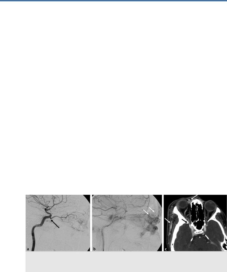
Fig. 7.1 Left ICA angiogram in lateral view in early (a) and late (b) phases shows that the ophthalmic artery originates at the junction between the horizontal cavernous and the clinoid segment (arrow), within the region of the ILT, and supplies the facial compartment of a diffuse AVM (white arrows in b). Contrast-enhanced computed tomography (c) demonstrates the abnormal vessels over the roof of the nose and along the right side of the face (white arrows). The ophthalmic artery enters the orbit through the superior orbital fissure (black arrow in c).
31
The Dorsal and Ventral Ophthalmic Arteries
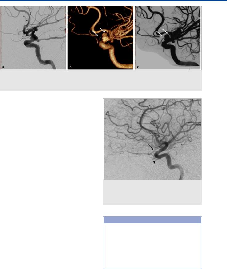
Fig. 7.2 Left ICA angiogram in lateral view (a) shows a broad-necked aneurysm in the distal supraclinoid segment in a 54-year-old man who presented with subarachnoid hemorrhage. Note the absence of an OA in the expected location at the junction of the clinoid and supraclinoid ICA segments. Left ICA 3D rotational reconstruction (b) and maximum-intensity projection image (c) demonstrate an artery arising from the proximal left A1 segment (white arrows) coursing inferiorly, entering the orbit and supplying the orbital components, corresponding to a primitive ventral ophthalmic artery.
●A DOA may coexist with an adult-type OA (i.e., dual origin of the OA) or, less frequently, a PVOA. If an intraorbital anastomosis between both branches exists, both arteries will supply the oculosensory structures. In the absence of an intraorbital anastomosis, the central retinal artery will arise from the most distal (ventral) vessel.
●If the PVOA is not annexed by the supraclinoid ICA, the former will represent the main supply to the oculosensory structures (the so-called persistent PVOA). In this case, the retinal artery will be supplied by the PVOA, which will originate from the A1 segment of the ACA and enter the orbit through the optic canal. This variant can be seen with both an adult-type OA or a DOA, which will supply the rest of the orbital elements. Rarely, a persistent PVOA will be present as the sole supply to the orbit.
●In cases in which the A1 segment disappears, the PVOA may give rise to the ACA in a variant known as the infraoptic course of the ACA (see Case 10).
7.3 Clinical Impact
In patients with a DOA and a normal or PVOA, it will be the most distal artery that will supply the oculosensory structures. In the setting of coexistence of the DOA with a normal OA or PVOA, embolization through the DOA can be performed safely, if there are no anastomoses between the DOA and the PVOA or OA.
7.4 Additional Information and
Cases
See Fig. 7.2; Fig. 7.3; Fig. 7.4; Fig. 7.5; Fig. 7.6; andFig. 7.7.
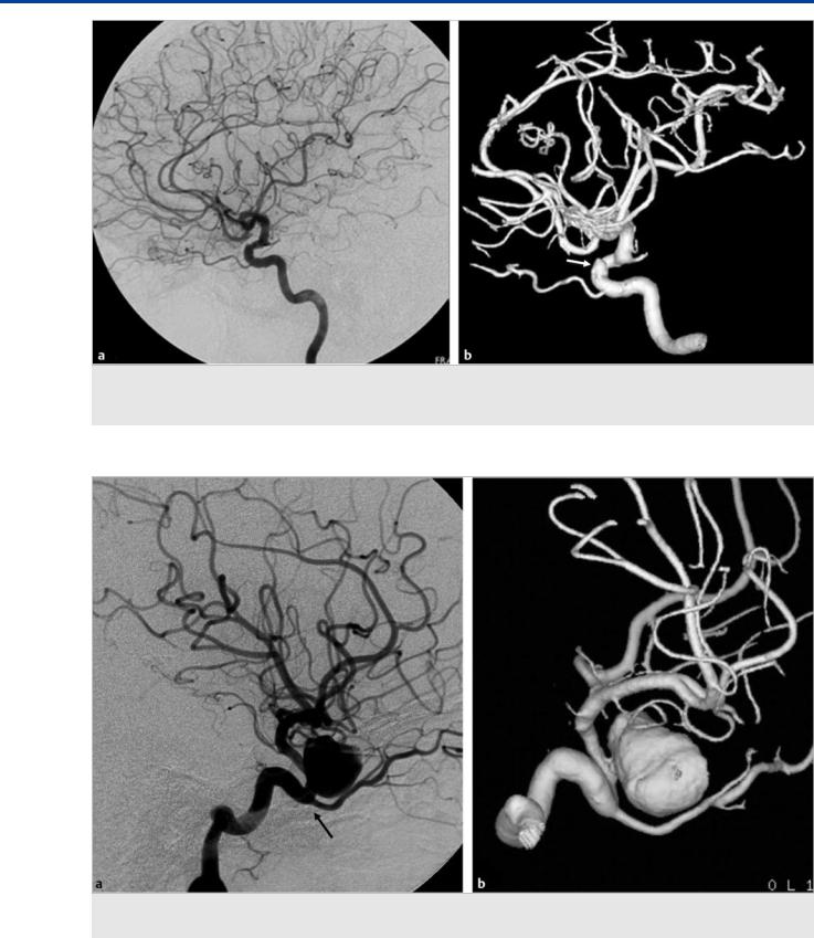
Fig. 7.3 Left ICA angiogram in lateral view demonstrates the persistence of the dorsal ophthalmic artery (arrowhead) with an adult-type ophthalmic artery (arrow) or so-called dual origin of the ophthalmic artery. In this case, the central retinal artery arises from the supraclinoid ophthalmic artery (the more distal or ventral artery).
Pearls and Pitfalls
●The remnant of the DOA in the adult is the ILT.
●If there is a DOA, it will enter the orbit through the superior orbital fissure and not through the optic canal, so all of its trajectory is extradural.
●A PVOA may be seen arising from the A1 segment. This artery will enter the orbit through the optic canal.
●In cases of a dual origin of the OA (i.e., a DOA and a PVOA or a normally located OA), it is the most distal vessel that provides the arterial supply to the oculosensory structures.
32
The Dorsal and Ventral Ophthalmic Arteries
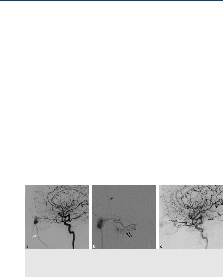
Fig. 7.4 Left ICA angiogram in lateral view (a) and 3D rotational reconstruction (b) demonstrate persistence of the dorsal ophthalmic artery, as the supraclinoid ICA has failed to annex the primitive ventral ophthalmic artery, which has regressed. Note the presence of a carotico-ophthalmic aneurysm (white arrow) in this patient, who presented with subarachnoid hemorrhage.
Fig. 7.5 Right ICA angiogram in lateral view (a) and 3D rotational reconstruction (b) image demonstrate a dorsal ophthalmic artery arising from the right ILT region (arrow) in a patient with a large carotico-ophthalmic aneurysm.
33

The Dorsal and Ventral Ophthalmic Arteries
Fig. 7.6 Left ICA angiogram in lateral view in arterial (a) and capillary (b) phases shows a dorsal ophthalmic artery arising from the ILT region of the cavernous left ICA. The choroidal blush seen on the capillary phase (arrow) is an indicator of the origin of the central retinal artery.
Fig. 7.7 Right ICA angiogram in lateral view (a) and 3D rotational reconstruction (b,c) images demonstrate a case with persistence of the dorsal ophthalmic artery and an adult-type ophthalmic artery or the so-called dual origin of the ophthalmic artery.
Further Reading
[1]Agarwal N, Singh PL, Karimi RJ, Gandhi CD, Prestigiacomo CJ. Persistent vestige of dorsal ophthalmic artery: a case report. J Neurointerv Surg 2013; 5: e25
[2]Hayreh SS. Orbital vascular anatomy. Eye (Lond) 2006; 20: 1130–1144
[3]Komiyama M. Letter to the editor – embryology of the ophthalmic artery: a revived concept. Interv Neuroradiol 2009; 15: 363–368
[4]Lasjaunias P, Berenstein A, ter Brugge KG. Surgical Neuroangiography. Vol. 1. 2nd ed. Berlin: Springer; 2006
[5]Willinsky R, Lasjaunias P, Berenstein A. Intracavernous branches of the internal carotid artery (ICA). Comprehensive review of their variations. Surg Radiol Anat 1987; 9: 201–215
34
The Branches of the Ophthalmic Artery
8 The Branches of the Ophthalmic Artery
8.1 Case Description
8.1.1 Clinical Presentation
A 54-year-old man presented with a long-standing pulsatile mass in the left medial periorbital region.
8.1.2 Radiologic Studies
See Fig. 8.1.
8.1.3 Diagnosis
Medial orbital arteriovenous malformation (AVM) with main supply derived from the ophthalmic artery (OA).
8.2 Anatomy
The blood supply of the orbit depends mainly on the OA, with minor contributions from the external carotid artery (ECA). The intraorbital segment of the OA gives rise to numerous arterial branches that supply the orbital structures. The branching pattern of the OA shows a high variability in the order and site of origin of the di erent branches. Conceptually, these branches can be divided into four groups: the ocular group (central retinal and ciliary arteries), the orbital group (lacrimal and muscular arteries), the extraorbital group (ethmoidal, supraorbital, supratrochlear, palpebral, and dorsal nasal arteries), and the dural group (recurrent deep and superficial arteries). Typically, these branches are annexed by the OA and form the “adult” pattern of the OA.
8.2.1 Ocular Group
The ocular group originates from the embryonic internal carotid artery (ICA), via the ventral OA (hyoid branch) and shows
little variability in its branching pattern. The order in which the ocular group branches originate is independent of the OA origin but depends on whether the artery courses over (83% of cases) or under (17% of cases) the optic nerve (ON). When the OA courses over the ON, the first branch is the central retinal artery (CRA), followed by a long posterior ciliary artery. When the OA courses under the ON, the first branch is the long lateral posterior ciliary artery and the second is the CRA. The CRA is a terminal branch, with no well-established anastomoses. Therefore, compromising this artery during embolization procedures will invariably result in retinal ischemia and monocular blindness. The long posterior ciliary arteries may arise separately or as part of a musculociliary trunk. They supply the choroid and are responsible for the choroidal blush, which serves as a surrogate marker for the CRA, as the ciliary arteries and the CRA arise closely together from the OA.
8.2.2 Orbital Group and Lacrimal Artery
The third branch typically corresponds to the lacrimal artery. The lacrimal artery typically arises from the first bend, lateral to the ON, and follows an oblique lateral course along the lacrimal nerve to reach a superior and medial position with respect to the lateral rectus to finally reach the lacrimal gland. The recurrent meningeal branch of the middle meningeal artery joins the lacrimal artery through the superior orbital fissure, serving as a potential anastomosis between ECA and ICA. Alternatively, the lacrimal artery can arise from the middle meningeal artery (meningolacrimal variant; see Case 25) and enter the orbit through the Hyrtl foramen. The lacrimal artery finally divides into muscular, zygomatic, and glandular branches. The latter gives rise to the lateral palpebral artery (see following).
Fig. 8.1 Left ICA angiogram in lateral view (a) shows an AVM nidus in the medial orbit, supplied mainly by the medial palpebral arteries, draining via the angular vein to the common facial vein (white arrow). Superselective microcatheter injection in the OA (b) proximal to the characteristic OA bend shows opacification of the long ciliary arteries (black double arrows) and the choroidal blush, meaning the catheter is proximal to the CRA, which is thus an unsafe position for embolization (see following). Note the anterior falcine artery (black arrowhead), which is a distal branch of the anterior ethmoidal artery. After a more distal catheterization and liquid embolic embolization, the left ICA angiogram in lateral view (c) shows no residual AV shunting and persistent that choroidal blush, indicating that the CRA was preserved.
35

The Branches of the Ophthalmic Artery
8.2.3 Extraorbital Group
The ethmoidal arteries supply blood to the ethmoid sinuses, nasal cavity, and nasal septum. The anterior ethmoidal artery courses medial and inferior to the superior oblique muscle and enters the skull through the anterior ethmoidal foramen to become the anterior falcine artery. The size of this artery will depend on the extent of dural supply to the falx and anterior cranial fossa, which is highly variable. The posterior ethmoidal artery is an inconstant branch. It enters the skull through the posterior ethmoidal foramen and gives variable dural supply to the posterior and medial third of the anterior cranial fossa. At least one of the ethmoidal arteries will invariably be involved in the supply to anterior fossa meningiomas or dural AV fistulas. Nasal septal supply can be prominent and can explain failed ECA embolizations for treatment of refractory epistaxis.
The supraorbital artery usually arises from the third segment of the OA (after the bend around the ON). It courses under the roof of the orbit to exit through the supraorbital foramen and supplies the upper eyelid and scalp, where it anastomoses with superficial temporal artery branches.
The palpebral arteries are composed of the medial group (superior and inferior), distal branches of the OA, and the lateral palpebral branch of the lacrimal artery. There is a rich anastomotic network between these arteries within the eyelids.
The supratrochlear and dorsal nasal arteries are terminal branches of the OA. They leave the orbit at the medial corner of the orbit. The supratrochlear artery has a rich anastomotic network with its contralateral homolog and with multiple ECA branches that supply the skin in this region. The dorsal nasal artery anastomoses with its contralateral homologue over the root of the nose and with the angular branch of the facial artery ( Table 8.1).
8.3 Clinical Impact
Knowing the anatomy of the orbital segment of the OA is fundamental for performing a safe embolization of orbital lesions. The distal branches of the OA are safe to embolize, as long as the microcatheter tip is located beyond the origin of the CRA (arterial bend seen in lateral angiograms) and reflux is avoided.
Table 8.1 Summary of the major anastomoses between the OA and ECA.
OA Branch |
Connecting Artery |
ECA Branch |
|
|
|
Lacrimal Artery |
Recurrent Meningeal Artery |
Middle Meningeal Artery |
|
|
|
|
Transverse Facial Artery |
Superficial Temporal Artery |
Ethmoidal Arteries |
Nasal Septal Arteries |
Sphenopalatine Artery (Internal Maxillary Artery) |
|
|
|
Supraorbital Artery |
Frontal Branch |
Superficial Temporal Artery |
|
|
|
Palpebral Arteries |
Zygomatic Branch |
Superficial Temporal Artery |
|
Infraorbital Artery |
Internal Maxillary Artery |
|
|
|
|
Angular Artery |
Facial Artery |
|
|
|
Supratrochlear Artery |
Frontal Branch |
Superficial Temporal Artery |
|
|
|
Dorsal Nasal Artery |
Angular Artery |
Facial Artery |
|
Lateral Nasal Artery |
Facial Artery |
|
|
|
|
|
|
|
|
|
Fig. 8.2 A 19-year-old man presented with a partially treated large orbital AVM. Axial CTA image (a) shows a superior palpebral mass with multiple tortuous vessels; pathognomonic findings for a facial region AVM (white arrow). Right ICA angiogram in lateral view (b) shows prior OA ligation (black arrow) with a preserved choroidal blush (not shown). Right ECA angiogram in lateral view (c) shows how the AVM has recruited supply from various superficial temporal artery and internal maxillary artery branches.
36