

Книги по МРТ КТ на английском языке / Neurovascular anatomy in interventional neuroradiology Krings et al 2015
.pdfThe Aberrant Subclavian Artery
other congenital abnormalities, such as hypoplastic left heart syndrome, coarctation of the aorta, and atrioventricular canal defects.
The presence of an ARSA increases the di culty of interventional procedures requiring catheterization of the right subclavian artery or right vertebral artery (VA). It can be readily iden-
Fig. 2.2 MRA in anteroposterior (AP) view (a) and coronal MRA (b) demonstrate an aberrant right subclavian artery resulting from formation from the C7 segmental artery. Note the separate origins of the RCCA and LCCA in this case, rather than a common origin.
tified on cross-sectional studies, such as CTA and magnetic resonance angiography. Its presence can also be inferred from barium swallow studies performed for dysphagia, in which findings include anterior displacement of the esophagus with an abnormal indentation on the posterior wall of the esophagus in the upper mediastinum. On catheter angiography, the absence of filling of the right subclavian artery on right brachiocephalic injection should raise the suspicion of an ARSA if it has been not already identified from diagnostic cross-sectional imaging. The origin of the vessel distal to the left subclavian artery can then be sought, and an arch aortogram can be performed if necessary.
In the setting of an ARSA, where possible, posterior circulation endovascular procedures may have to be performed through the left VA, as right vertebral catheterization from a femoral approach can be problematic. Alternatively, a right radial or brachial approach can be considered for VA catheterization; however, this approach can cause di culties for coronary catheterization. For example, the angular course of the aberrant vessel to the aorta can impose di culty in passing a guidewire into the ascending aorta.
Treatment of the anomaly is not required in asymptomatic patients. However, in symptomatic patients or in the setting of aneurysm formation, intervention may be considered with various surgical or combined surgical and endovascular approaches. One possible combined approach is the endovascular occlusion of the aortic origin of the aberrant artery combined with surgical subclavian artery transposition and distal prevertebral occlusion. Occlusion of the proximal right subclavian artery without subclavian artery reconstruction can result in right arm ischemia or subclavian steal syndrome.
Fig. 2.3 3D CTA reformats of the thoracoabdominal aorta in AP (a) and oblique (b) views in a 73-year-old woman being worked up for an abdominal aortic aneurysm. In this case, the first vessel to arise from the aortic arch is the RCCA (white arrow). The right subclavian artery is the last vessel to arise from the aortic arch (white arrowhead), after the left subclavian. The aberrant subclavian artery will cross the midline behind the thoracic esophagus, after which it will resume its normal path (not shown).
7

The Aberrant Subclavian Artery
Fig. 2.4 Contrast-enhanced axial CT (a–d) and 3D CTA reconstruction images in AP (e) and oblique (f) views demonstrating an aberrant right VA (white arrows), originating directly from the aortic arch (lusoria origin), related to development of the right VA from the C8 segmental artery, rather than the C6 or C7 levels.
Fig. 2.5 CTA in axial cut (a) and 3D reformats in AP (b) and superior (c) views demonstrating an aberrant subclavian artery (asterisks in a,b,c). 1, right VA; 2, RCCA; 3, nasogastric tube; 4, LCCA; 5, left VA.
2.4 Additional Information and
Cases
Occasionally, an ARSA arises from a common trunk with the left subclavian artery. This can occur in combination with a shared origin of the CCAs, forming an uncommon type of vascular ring, sometimes called a bitruncus arch. Only two great vessels arise from the arch, and compression of the trachea by the bicarotid truncus in front and the bisubclavian truncus behind may cause stridor.
Isolation of the right subclavian artery occurs when there are two breaks in the primitive aortic arch, leaving the subclavian artery separated from the aortic arch. Patients with this anomaly can present with subclavian steal syndrome as a result of
retrograde VA flow supplying the upper limb. This anomaly generally occurs on the side contralateral to the arch and so is more commonly associated with a right aortic arch ( Fig. 2.3;Fig. 2.4; Fig. 2.5; Fig. 2.6).
Pearls and Pitfalls
●The presence of an aberrant right subclavian artery is usually asymptomatic; however, tortuosity and dilatation of the aberrant artery or aneurysmal dilatation of the Kommerell diverticulum can result in compression of the esophagus, resulting in dysphagia (known as dysphagia lusoria in some patients).
●Endovascular procedures involving the right VA and right subclavian artery may be di cult in patients with an ARSA.
8

The Aberrant Subclavian Artery
Further Reading
[1] Kau T, Sinzig M, Gasser J et al. Aortic development and anomalies. Semin Intervent Radiol 2007; 24: 141–152
[2] Müller M, Schmitz BL, Pauls S et al. Variations of the aortic arch – a study on the most common branching patterns. Acta Radiol 2011; 52: 738–742
[3] Osborn AG. The aortic arch and great vessels. In: Diagnostic Cerebral Angiography. 2nd ed. Philadelphia, PA: Lippincott Williams & Wilkins; 1999:3–29
[4] Sadler TW. Cardiovascular system. In: Langman’s Medical Embryology. 11th ed. Philadelphia, PA: Wolters Kluwer/Lippincott Williams & Wilkins; 2010:16;5–196
[5] Shaw JA, Gravereaux EC, Eisenhauer AC. Carotid stenting in the bovine arch. Catheter Cardiovasc Interv 2003; 60: 566–569
[6] Stojanovska J, Cascade PN, Chong S, Quint LE, Sundaram B. Embryology and imaging review of aortic arch anomalies. J Thorac Imaging 2012; 27: 73–84
Fig. 2.6 CTA 3D reconstructions in AP (a,b), posterior-anterior (c), and oblique (d) views demonstrate separate origins of the right external and internal carotid arteries related to persistence of the ductus caroticus, with complete involution of the third aortic arch, resulting in absent migration of the external carotid artery toward the carotid axis.
9

Section II
Internal Carotid Artery
II

The Carotid Segments, the Aberrant ICA, and the Persistent Stapedial Artery
3 The Carotid Segments, the Aberrant ICA, and the Persistent Stapedial Artery
3.1 Case Description
3.1.1 Clinical Presentation
A 32-year-old man presented with pulsatile tinnitus and a retrotympanic vascular mass.
3.1.2 Radiologic Studies
See Fig. 3.1; Fig. 3.2.
3.1.3 Diagnosis
Aberrant right internal carotid artery (ICA).
3.2 Anatomy
The aberrant ICA occurs as a hemodynamic response to a segmental agenesis of the first (i.e., cervical) segment of the ICA secondary to disturbed di erentiation of the third branchial arch. It represents a collateral pathway between the external
carotid artery (ECA) and the petrous ICA via the ascending pharyngeal artery (APhA).
The ICA segments have been classified by various methods, most of which are based on anatomic or surgical criteria. However, to understand the occurrence of specific variations like the one described in the present case, a model based on embryologic considerations, such as that proposed by Santoyo-Vazquez and Lasjaunias, may be more helpful. In this model, the ICA is formed by seven segments, the boundaries of which are defined by transient embryonic vessels.
As previously described, during formation of the craniocervical vessels and the arch, the ventral aorta and both dorsal aortas (DAos) are connected by six aortic arches (AAs). The lower three arches (IV–VI) participate in the development of the aortic arch and supra-aortic trunks, whereas arches I–III are involved in ICA development. Cranial to the AA I, at this stage, the DAo will also give origin to other embryonic vessels. In caudocranial order, these are the primitive maxillary artery (PMI), the dorsal ophthalmic artery (DO), and the ventral ophthalmic artery.
Bearing these considerations in mind, the future segments of the adult ICA are defined by the following embryonic vessels:
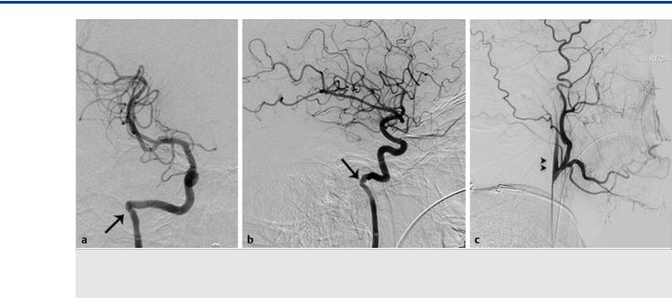
Fig. 3.1 3D CTA in anteroposterior (AP) view (a) demonstrates an aberrant course of the right ICA (white arrow), running more laterally compared with the contralateral left side. Contrast-enhanced axial CT images in bone (b,c) and soft tissue (d,e) windows reveal a vascular structure corresponding to the right ICA within the right middle ear.
12
The Carotid Segments, the Aberrant ICA, and the Persistent Stapedial Artery
Fig. 3.2 Right ICA angiogram in AP (a) and lateral (b) views confirms the lateral course of the right ICA. The right ECA angiogram in lateral view (c) reveals the ascending pharyngeal origin of the right ICA (double arrowheads). Note the focal narrowing of the “aberrant ICA” as it enters the skull base, marking the entry point into the inferior tympanic canaliculus (arrows).
●segment 1: AA III,
●segment 2: the DAo between AAs III and II,
●segment 3: the DAo between AAs II and I,
●segment 4: the DAo between AA I and the PMI,
●segment 5: the DAo between the PMI and the DO,
●segment 6: the DAo between the DO and the opening of the ophthalmic artery (OA), and
●segment 7: the DAo between the opening of the OA and the bifurcation of the ICA into a rostral (telencephalic) and a caudal division.
Cranially to the seventh segment, there are no additional ICA segments, as all other future arteries that will develop further distally will have a di erent temporal or phylogenetic origin. During embryonic life, the aortic arches and the embryonic vessels will undergo regression and significant modifications that will give shape to the ICA as it is seen in adults.
However, the remnants of these embryonic vessels will be relevant in defining the boundaries of the di erent ICA segments: AA III will persist and evolve into the cervical ICA distal to the carotid bulb. The carotid bulb is, from an embryological point of view, therefore a separate structure from the ICA. AA II regresses rapidly, and the only portion remaining forms the stapedial and hyoid arteries. These will be annexed by the ventral pharyngeal artery (originating from the ventral aorta) to form the future ECA and internal maxillary artery. The remnant of AA II is the caroticotympanic artery (CTymA). AA I will regress and contribute to the development of the ECA, together with the ventral aorta. The remnant of AA I will be the mandibulovidian artery. The PMI will regress to form the posterior hypophyseal artery and contribute to form the meningohypophyseal trunk. In most adults, the DO will regress and contribute to form the inferolateral trunk. The caudal division of the ICA reaches the cephalic end of the ipsilateral ventral neural artery to constitute the future PcomA. The rostral or telencephalic division will evolve initially into the anterior choroidal artery and anterior cerebral artery, which later will give rise to the middle cerebral arteries.
In cases of segmental agenesis of the ICA, each of these embryonic vessels represents the potential point of vascular reconstitution of flow into the other distally preserved ICA segments. Most of these unusual flow patterns have been improperly described as aberrant courses of the ICA. After a complex process of regression and annexation of the di erent vascular structures involved, the ICA develops into its final appearance, resembling a continuous vessel, with its characteristic path and curves. Still, the seven embryonic segments can be distinguished by the origin of the embryonic arteries’ remnants:
1.cervical segment: immediately distal to the bulb to the entry point of the of the carotid canal,
2.ascending intrapetrous segment: from the entry point of the carotid canal to the origin of the CTymA,
3.horizontal intrapetrous segment: from the origin of the CTymA to the origin of the mandibulovidian artery,
4.segment ascending foramen lacerum: from the origin of the mandibulovidian artery to the origin of the meningohypophyseal trunk,
5.horizontal cavernous segment: from the origin of the meningohypophyseal trunk to the origin of the inferolateral trunk,
6.clinoid segment: from the origin of the inferolateral trunk to the origin of the OA, and
7.supraclinoid ICA segment: from the origin of the OA.
Each of the segments, because of their phylogenetic di erences, has a di erent heritage and vulnerability, which explains the specific involvement of certain segments and the preservation of adjacent ones in certain disease processes.
In normal subjects, the ICA enters the petrous bone medial to the styloid process via the carotid canal. The vertical petrous segment runs anterior to the cochlea and is separated from the middle ear by a thin plate of bone. Then, the ICA turns anteriorly to run inferior and posteromedial to the Eustachian tube, traverses through the foramen lacerum, and enters the cavernous sinus.
In patients with an aberrant ICA, the inferior tympanic branch of the APhA enlarges in response to segmental ICA agen-
13
The Carotid Segments, the Aberrant ICA, and the Persistent Stapedial Artery
esis and anastomoses with the CTymA (remnant of the hyoid artery) to reconstitute the ICA in the horizontal petrous segment. The inferior tympanic artery enters the skull through the inferior tympanic canaliculus (Jacobsen canal), which explains the focal narrowing of the ICA seen on digital subtraction angiography and computed tomographic angiography at this level.
Therefore, in cases of an aberrant course of the ICA, the “cervical ICA” is actually composed of the APhA, its inferior tympanic branch, and the caroticotympanic branch of the ICA, and not by the first ICA segment.
Clinical diagnosis of aberrant ICA is di cult because symptoms and signs are often nonspecific or absent. Symptoms like pulsatile tinnitus, conductive hearing loss, and a pulsatile retrotympanic mass can be seen. The clinical picture can be interpreted as otosclerosis, glomus tumor, or other vascular malformation. To avoid an arterial injury during surgery, a temporal bone computed tomography (CT) scan should be performed when dealing with a “vascular” retrotympanic mass.
On CT, the aberrant ICA runs posterior to the jugular bulb, with a focal stenosis as it enters the skull base. There will be a deficient bony plate along the tympanic portion of the ICA, and the vertical segment of the carotid canal will be absent. On digital subtraction angiography, the aberrant ICA will show a characteristic lateral swing beyond the vestibular line (also called the line of Lapayowker) within the petrous bone and will then turn medially into the middle ear cavity.
The stapedial artery is a transient embryonic vessel that arises at 4 to 5 weeks of fetal life. It is a branch of the hyoid artery, which derives from the second branchial arch. It extends cranially and passes through the mesenchymal primordium of the stapes to form the obturator foramen. The stapedial artery gives rise to two intracranial arteries: the upper or supraorbital branch, which later becomes the middle meningeal artery (MMA), and the lower or maxillomandibular division, which divides into two branches (a mandibular branch and an infraorbital branch) that will later become the inferior alveolar and infraorbital arteries, respectively. The maxillomandibular division
leaves the cranial cavity through the foramen spinosum and anastomoses with the ventral pharyngeal artery at this level. This anastomosis triggers the involution of the stapedial artery, which occurs during the 10th week of fetal life. The hyoid artery persists as the caroticotympanic branch of the ICA. If the stapedial artery persists (PSA), the connection between the inferior division is annexed by the ventral pharyngeal artery, and the superior division is supplied by the PSA. As the connection between both divisions is through the foramen spinosum, in cases of PSA, this foramen is absent or hypoplastic. The end result will be that the MMA will be supplied by the ICA via the PSA.
The PSA has a prevalence of ~0.5% in the general population and presents as a pulsatile mass in the middle ear cavity, with or without pulsatile tinnitus. Occasionally, it may show conductive hearing loss as a result of stapes ankylosis. A PSA can be seen as an isolated variant or in association with an aberrant ICA. In the latter cases, it arises from the petrous ICA, enters the anteromedial hypotympanum via the Jacobsen canal, crosses over the cochlear promontory, passes between the crura of the stapes (obturator foramen) to enter the facial nerve (fallopian) canal, or runs parallel to it in its own canal. Then the PSA continues superiorly, as an enlarged superior tympanic or superficial petrosal artery, to form the MMA. When the PSA occurs in isolation, the abnormal artery is formed by enlargement of the more distal caroticotympanic artery arising from the vertical petrous ICA and then follows the same course as described earlier. Therefore, the foramen spinosum is absent or hypoplastic, as the MMA does not arise from the proximal internal maxillary artery, as typically seen.
In cases with a PSA, CT findings include a small canaliculus exiting the carotid canal (in cases of isolated PSA), a linear soft tissue density structure crossing the middle ear over the promontory, an enlarged facial nerve canal, or a separate parallel bony canal. The foramen spinosum will be absent, as previously mentioned. Of note, ~3% of the population may have an absent foramen spinosum without a PSA, so it is an indirect sign of PSA, and the diagnosis has to be made based on the other CT findings.
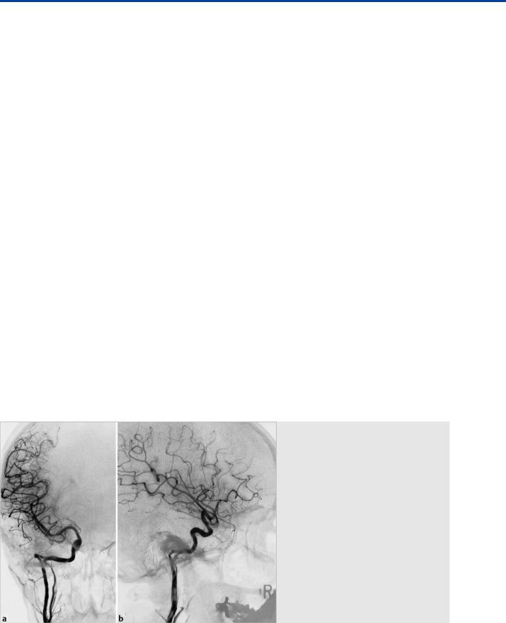
Fig. 3.3 A 28-year-old woman presented with conductive hearing loss and retrotympanic mass. Right ICA angiograms in AP (a) and lateral (b) views demonstrate a double origin of the right ICA. The cervical segment of the “normal” right ICA appears hypoplastic and joins the “ascending pharyngeal origin” ICA at the petrous segment.
14
The Carotid Segments, the Aberrant ICA, and the Persistent Stapedial Artery
3.3 Clinical Impact
The aberrant ICA is a well-recognized although rare vascular anomaly. Clinically it can mimic glomus tumors, other vascular malformations (such as aneurysms and hemangiomas), or otosclerosis. Misdiagnosis of this anomaly may lead to serious morbidity resulting from massive bleeding or stroke. If there is uncontrolled bleeding, endovascular treatment has to be performed and may require parent vessel sacrifice, stent grafts, or overlapping flow diverters.
3.4 Additional Information and
Cases
An important clinical di erential diagnosis is the tympanic or jugulotympanic paraganglioma, the most common middle ear vascular tumor. Although clinically it may be challenging to differentiate between an aberrant ICA, PSA, or paraganglioma, temporal bone CT will easily distinguish these entities. It will show a small soft tissue mass over the promontory in cases of tympanic paragangliomas, and a permeative vascularized mass centered in the jugular foramen, extending into the middle ear, in cases of jugulotympanic paragangliomas. Another di erential diagnosis is the dehiscent jugular bulb, for which temporal bone CT will show a focal dehiscence of the bony plate covering the jugular bulb, with a small portion of the internal jugular vein protruding into the middle ear ( Fig. 3.3; Fig. 3.4;Fig. 3.5; Fig. 3.6).
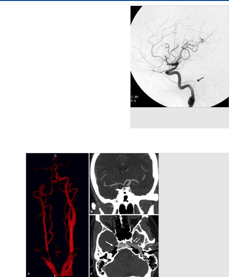
Fig. 3.4 A 27-year-old man presented with pulsatile mass seen during otoscopy. Left ICA angiogram reveals a vessel arising from the petrous ICA (black arrow), supplying the MMA, which is the classic angiographic appearance of a PSA.
Fig. 3.5 3D CTA (a) and contrast-enhanced CT in coronal (b) and axial (c) images demonstrate a congenital absent right ICA. Note the absence of the right carotid canal (white arrow) and prominent size of the left ICA (white double arrows) supplying the right cerebral hemisphere through the anterior communicating artery.
15

The Carotid Segments, the Aberrant ICA, and the Persistent Stapedial Artery
Fig. 3.6 3D CTA in AP (a) and oblique (b) views demonstrates the absence of the left ICA. The left ACA is supplied through the anterior communicating artery and the left middle cerebral artery through the left posterior communicating artery. Contrast-enhanced axial CT (c,d) images show the absence of the left carotid canal (black arrow) and the prominent sizes of the left ECAs (white arrow) and left vertebral arteries (double black arrowheads).
Pearls and Pitfalls
●In the “aberrant ICA,” there is agenesis of the first ICA segment (cervical segment).
●The “aberrant ICA” actually is composed by the APhA, its inferior tympanic branch, and the caroticotympanic branch of the ICA, which serve as a collateral route to reconstitute the ICA in the horizontal petrous segment.
●The PSA may occur in isolation or associated with an aberrant ICA. In either case, the foramen spinosum will be absent or hypoplastic and the MMA will originate from the PSA and not from the ECA.
Further Reading
[1]Lasjaunias P, Santoyo-Vazquez A. Segmental agenesis of the internal carotid artery: angiographic aspects with embryological discussion. Anat Clin 1984; 6: 133–141
[2]Roll JD, Urban MA, Larson TC, III, Gailloud P, Jacob P, Harnsberger HR. Bilateral aberrant internal carotid arteries with bilateral persistent stapedial arteries and bilateral duplicated internal carotid arteries. AJNR Am J Neuroradiol 2003; 24: 762–765
[3]Sauvaget E, Paris J, Kici S et al. Aberrant internal carotid artery in the temporal bone: imaging findings and management. Arch Otolaryngol Head Neck Surg 2006; 132: 86–91
[4]Silbergleit R, Quint DJ, Mehta BA, Patel SC, Metes JJ, Noujaim SE. The persistent stapedial artery. AJNR Am J Neuroradiol 2000; 21: 572–577
[5]Thiers FA, Sakai O, Poe DS, Curtin HD. Persistent stapedial artery: CT findings. AJNR Am J Neuroradiol 2000; 21: 1551–1554
[6]Willinsky R, Lasjaunias P, Berenstein A. Intracavernous branches of the internal carotid artery (ICA). Comprehensive review of their variations. Surg Radiol Anat 1987; 9: 201–215
16