

Книги по МРТ КТ на английском языке / Neurovascular anatomy in interventional neuroradiology Krings et al 2015
.pdfThe “Dangerous” Anastomoses III: Upper Cervical Anastomoses
27 The “Dangerous” Anastomoses III: Upper Cervical
Anastomoses
27.1 Case Description
27.1.1 Clinical Presentation
A 65-year-old woman with a past history of coronary and peripheral vascular arterial disease, hypertension, and type 2 diabetes presented with dizziness and one episode of blackout. Vertebro-basilar insu ciency was suspected.
27.1.2 Radiographic Studies
See Fig. 27.1.
27.1.3 Diagnosis
Right ECA occlusion with retrograde reconstitution of the ECA through the upper cervical anastomoses from the VA.
27.2 Embryology and Anatomy
The ascending pharyngeal artery (APhA) corresponds to the remnant of the embryonic hypoglossal artery (HA). The HA originates from the third aortic arch and contributes to the development of the most proximal aspect of the cervical ICA. It is one of the embryonic carotid–vertebrobasilar anastomoses,
originating from the proximal cervical ICA, slightly distal to the carotid bulb, and enters the posterior cranial fossa via the hypoglossal canal. The relationship between the third aortic arch and the HA can result in various anastomoses between their adult homologs, the ICA, and the APhA, with the most extreme of the spectrum being the persistent HA. See also Case 4.
In the adult, the APhA forms part of the pharyngo-occpital system, a vascular network in the suboccipital region through which the cervical vertebral and carotid arteries are connected by four routes: the occipital, ascending pharyngeal, vertebropharyngeal (C3), and C4-collateral routes.
The APhA originates most commonly from the posterior wall of the proximal ECA and anastomoses with the VA, typically at two levels. The more proximal one occurs laterally between the musculospinal branch of the APhA and the C3 radicular anastomotic branch of the VA. The distal one is the prevertebral branch via the odontoid arch, which usually arises from the HA and follows the same path as the persistent HA, serving as one of the collateral routes in cases of proximal ECA or VA occlusion, as seen in the index case.
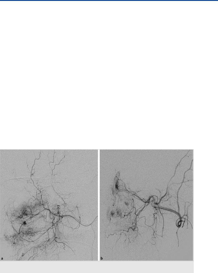
The occipital artery is a remnant of the embryologic type 1 and type 2 proatlantal arteries, which correspond to the C1 and C2 segmental arteries, respectively. In an adult, the occipital artery still retains its connection from the ECA to the VA through the posterior anastomotic radicular routes via the musculocuta-
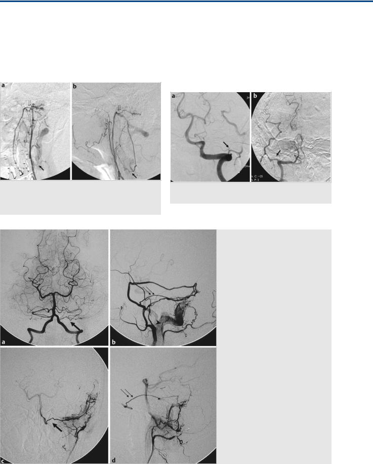
Fig. 27.1 MRA of the neck (a) shows a highgrade stenosis of the proximal intracranial right VA or V4 segment (white arrow). Incidentally, there is a right ECA occlusion (asterisk). Right VA angiogram in anteroposterior (AP) (b) and lateral
(c) views and a three-dimensional reconstruction image (d) show contrast filling of both the occipital artery and the APhA through an anastomosis via the artery of the hypoglossal canal (arrows) and musculocutaneous branches from the C1 segment. There is retrograde flow down to the origin of the ECA and opacification of the facial artery (arrowheads).
137
The “Dangerous” Anastomoses III: Upper Cervical Anastomoses
neous branches, which arise from the horizontal portion of the occipital artery at the C1 and C2 levels. These anastomotic channels are rather large and can be seen during selective injections of the occipital artery. They serve as the major collaterals from the VA in case of common carotid artery or ECA ligation or occlusion. The stylomastoid artery, originating either from the occipital or posterior auricular arteries, supplies the meninges of the posterior fossa and anastomoses with other meningeal
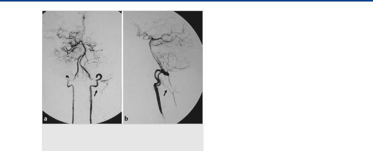
Fig. 27.2 Left APhA angiograms in AP (a) and lateral (b) views demonstrate anastomoses of the musculospinal branch at the C3 level (arrows), with the VA with additional contributions from the odontoid arch (thin double arrows).
arteries, including the posterior meningeal artery from the distal cervical, i.e. suboccipital VA. For further information regarding the meningeal supply refer to Case 31.
The ascending and deep cervical arteries anastomose with the VA at the C2 to C4 levels. Both arise from the subclavian artery: the ascending cervical artery from the thyrocervical trunk and the deep cervical artery from the costocervical trunk. These arteries typically enlarge and serve as major collaterals in case of proximal VA occlusion.
Fig. 27.3 Left (a) and right (b) VA angiograms in AP view demonstrate C1 collaterals to the occipital arteries bilaterally (arrows).
Fig. 27.4 Left VA angiogram in AP view (a), left ECA angiogram in lateral view (b), and left occipital artery angiograms in AP (c) and lateral
(d) views postembolization of a left transversesigmoid sinus dural arteriovenous fistula demonstrate the transmeningeal anastomoses to a posterior meningeal branch of the VA (arrows) and through the facial arcade to the petrous branch of the middle meningeal artery (thin double arrows) from the stylomastoid branches of the occipital artery (arrowhead).
138
The “Dangerous” Anastomoses III: Upper Cervical Anastomoses
Fig. 27.5 Left VA angiograms in AP (a) and lateral (b) views demonstrate the anastomoses between the VA and deep cervical artery at the C2 level (arrows).
27.3 Clinical Impact
The APhA has multiple anastomoses with the intracranial circulation, via internal carotid and vertebral arteries, as discussed in Case 30. The main complications, if the anastomoses are not promptly recognized, are seen in embolization procedures with small particles or liquid embolic materials. In the specific case of the VA/APhA anastomosis via the hypoglossal canal artery, an external artery particle injection may result in embolic strokes in the posterior circulation. The use of liquid embolic material could result in occlusion of the vasa nervorum of the hypoglossal nerve, supplied by the artery of the hypoglossal canal, with subsequent 12th cranial nerve palsy. As the anastomosis is
located proximal to the origin of the anterior spinal artery, a potential risk of anterior spinal artery ischemia exists.
The occipital and cervical arteries also have several anastomoses with the vertebral arteries. Caution must be used when embolizing within these territories with small particles and liquid embolic materials to avoid posterior fossa infarctions. Occasionally, the anterior spinal artery may arise from the ascending and deep cervical arteries.
27.4 Additional Information and
Cases
See Fig. 27.2, Fig. 27.3, Fig. 27.4, and Fig. 27.5.
Further Reading
[1]Cavalcanti DD, Reis CV, Hanel R et al. The ascending pharyngeal artery and its relevance for neurosurgical and endovascular procedures. Neurosurgery 2009; 65 Suppl: 114–120, discussion 120
[2]Geibprasert S, Pongpech S, Armstrong D, Krings T. Dangerous extracranial-in- tracranial anastomoses and supply to the cranial nerves: vessels the neurointerventionalist needs to know. AJNR Am J Neuroradiol 2009; 30: 1459–1468
[3]Hacein-Bey L, Daniels DL, Ulmer JL et al. The ascending pharyngeal artery: branches, anastomoses, and clinical significance. AJNR Am J Neuroradiol 2002; 23: 1246–1256
[4]Houseman ND, Taylor GI, Pan WR. The angiosomes of the head and neck: anatomic study and clinical applications. Plast Reconstr Surg 2000; 105: 2287–2313
[5]Lasjaunias P, Berenstein A, ter Brugge KG. Surgical Neuroangiography. Vol. 1. 2nd ed. Berlin: Springer; 2006
[6]Lasjaunias P, Théron J, Moret J. The occipital artery. Anatomy—normal arteriographic aspects—embryological significance. Neuroradiology 1978; 15: 31– 37
[7]Strub WM, Leach JL, Tomsick TA. Left vertebral artery origin from the thyrocervical trunk: a unique vascular variant. AJNR Am J Neuroradiol 2006; 27: 1155–1156
139

The Cranial Nerve Supply
28 The Cranial Nerve Supply
28.1 Case Description
28.1.1 Clinical Presentation
An 82-year-old, right-handed woman presented with a severalmonth history of falls and gait ataxia, worse on the left side, which prompted further imaging.
28.1.2 Radiologic Studies
See Fig. 28.1, Fig. 28.2.
28.1.3 Diagnosis
Dural arteriovenous fistula (dAVF) of the petrous ridge with cortical venous reflux. The dAVF is fed by the middle meningeal
artery (MMA), including its petrosal branch and the stylomastoid branch of the posterior auricular artery (i.e., the facial arcade).
28.2 Anatomy
Under normal conditions, the caliber of the arteries that supply the cranial nerves (the vasa nervorum) is between 100 and 300 μm, and they are, therefore, in most instances only visible on conventional angiography during superselective injections. Nevertheless, knowledge of their origin, their anastomoses, and their potential variations is paramount for safe embolization of a variety of skull base lesions, including dAVF and hypervascularized tumors.
Fig. 28.1 MRI axial fluid-attenuated inversion recovery (a), T1 contrast-enhanced (b), and susceptibility (c) weighted images demonstrate a vascular pouch in the left inferior cerebellum with significant perifocal edema, as well as abnormal sulcal vessels. Left vertebral artery angiogram in lateral view
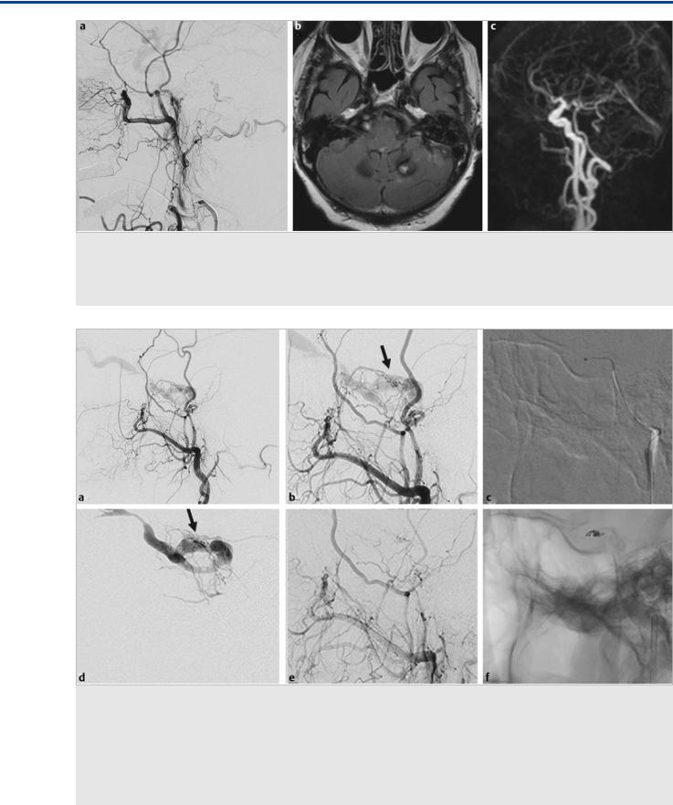
(d) reveals a cerebellar arteriovenous malformation, the imaging characteristics of which cannot explain the visualized pouch on MR. Subsequent injection into the left ECA (e) demonstrates a dural arteriovenous fistula along the petrous ridge, with arterial filling via the meningohypophyseal trunk and the facial arcade and multiple venous pouches and venous stenoses. After embolization of the distal MMA with glue deposition into the venous segment, minimal residual flow was visualized via the petrosal branch of the MMA (injection in f). From this vessel, transient opacification of a normal distal branch was seen that connected to the stylomastoid branch of the posterior auricular artery (arrow). As glue had been deposited in the foot of the vein, we only ligated the petrosal branch of the MMA with concentrated glue to ensure that the supply to the facial nerve was not compromised and the flow to the fistula was further reduced, which was supposed to facilitate further thrombosis. Case continued in Fig. 28.2.
140
The Cranial Nerve Supply
Fig. 28.2 Immediate control angiogram (a) demonstrated residual very slow filling of the shunt via the stylomastoid branch of the posterior auricular artery. Upon follow-up MR 2 weeks later (b), the patient had thrombosed her venous pouch, the edema was no longer visualized, and only the arteriovenous shunting related to the cerebellar arteriovenous malformation was noted on the dynamic contrast-enhanced magnetic resonance angiography, with no further shunting visualized from the dural branches. (c) Clinically, she did very well, with no new neurological deficits and reversal of her presenting symptoms.
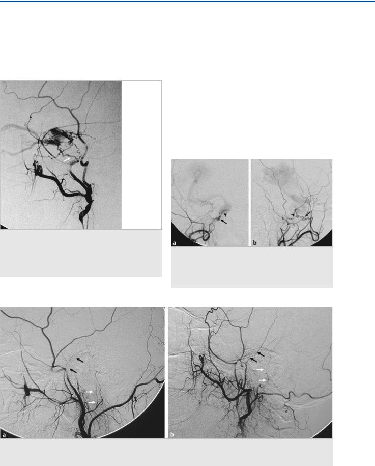
Fig. 28.3 This patient had a dural arteriovenous fistula of the cavernous sinus that was supplied by multiple interconnecting internal maxillary branches that all converged into a single artery (arrows) at the superior roof of the cavernous sinus, as seen on the left ECA angiogram in lateral view (a,b). We opted for a transvenous approach through the inferior petrosal sinus (c), and the microcatheter could be placed via the cavernous sinus retrogradely into the feeding artery. Contrast injection (d) to verify the position shows both the cavernous sinus with its outflow and, retrogradely, the arterial network converging into the single fistulous zone. The fistula could be treated by a few coils that were deposited transvenously into the arterial common trunk, followed by retrograde coiling into the superior portion of the cavernous sinus (e,f). This case indicates the rich anastomotic network of dural branches that supply the cranial nerves of the cavernous sinus arising from the ECA and anastomosing with the ICA. Treating these fistulas either transarterially or transvenously with a liquid embolic agent that penetrates deep into this anastomotic network puts the patient at risk for ophthalmoplegia or embolic events in the ICA territory.
141
The Cranial Nerve Supply
28.2.1 Cranial Nerves I and II
Being outpouchings of the brain, rather than true cranial nerves, the olfactory and optic nerves are not considered peripheral nerves and will be described only briefly: the olfactory nerve is supplied by the olfactory artery and ethmoidal branches of the ophthalmic artery, and the optic nerve is supplied by the proximal ophthalmic artery.
Fig. 28.4 The branches of the trigeminal nerve are supplied as they course through the foramina of the skull base by the artery of the foramen ovale (mandibular branch; white arrow) from the MMA or accessory meningeal artery, whereas the maxillary nerve is supplied by the artery of the foramen rotundum (black arrow), a branch of the distal internal maxillary artery. Both branches anastomose with the ILT.
28.2.2 Cranial Nerves III, IV, and VI
In its cisternal segment, the oculomotor nerve receives arterial supply in the vicinity of the posterior perforating substance from the basilar or the posterior cerebral artery.
Once it extends forward from the brainstem, the trochlear nerve is supplied by the superior cerebellar artery and additional circumferential arteries of the P1 segment of the posterior cerebral artery, with which it courses through the ambient cistern.
The abducens nerve is supplied by the clival dural network along its course anterior to the pons, including the medial and lateral clival arteries from the hypoglossal and jugular branch of the neuromeningeal trunk of the ascending pharyngeal artery and, more cranially, the medial and lateral clival arteries from the meningohypophyseal trunk.
These dural arteries also contribute to the supply of cranial nerves III, IV, and V in their dural and transosseous course. The
Fig. 28.5 ECA injection, anteroposterior (a) and lateral (b) views, in a patient with a pial brain arteriovenous malformation of the fistulous type with significantly increased flow through the ICA. The ECA, with its artery of the foramen rotundum (arrow) that connects to the ILT (arrowhead) of the ICA, is “sumped” into the ICA.
Fig. 28.6 The “facial arcade” supplying the facial nerve is demonstrated in two patients. Left ECA angiograms (a,b) in lateral view demonstrate the facial arcade supply through the petrous branch of the MMA (black arrows) and the stylomastoid branch (white arrows), which arose from the posterior auricular artery in one patient (a) and from the occipital artery in the other patient (b).
142

The Cranial Nerve Supply
Fig. 28.7 MRI with T2-weighted (a) and susceptibility-weighted (b) images demonstrates a dilated and arterialized lateral mesencephalic vein (bright signal on susceptibility-weighted imaging), indicating an arteriovenous shunt (arrows). Left ECA angiograms in anteroposterior (c) and lateral (d,e) views verify the shunt that is supplied by the facial arcade through the petrous branch of the MMA (thin black arrows) and the stylomastoid branch of the posterior auricular artery (thin white arrows). After complete obliteration of the fistula, the follow-up digital subtraction angiography (f) demonstrates persistent filling of the facial arcade, which is now of normal size.
third and fourth cranial nerves are supplied by the artery of the free margin of the tentorium on the roof of the cavernous sinus. This artery may arise from the meningohypophyseal trunk, directly o the internal carotid artery (ICA), but also from the MMA, the ophthalmic or lacrimal artery, or the inferolateral trunk (ILT). Further distally (i.e., in the cavernous sinus and the superior orbital fissure), the anteromedial branch of the ILT supplies nerves III, IV, and VI ( Fig. 28.3).
28.2.3 Cranial Nerve V
After the trigeminal nerve exits from the pons, it is supplied by the vestigial artery of the trigeminal artery that arises from the basilar artery. After the trigeminal nerve enters Meckel’s cave, the Gasserian ganglion is supplied by the posteromedial and posterolateral branches of the ILT and may receive additional
supply from cavernous branches from the MMA. V2 is supplied by the artery of the foramen rotundum, which, in turn, is fed by the anterolateral branch of the ILT that arises from the cavernous horizontal segment of the ICA and by the artery of the foramen rotundum of the distal maxillary artery. V3 and the motor root of the trigeminal nerve have a common course through the foramen ovale and are supplied by the accessory meningeal artery, which anastomoses with the posteromedial branch of the ILT and the cavernous branches from the MMA ( Fig. 28.4; Fig. 28.5).
28.2.4 Cranial Nerves VII and VIII
In their cisternal course from the brainstem to the medial part of the internal acoustic meatus, the facial and vestibulocochlear nerves are supplied by the internal auditory artery of the cere-
143

The Cranial Nerve Supply
Fig. 28.8 The labyrinthine artery that supplies the vestibulocochlear nerve arises from the distal anterior inferior cerebellar artery. This patient had an acute onset of headache followed by unilateral hearing loss and ataxia. On exam, he presented with a nonfunctioning vestibulocochlear nerve on the left. MRI, including heavily weighted T2 (a), and contrast-enhanced T1 with fat suppression (b) and axial CTA (c) revealed what was believed to be a dissecting (i.e., fusiform) aneurysm from the left anterior inferior cerebellar artery that, as it had not to hemorrhage, was followed conservatively and regressed over the course of the next 2 months (d,e). The eighth cranial nerve, however, did not regain its function.
bellolabyrinthine branch arising from the anterior inferior cerebellar artery. The internal auditory artery is also supplying the first labyrinthine segment of the facial nerve, proximal to the geniculate ganglion.
The second (tympanic) and third (mastoid) segments of the facial nerve are supplied by the “facial arcade” (i.e., an anastomotic circle that derives its supply anteriorly from the petrosal branch of the MMA and posteriorly from the stylomastoid branch of the posterior auricular artery). The stylomastoid branch can also arise from a common trunk of the posterior auricular artery and occipital artery, a variation seen in 50% of patients. The petrosal branch originates from the MMA distal to the foramen spinosum and courses posteriorly along with the superficial petrosal nerve to join the geniculate ganglion. The arcade toward the stylomastoid artery follows the intrapetrous facial nerve in the facial canal, horizontally at the tympanic portion, and then vertically at the mastoid portion.
The petrous branch of the MMA represents the dominant supply in the vast majority of cases; however, in some cases of dAVF in this region, both arteries are able to supply the facial nerve (refer also to Case 5’s Fig. 5.4 for an example). Other arterial supply to the tympanic cavity may anastomose with the facial arcade and may be involved in dural arteriovenous shunts along the petrous ridge, including the infratympanic artery (branch of the ascending pharyngeal artery), the anterior tympanic artery (branch of the maxillary artery), and the carotico-
tympanic artery (branch of the ICA). Classically, there is no anastomosis between the internal auditory artery and the petrous branch of the MMA ( Fig. 28.6; Fig. 28.7; Fig. 28.8).
28.2.5 Cranial Nerves IX, X, XI, and XII
In their intraforaminal course, the nerves coursing through the pars nervosa of the jugular foramen (IX and X) are supplied by the jugular branch of the neuromeningeal trunk of the ascending pharyngeal artery, which anastomoses with the lateral clival artery of the meningohypophyseal trunk. The accessory spinal nerve is supplied from the musculospinal branch from the neuromeningeal trunk that originates before entering the foramen magnum, and proximal to the odontoid arch. At the level of the hypoglossal canal, the twelfth cranial nerve is supplied by the hypoglossal branch of the neuromeningeal trunk of the ascending pharyngeal artery. This artery anastomoses with the arcade of the dens that derives its supply from the C3 portion of the vertebral artery ( Fig. 28.9).
28.3 Clinical Impact
Particles and liquid embolic materials are the two most commonly used embolization materials employed in the external carotid artery (ECA) system. While the penetration capacity of particles (the most commonly used are Gelfoam, Gelfoam
144

The Cranial Nerve Supply
Fig. 28.9 The lower cranial nerves are supplied by the ascending pharyngeal artery though its posteriorly directed neuromeningeal trunk (white arrow) that anastomoses with the arcade of the dens (black arrow) at the level of the hypoglossal canal.
powder, and polyvinyl alcohol particles) is dependent on their respective sizes, the liquid embolic materials can open and enter not only the small anastomotic channels that are not visualized on the initial angiograms but also the vasa nervorum that supply the cranial nerves, as outlined here. They are
often larger than the potential anastomoses and range from 100 to 300 μm, and may therefore be seen on superselective injections.
Few pathologies may lead to specific involvement of the vascularization of the cranial nerves. Diabetes or viral infections may be responsible for vasculitis of these small vessels and subsequent nerve ischemia and deficit. This is described, for example, in Bell palsy, where the viral involvement is responsible for a breakdown of the hematonervous barrier, resulting in gadolinium enhancement on MRI.
Pearls and Pitfalls
●Vasa nervorum are larger than most of the “dangerous” ex- tracranial–intracranial anastomoses and may be visualized on superselective arterial injections. The major risk is that most of the cranial nerves are supplied by networks of vessels that are in hemodynamic equilibrium, the best example of which is the facial arcade. Embolization of one of the supplying vessels may still result in su cient blood flow to the cranial nerve via the other channel, unless an embolic agent is used that polymerizes slowly, and therefore penetrates deeply into the entire network of vessels, potentially supplying a cranial nerve.
●Transarterial embolization of lesions in the cavernous sinus region requires exquisite knowledge of the arterial supply to cranial nerves III, IV, V, and VI to avoid cranial nerve palsies after liquid embolic embolization in this region.
Further Reading
[1]Blunt MJ. The blood supply of the facial nerve. J Anat 1954; 88: 520–526
[2]El-Khouly H, Fernandez-Miranda J, Rhoton AL, Jr. Blood supply of the facial nerve in the middle fossa: the petrosal artery. Neurosurgery 2008; 62 Suppl 2: ONS297–ONS303, discussion ONS303–ONS304
[3]Geibprasert S, Pongpech S, Armstrong D, Krings T. Dangerous extracranial-in- tracranial anastomoses and supply to the cranial nerves: vessels the neurointerventionalist needs to know. AJNR Am J Neuroradiol 2009; 30: 1459–1468
[4]Krisht A, Barnett DW, Barrow DL, Bonner G. The blood supply of the intracavernous cranial nerves: an anatomic study. Neurosurgery 1994; 34: 275–279, discussion 279
[5]Ozanne A, Pereira V, Krings T, Toulgoat F, Lasjaunias P. Arterial vascularization of the cranial nerves. Neuroimaging Clin N Am 2008; 18: 431–439xii.
145
The Vascular Anatomy of the Nose
29 The Vascular Anatomy of the Nose
29.1 Case Description
29.1.1 Clinical Presentation
A 23-year-old patient with a known family history of recurrent epistaxis presented to the otolaryngology department with recurrent epistaxis. Within the last 24 hours, his hemoglobin level had dropped from 14.6 to 8.5 g/dL despite posterior packing at an outside institution. Given the significant blood loss, an emergency angiography was performed with the intent to embolize.
29.1.2 Radiologic Studies
See Fig. 29.1.
29.1.3 Diagnosis
Nosebleeds resulting from multiple mucocutaneous telangiectasias of the distal sphenopalatine arteries, suggestive of hereditary hemorrhagic telangiectasia (HHT).
29.2 Anatomy
The vascular supply to the nasal fossa involves both external and internal carotid arterial contributions, with four major contributing vessels involved on each site.
First, the sphenopalatine artery is the major contributor for the posteromedial and posterolateral supply to the nasal fossa.
This vessel is the most important source of supply to the mucoperiosteum of the nose. It originates from the pterygopalatine segment of the maxillary artery. The sphenopalatine artery exits from the superomedial aspect of the pterygopalatine fossa via the sphenopalatine foramen and enters the nasal fossa behind and slightly above the middle concha. It has two major groups of branches: posterior lateral nasal and posterior medial (or septal) branches. The posterior lateral nasal arteries supply the conchae and can anastomose with the anterior and posterior ethmoidal arteries superiorly. After the posterior lateral nasal artery has branched o the sphenopalatine artery, its main trunk courses medially along the posterior roof of the nasal cavity. Once the vessel has reached the septum, it gives origin to the posterior medial (or septal) branches that run anteriorly along the midline. The inferior septal branch courses as the nasopalatine artery through the incisive canal to anastomose with the greater palatine artery. Superiorly, small branches run toward the cribriform plate and anastomose with nasal branches of the ethmoidal arteries ( Fig. 29.2).
Second, the anterior and posterior ethmoidal arteries supply the superior aspect of the nasal fossa. Both arteries arise from the ophthalmic artery and send numerous small branches through the cribriform plate that anastomose medially and laterally with nasal branches of the sphenopalatine artery and provide a potential collateral pathway between the internal and external carotid circulations, which may be a cause for failed control of epistaxis after external carotid artery (ECA) embolization.
Fig. 29.1 Selective sphenopalatine injection in lateral (a) and anteroposterior (AP) (b) views demonstrates multiple telangiectasias of the nasal mucosa, as classically seen in HHT. They are fed by the medial (septal) and lateral (conchal) arteries of the distal sphenopalatine artery.
146