

80 6 Intraoperative Complications
Minor mucosal lacerations or burns generally carry a good prognosis, given the ability of the mucosa to heal without scarring. Minor burns may be managed conservatively, with observation five days post-event to assess healing. Superficial mucosal lacerations that are amenable to closure may be sutured using a fast resorbable suture, to reapproximate edges and promote healing.
Larger mucosal burns or burns and lacerations involving the vermillion border or external facial skin require tertiary referral to a specialist oral and maxillofacial surgeon. Referral must be made promptly, so that any required treatment can be provided without delay.
When this complication is encountered, communication with the patient is key. Damage to the lips should be clearly listed and discussed in the preoperative setting. Post-injury, patients must be informed regarding the nature of the incident, tissues involved, likely prognosis, and disposition for advanced surgical care, if needed.
6.2 Damage to Adjacent Teeth or Restorations
Elevator instruments are designed to be wedged between a tooth being extracted and the surrounding alveolar bone. Use of elevators therefore applies force in two directions: against the tooth being extracted, but also against the bone.
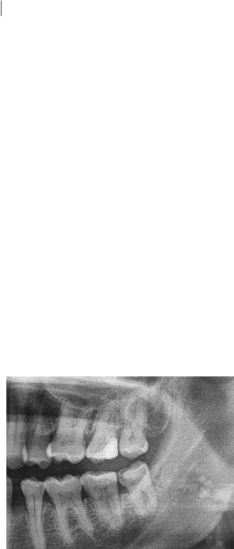
Improper use of elevators may see them being placed between the tooth being extracted and an adjacent tooth, exposing the latter to extraction forces. This can lead to either luxation of the entire adjacent tooth or, more likely, damage to the structural integrity of that tooth’s crown or restorations. Situations where this is likely are usually predictable during the preoperative assessment, through examination of the radiograph for the periodontal integrity of the adjacent teeth or for the presence of large restorations or crowns (Figure 6.1).
Luxation of adjacent teeth is a difficult problem to manage. As soon as there is any clinical suspicion that an adjacent tooth has been mobilised, the procedure should be stopped, and the area examined to ensure there is no associated dentoalveolar complex fracture. Once an adjacent tooth has been mobilised, elevator instruments become unsuitable for continuation of the extraction;
Figure 6.1 Upper second molar showing heavy restoration, putting it at high risk of iatrogenic injury if care is not taken during removal of the upper third molar.
https://t.me/DentalBooksWorld

6.4 Tooth Aspiratio n orngestio 81
either forceps only should be utilised or surgical methods must be adopted. Once the tooth marked for extraction has been removed, standard trauma management of a luxated or avulsed tooth should be applied.
Where the adjacent teeth have been previously restored, this restoration is susceptible to dislodgement or breakage, as it does not carry the structural strength of a native, unrestored tooth. Dislodgement of large restorations creates an additional intraoperative problem that must be addressed. Any broken restoration material or fractured enamel should be removed from the surgical site, as this may pose an airway risk or elicit a foreign-body reaction if inadvertently pushed into the soft tissues. The extraction must be completed carefully, without the use of elevators near the broken tooth. The patient must be informed of the incident, and of the need for temporisation of the previously restored area. This should occur immediately, with a future definitive restorative plan arranged after the immediate postoperative period.
6.3 Mandible Fracture
If exerted inappropriately on mandibular teeth, the forces of extraction may lead to fracture of the mandible. This serious complication is more often than not the result of a failure to recognise the reason why a tooth may not be mobilising, and the use of excessive force in a direction of weakness of the mandibular bone. It commonly occurs in patients with atrophic mandibles or short mandibular body height, or where attempts are made to remove an impacted tooth without use of surgical techniques by a novice practitioner. Fractures may also be encountered in patients with osteoporosis or abnormal pathologic processes in the area, such as cysts or tumours.
Mandible fracture may be suspected if there is a loud ‘crack’ heard, associated with increasing pain, and loss of continuity of the mandibular teeth with stepping or diastemas. Gingival lacerations may be evident, with surrounding haematomas.
If clinically suspected, mandible fracture must be ruled out with radiograph prior to proceeding with extraction. The radiographic appearance of a radiolucent, irregular line passing obliquely or perpendicularly to the mandible bone is diagnostic of fracture. If evident, attempts to remove the tooth should cease immediately. This complication requires emergency treatment under general anaesthetic by a specialist oral and maxillofacial surgeon, and the patient should be referred to the nearest tertiary referral hospital for inpatient management.
6.4 Tooth Aspiration or Ingestion
Loss of a tooth into the oropharynx is a medical emergency and should be treated as such until proven otherwise. In the worst-case scenario, a tooth can enter the tracheobronchial tree, causing airway obstruction and cardiorespiratory arrest. More likely, however, is that the patient’s gag reflex is activated, triggering either a swallow – causing the tooth to be ingested into the gastrointestinal tract – or a vomit – expelling the contents of the stomach and elevating the tooth out of the upper airway.
If a tooth is lost into the oropharynx, the patient should be managed according to basic life support protocols. If there are signs of impending respiratory obstruction, the patient requires immediate ambulance transfer to the nearest hospital for bronchoscopy and removal of foreign body. Their airway must be secured, and high-flow oxygen applied via mask. If the patient is unresponsive or not breathing, cardiopulmonary resuscitation should be commenced until the arrival of paramedic personnel.
https://t.me/DentalBooksWorld

82 6 Intraoperative Complications
If the patient is clinically stable, transfer to a local emergency department is still required, to confirm the location of the lost tooth with a chest x-ray; this is imperative, as patients may still be clinically stable even if tooth fragments are present in the tracheobronchial tree. If the tooth has been swallowed and is in the stomach, it will most likely pass through the alimentary system and excreted in faeces.
6.5 Instrument Fracture
Instrument fractures only occur when there is a pre-existing defect in the instrument being used or when fine instruments are used for a purpose for which they are not designed. This is most commonly seen with the use of a fine periotome to simultaneously disrupt the periodontal ligament of a tooth and elevate a large tooth. Periotomes have a small, sharp end that is an efficient cutting instrument, but is not designed to withstand rotational force along its axis.
If instrument fracture occurs, it is imperative that the fractured components be removed carefully. Fractured instrument components may act as a nidus of infection or elicit a foreign body granulomatous reaction that can culminate in acute osteomyelitis of the bone. A surgical approach may be necessitated to remove the fractured segments, possibly requiring deeper surgical exploration of the surgical site once the tooth has been extracted.
6.6 Intraoperative Bleeding
During or after extraction of a tooth, bleeding is a common complication. Whilst it generally stops spontaneously, failure to manage intraoperative bleeding may lead to life-threatening postoperative bleeding, which can result in haematoma or ecchymosis, and require blood transfusion or hospitalisation.
Intraoperative bleeding may result from trauma to the soft tissues, causing laceration of blood vessels, or from bone trauma, resulting in bleeding from nutrient canals or central bone vessels. It can be exacerbated by the presence of inflammation or infection, due to the vasodilatory effect of inflammatory mediators. A number of medical conditions can contribute to increased bleeding risk, and these require specific assessment and management prior to any surgical procedures.
Intraoperative bleeding during dental extractions that is unexpected must be stabilised prior to patient discharge. Whilst a number of methods and materials are available for haemostasis, use of the principles of pressure, packing, and patience is the first (and often only) method required to obtain sufficient haemostasis. In nearly all cases, the best course of action is to remove the tooth first, prior to application of haemostatic measures; the exception is in situations where bleeding is profuse and significant, where the tooth may be utilised as a well-adapted pressure dressing (e.g. where a high-flow arteriovenous malformation is encountered).
Immediate pressure to the area using a gauze bite pack for approximately 5–10 minutes provides enough time for the surgeon to prepare for additional haemostatic agents, if required (Table 6.1), and for the tissues and vessels to progress through vasospasm and platelet plug formation. A reduced flow of haemorrhage allows for a thorough examination of the surgical site in a systematic manner, commencing with the soft tissues surrounding the extraction site, through to all bony windows of the tooth socket, and finally to the apex of the socket. Occasionally, a single bleeding point is identifiable; more commonly, the intraoperative haemorrhage is a general ooze from all of the tissues. If a single bleeding point is found, specific local measures may be used to control the
https://t.me/DentalBooksWorld

6.7 Oroantra l Cmmunicatio 83
Table 6.1 Haemostatic agents that may be used to assist with control of bleeding after dental extraction.
Local anaesthetic infiltration
Suturing
Gauze pressure ± impregnation
Cellulose
Gelatin foam
Thrombin
Fibrin
Tranexamic acid mouthwash
Calcium alginate
Bone wax
bleeding in the area. Bone wax is a useful tool for this purpose, where intrabony vessels require plugging.
If generalised oozing is present, a simple method of haemostasis is to pack the socket with an absorbable haemostatic agent of choice and then place a retention suture over it. Additional pressure with haemostatic gauze soaked in 0.2% tranexamic acid solution or 1 : 100 000 adrenaline for 30 minutes increases the haemostatic effect.
Patients with significant intraoperative bleeding that cannot be controlled may require urgent referral to the closest emergency department for further investigation and haemorrhage control. This should be organised promptly in discussion with the local oral and maxillofacial surgery service.
6.7 Oroantral Communication
An oroantral communication is a direct hole between the oral cavity and the maxillary sinus. The most common cause is extraction of a lone-standing posterior maxillary tooth, with an associated fracture of the thin alveolar bone that separates the two cavities. This usually occurs in the context of lone-standing solitary upper first molars or high-impacted upper third molars; however, any tooth that has roots anatomically close to the sinus cavity may be at risk. Occasionally, oroantral communications can be associated with displacement of a tooth root or fragment into the maxillary sinus.
The likelihood of oroantral communication from an upper first molar may be precisely determined preoperatively on the panoramic radiograph, based on the relationship of the sinus floor to the root of the tooth. If the floor of the antrum lies below more than one-quarter of the distance between the apex and the cementoenamel junction, the risk of oroantral communication is considered high.
The surgeon may only be alerted to the presence of an oroantral communication after tooth extraction has been completed. Large oroantral communications will be visible on examination of the socket; irrigation with normal saline will produce the sensation of water trickling down the back of the patient’s nose, as the fluid passes directly into the maxillary sinus and on to the nasal cavity. Smaller ones can be identified by asking the patient to Valsalva whilst holding the nares shut; airflow or bubbles in the socket may be appreciable. At no stage should the socket be instrumented, as this has the potential to either create or worsen an oroantral communication.
https://t.me/DentalBooksWorld

84 6 Intraoperative Complications
Oroantral communications may close spontaneously when the defect has a width of less than 5 mm. Defects over this size are unlikely to close spontaneously and require surgical repair. This should occur promptly, as, if untreated, 50% of patients will develop acute bacterial sinusitis after 48 hours. Partial oroantral communications, where the bony sinus floor is breached but the maxillary sinus membrane is intact, generally do not require surgical intervention and will heal uneventfully.
If an oroantral communication is not detected intraoperatively, patients may present within one to two weeks postoperatively with symptoms of regurgitation of liquids between the nose and mouth, air passage between the two cavities, altered vocal resonance, or symptoms suggestive of sinusitis.
Repair of oroantral communication and removal of displaced root fragments from the maxillary sinus is an advanced surgical skill that is performed by specialist oral and maxillofacial surgeons. The patient should be appropriately informed of this complication, and a semi-urgent outpatient referral to a specialist service is necessary. Given the propensity for these patients to develop acute bacterial sinusitis, a course of preoperative empirical oral antibiotics with sinus coverage should be prescribed, along with saline nasal sprays for lavage.
6.8 Dentoalveolar Fracture
Lone-standing teeth that have been under repetitive, heavy cycles of load are more likely to be ankylosed to the underlying bone. Clinically, these teeth will often respond differently to standard approaches to elevation and delivery using dental instruments. In situations where a tooth is ankylosed, dentoalveolar fracture is the unintended consequence of tooth extraction.
Dentoalveolar fractures may range in presentation from a small piece of alveolus attached to the root surface to large dentoalveolar fragments where there is separation of a large complex of bone with attached teeth from the maxilla or mandible (Figure 6.2). In larger fragments, there may be involvement of surrounding structures, which may need to be addressed. For example, large dentoalveolar segment fractures of the posterior maxilla are a frequent cause of later oroantral communication.
The surgeon should be suspicious of dentoalveolar fracture in cases where abnormally high amounts of force are required to mobilise a tooth, or if there is abnormal or unexpected movement of the adjacent teeth or under the soft tissues. A large ‘crack’ may or may not be associated with creation of a dentoalveolar fracture. Patients may experience pain associated with the fracture if it extends past the area of local anaesthesia.
If the presence of a small buccal alveolar bone fracture is noted, the surgeon’s aim should be to remove the tooth safely, preserving the gingival soft tissues. If the fractured segment only covers the root surface of the tooth being extracted, it is appropriate to use a periosteal elevator to gently separate the fractured bone from the gingiva, then remove the tooth and fractured segment as a single piece. Any soft tissue trauma should be addressed and sutured, where necessary.
If the fractured segment extends to the root surfaces of adjacent teeth, the surgeon should attempt to preserve as much soft tissue attachment to the piece of bone as possible, and to separate the tooth being extracted from the ankylosed bone segment. This requires gentle and careful luxation; if the tooth cannot be successfully released from the bony segment, the segment will have to be sacrificed. This comes at the cost of future periodontal problems for the adjacent teeth and should be avoided where possible. If the bone segment can be salvaged, a tight approximation of
https://t.me/DentalBooksWorld

6.8 entoalveolar ractmre 85
Small Dentoalveolar Fracture
Small bone segment of ankylosed tooth
Soft tissue trauma
Large Dentoalveolar Fracture
Bone fragment includes adjacent bone covering of lateral incisor and premolar
Figure 6.2 Small and large dentoalveolar fractures.
the soft tissues is essential in order to reposition this fractured segment, which is now essentially biologically identical to a free bone graft, and is susceptible to future necrosis and sequestration.
When the fractured segment is large and other teeth are attached to the fractured complex, repositioning and rigid splinting of the segment is the priority, to allow for bony healing and salvage of as many teeth as possible. Whilst fractures of this nature can appear disastrous, appropriate repositioning and splinting using traumatology principles carries an excellent prognosis. The tooth extraction procedure should be delayed as long as is safely possible until bone healing is complete, at approximately four weeks post-repositioning. A surgical approach is then necessary, in order to reduce the amount of force required for future attempts at tooth extraction.
https://t.me/DentalBooksWorld
https://t.me/DentalBooksWorld
87
7
Third Molar Surgery
This chapter introduces the practice of third molar surgery, including classification systems, difficulty assessment, indications, and surgical approach.
Extraction of third molar (‘wisdom’) teeth is a unique aspect of dentoalveolar surgery, and carries additional challenges in treatment planning and approach. The decision to remove wisdom teeth is not as straightforward as that to remove other teeth with pathology; in fact, in some centres worldwide, the prophylactic removal of asymptomatic, pathology-free third molars is advocated, for a number of reasons. This removal is not without risks and complications, and remains an area of controversy.
The main predictive factor underlying third molar impaction appears to be inadequate space between the distal part of the second molar and the ascending ramus. This can be influenced by a relatively small jaw, relatively large teeth, and the presence of crowding in the dentition, as well as the age of the patient. However, even amongst expert dentists and oral and maxillofacial surgeons, the ability to predict future impaction based upon these findings on panoramic radiography is poor, particularly in younger populations.
Third molar teeth are complex and difficult to remove, for a number of reasons:
●●The indications for their removal are different than those for general tooth extraction.
●●Third molars that require removal nearly always have some form of hard or soft tissue impaction, which needs to be addressed when planning for removal; that is, wisdom tooth removal is almost exclusively a surgical extraction, as opposed to a simple one.
●●Mandibular third molars may be in close proximity to the inferior alveolar nerve and the lingual nerve.
●●Maxillary third molars are closely related to the maxillary sinus, maxillary tuberosity, and related vascular structures.
●●Postoperative complications relating to infection can be worse than with other teeth, given the proximity of the third molar to surrounding deep neck spaces.
7.1 Treatment Planning of Impacted Third Molars
The list of indications for third molar extraction is extensive: extraction may be indicated for symptoms or pathology associated with the wisdom teeth themselves, their effects on the wider dentition, or as part of a wider comprehensive dental or medical treatment plan (Tables 7.1 and 7.2).
Principles of Dentoalveolar Extractions, First Edition. Seth Delpachitra, Anton Sklavos and Ricky Kumar. © 2021 John Wiley & Sons Ltd. Published 2021 by John Wiley & Sons Ltd.
Companion website: www.wiley.com/go/delpachitradentoalveolarextractions
https://t.me/DentalBooksWorld

887 Third Molar Surgery
Table 7.1 Indications for third molar extraction as per the National Institute for Health and Care Excellence (NICE) and the American Association of Oral and Maxillofacial Surgeons (AAOMS).
NICE guidelines (UK) |
AAOMS indications (USA) |
Recurrent history of infection (including pericoronitis)
Unrestorable caries
Nontreatable pulpal and/or periapical pathology
Cellulitis, abscess, and osteomyelitis Periodontal disease
Prophylactic removal in the presence of specific medical and surgical conditions
Facilitation of restorative treatment (including provision of prosthesis)
Internal and/or external resorption of tooth or adjacent teeth
Pain directly related to the third molar
Tooth in the line of fracture of the mandible
Fracture of the tooth
Disease of the dental follicle (including cyst and tumour)
Tooth or teeth impeding orthognathic or reconstructive surgery
Teeth involved in the field of tumour resection
Tooth suitable for use as a donor for transplantation
Orthodontic treatment (e.g. maxillary retraction)
Pain
Carious tooth Pericoronitis
Facilitation of the management of or limitation of progression of periodontal disease
Nontreatable pulpal or periapical lesion
Acute and/or chronic infection (e.g. cellulitis, abscess)
Ectopic position (malposition, supraeruption, traumatic occlusion)
Abnormalities of tooth size or shape precluding normal function
Facilitation of prosthetic rehabilitation
Facilitation of orthodontic tooth movement and promotion of stability of the dental occlusion
Tooth in the line of fracture complicating fracture management
Tooth involved in surgical treatment of associated cysts and tumours
Tooth interfering with orthognathic and/or reconstructive surgery
Preventive or prophylactic removal, when indicated, for patients with medical or surgical conditions or treatments (e.g. organ transplants, alloplastic implants, bisphosphonate therapy, chemotherapy, radiation therapy)
Clinical findings of pulp exposure by dental caries Clinical findings of fractured tooth or teeth Impacted tooth
Internal or external resorption of tooth or adjacent teeth Patient’s informed refusal of nonsurgical treatment options Anatomic position causing potential damage to adjacent teeth Use of the third molar as a donor tooth for tooth transplant Tooth impeding the normal eruption of an adjacent tooth Resorption of an adjacent tooth
Pathology associated with the tooth follicle
Table 7.2 Contraindications for third molar extraction.
Extremes of age
Complex medical history, which can lead to significant postoperative morbidity (e.g. a history of radiotherapy to the region)
Situations where there is a high risk of intraoperative complications requiring tertiary-level services or general anaesthetic (e.g. high risk of mandible fracture)
Situations where an acute problem impacts access or local anaesthesia (e.g. acute odontogenic infection with trismus)
https://t.me/DentalBooksWorld

7.2 Difficulty Assessmen and Preoperativ Worku 89
Table 7.3 Treatment options for third molar teeth based upon presence of symptoms and presence of pathology.
|
Symptoms |
No symptoms |
|
|
|
Pathology |
1) REMOVE |
1) REMOVE |
No pathology |
1) REMOVE |
1) MONITOR |
|
2) MONITOR |
2) REMOVE |
|
3) OPERCULECTOMY |
|
|
|
|
The presence of third molar teeth is most commonly alerted to the patient by routine panoramic radiography performed by the general dentist, or by the onset of symptoms related to eruption or impaction. Generally, third molar teeth can be classified into four categories based on the presence of symptoms and the presence of periodontal pathology (Table 7.3). Symptoms can include pain, recurrent facial infections, and pericoronitis with food impaction. Pathology includes the presence of clinical signs of periodontal disease.
1)Symptomatic With Clinical Signs of Disease. Patients with symptoms and clinical signs of disease will largely benefit from removal of the third molar. In most cases, complete removal of the tooth, rather than conservative or minor surgical methods (e.g. operculectomy), is required to remove the associated pathology.
2)Symptomatic Without Clinical Signs of Disease. In this category, patients present with symptoms of pain or swelling, without evidence of developing impaction. Observation with symptom control is an appropriate means of managing these cases until their clinical prognosis becomes clearer with time. If symptom control methods are inadequate to provide sufficient patient comfort, extraction may be indicated. Similarly, these teeth may be removed alongside other third molars as part of a whole-of-mouth treatment plan.
3)Asymptomatic With Clinical Signs of Disease. This group of impacted third molars is detected on clinical examination in patients who have never exhibited symptoms but are noted to have periodontal pocketing, dental caries, or the presence of an operculum. Prophylactic removal of these teeth is recommended, to prevent periodontal defects to the adjacent second molar.
4)Asymptomatic Without Clinical Signs of Disease. The prophylactic removal of asymptomatic, disease-free third molars in this category has been suggested, in order to prevent the need for future third molar extractions in older age groups with greater case difficulty and surgical risk. However, there is insufficient literature to support this practice. Management of teeth in this category should be determined on a case-by-case basis, considering the individual circumstances that may determine patient prognosis.
7.2 Difficulty Assessment and Preoperative Workup
The major challenge in planning wisdom tooth extraction is the broad diversity of anatomic impactions, clinical presentations, and indications, and the extensive number of factors which can influence the difficulty of an individual case. The difficulty assessment phase of treatment planning is the most crucial deciding factor in terms of the success versus failure of a procedure, and in determining whether the case should be referred to a specialist oral and maxillofacial surgeon. Great
https://t.me/DentalBooksWorld

90 7 Third Molar Surgery
Table 7.4 Local and general factors that increase the difficulty of third molar removal.
Local factors |
Depth of impaction and type |
|
Root formation |
|
Proximity to inferior alveolar canal |
|
Caries or periodontal disease |
|
Mouth opening |
|
Gag reflex |
General factors |
Patient psychological factors |
|
Age |
|
Sex |
|
Ethnicity |
|
Patient weight |
|
|
Figure 7.1 Winter’s classification of third molars.From left to right: vertical impaction,mesioangular impaction,horizontal impaction,distoangular impaction.Source: Freire,B.B.,Nascimento,E.H.L.,Vasconcelos, K.de F.,Freitas,D.Q.,& Haiter-Neto,F.(2019).Radiologic assessment of mandibular third molars: an ex vivo comparative study of panoramic radiography,extraoral bitewing radiography,and cone beam computed tomography.OP u ScPgsPty, OP u MsnDiDes, OP u lheuegty en OP u R nDeuegty,128(2),166–175.© 2019 Elsevier.
care must be taken when deciding which wisdom teeth fall within the scope of the practitioner, as failed extractions can lead to significant morbidity for the patient, and to serious medicolegal concerns.
A comprehensive difficulty assessment involves the assessment of local and systemic factors, as well as indications and contraindications, for each individual scenario (Table 7.4).
Whilst a number of methods have been described for the classification of wisdom tooth impaction, there is poor correlation between any of them and the overall operative difficulty; this requires a much more comprehensive assessment of the patient. However, these classification systems can still be utilised as a method of communication between practitioners and patients, and can help to guide the surgical approach.
Winter’s classification is based upon the orientation of the third molar tooth’s axis relative to the adjacent second molar (Figure 7.1). Mesioangular third molars have an axis that is tilted in a mesial direction towards the second molar. Distoangular third molars have an axis tilted in a distal direction away from the second molar. Vertical third molars have an axis parallel to the second molar. Horizontal third molars have an axis perpendicular to the second molar.
On the basis of this classification, the general surgical approach to each type of impaction will be outlined later in this chapter. Keep in mind that the surgical principles described in Chapter 6 must always be adhered to with regards to soft tissue access, judicious bone removal, and sectioning of the tooth, where appropriate, in order to successfully complete the extraction.
https://t.me/DentalBooksWorld