

2 Gastrointestinal imaging
Contents
Liver 88
Biliary imaging 100
Pancreas 107
Spleen 116
Esophagus 123
Stomach 131
Small bowel 136
Large bowel 144
Mesentery and peritoneum 149
87

Liver
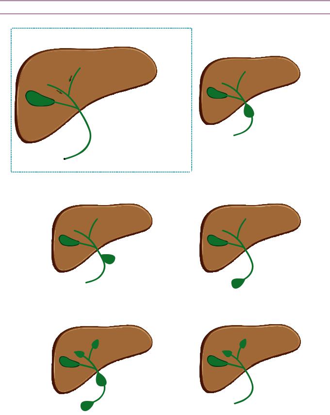
Liver anatomy
The Couinaud classification divides the |
iver nto eight segments Because each |
||
t s self-contained, |
individua |
segment |
be completely resected without |
disturbing the other segments |
|
|
|
Numbering of hepatic segments s clockwise when |
ooking at frontal/coronal view |
||
|
|
|
1 (caudate) |
|
|
|
not visible in frontal view |
*typically segments 6 and 7 are posterior and not visible on the frontal view.
8 |
4a |
2 |
|
|
|
7* |
4 |
|
|
|
|
5 |
|
3 |
|
4b |
|
6*
The portal veins divide the superior from inferior segments,
while the hepatic veins form the segmental borders in the axial plane.
The middle hepatic vein divides the left (2-4) from right (5-8) lobes of the liver.
axial view |
coronal view |
|
above the portal veins |
||
|
middle |
|
|
vein |
|
2 |
|
|
hepatic |
|
||
hepatic |
|
|
|
||
|
|
|
|
||
|
|
left |
|
|
|
|
vein |
|
|
|
|
|
|
|
|
|
|
right |
vein |
|
|
left |
liver |
|
|
|
|
||
hepatic |
|
|
|
|
|
|
|
right |
liver |
|
|
|
|
|
|
|
|
4
5
6*
right liver |
left liver |
|||
|
|
|
|
|
axial view |
coronal view |
below the portal veins |
vein hepatic right 
|
vein |
|
hepatic |
|
left |
right |
liver |
|
3
left |
liver |
|
6*
left liver
88

•Each hepatic segment features its own:
Central portal triad including branches of the portal vein, hepatic artery, and bile duct.
Peripheral venous drainage to the hepatic veins and ultimately the IVC.
•Mnemonic for remembering the segments:
Superior segments, from left to right: 2, 4, 8, 7, 1 (caudate). 2 doubled is 4; 4 doubled is 8; 8 minus 1 is 7.
Inferior segments, from left to right: 3, 4, 5, 6.
•Segments 2, 3, and 4 are in the left lobe of the liver.
•Segments 5, 6, 7, 8 are in the right lobe of the liver.
•Theleftandrightmainportalveinsdividethesuperiorfromtheinferiorsegmentsand continuetobranchsuperiorlyandinferiorlybeforeterminatinginthecenterofeachsegment.
•The branching of the portal veins is variable. The most common pattern is a bifurcation into right and left main portal veins, with the right main portal vein then branching into anterior and posterior branches.
•Small hepatic vein tributaries mark the peripheral margins of each segment.
•The caudate lobe drains directly to the IVC, not into the hepatic veins. The caudate lobe is spared in early cirrhosis since the direct drainage to the IVC spares the caudate from increased venous pressures due to portal hypertension. This leads to compensatory hypertrophy of the caudate lobe, which is a typical morphologic change of early cirrhosis.
Similarly, direct venous drainage to the IVC allows the caudate lobe to bypass the increased hepatic venous pressures seen in Budd–Chiari syndrome. Compensatory hypertrophy of the caudate lobe may preserve liver function in these patients.
CT and MRI of the liver
Liver CT
•A“routine”contrast-enhancedabdominalCTisacquiredintheportalvenousphaseof enhancement,typicallyobtained70secondsfollowingintravenouscontrastadministration.
•A portal venous phase CT reveals characteristic attenuation alterations and/or morphologic changes of diffuse liver disease, such as hepatic steatosis and cirrhosis. Most metastatic tumors are not hypervascular (although a few notable exceptions will be subsequently discussed) and can also generally be detected on the portal venous phase. Of note, rarely some breast cancers may be isoattenuating on the portal venous phase and more conspicuous on unenhanced CT.
•Most benign and malignant primary liver masses are hypervascular and thus most conspicuous in the arterial phase of enhancement. The arterial phase begins approximately 20–25 seconds after intravenous contrast injection. Many authors advocate that optimal conspicuity of a hypervascular liver lesion is obtained in the late arterial phase, which is between 9 and 16 seconds after abdominal aortic enhancement, or approximately 35 seconds after intravenous injection.
Liver MRI
•Compared to CT, MRI of the liver displays the same patterns of contrast enhancement but has superior lesion-to-liver contrast. MRI also does not impart ionizing radiation.
•MRI is able to obtain dynamic post-contrast images in multiple phases without any penalty in radiation exposure. Inand out-of-phase gradient imaging allows detection of intracytoplasmic lipid, which is seen in hepatic steatosis.Additionally, advanced techniques such as diffusion-weighted imaging may show potential for clinical use.
89
Hepatic metabolic disorders (diffuse liver disease)
Fatty liver (hepatic steatosis)
•Nonalcoholic fatty liver disease can be divided into steatosis and steatosis with associated inflammatory activity (steatohepatitis). Overall, greater than 15% of the population is afflicted with nonalcoholic fatty liver disease (NAFLD), which is a
component of the metabolic syndrome of obesity, insulin resistance, and dyslipidemia. Ultimately, steatohepatitis may progress to cirrhosis.
•CT can determine if steatosis is present and can provide a rough gauge as to its severity. Inand out-of-phase MRI imaging can more accurately quantify the degree of steatosis, although liver biopsy is the gold standard and best evaluates for the presence of inflammatory and early fibrotic change. By the time imaging can detect morphologic changes of cirrhosis, the changes may be irreversible.
•OnunenhancedCT,thelivershouldbeslightlyhyperattenuating relativetothespleen.The traditionalteachingisthatsteatosisispresentiftheliverattenuatesatleast10Hounsfield units(HU)lessthanthespleen,althoughnewworksuggeststhatevenasingleHUof relativehypoattenuation comparedtothespleenmayrepresenthepaticsteatosis.
•On contrast-enhanced CT, evaluation of hepatic steatosis is much less reliable compared to unenhanced CT due to different contrast uptake rates of the liver and the spleen. However, the liver is considered diffusely hypoattenuating if it attenuates at least 25 HU less than the spleen in the portal venous phase.
•Inand out-of-phase GRE MRI is a sensitive imaging technique to evaluate for the presence of (and to quantify the degree of) hepatic steatosis.
Diffuse hepatic steatosis: in- (left image) and out-of-phase (right image) images demonstrate near complete signal loss of the entire liver on out-of-phase images. Note the india-ink artifact at the periphery of organs that abut fat (e.g., spleen; arrows) in the out-of-phase images.
Case courtesy Cheryl Sadow, MD, Brigham and Women's Hospital.
When water-protons and fat-protons are present in the same MR voxel, the fat and water signals are summed in the in-phase images and subtracted in the out-of-phase images.
Relatively decreased signal on in-phase images suggests hepatic iron overload due to the longer TE of the in-phase images, which allows a longer dephasing time, exaggeration of T2* effect, and loss of signal.
•Variationsinportalvenoussupplymaycausegeographicregionsthataremoreorless affectedbyfattychange.Focalfatdoesnothaveanymasseffect,vesselscharacteristically runthroughit,andittendstooccurinthefollowingtypicallocationsanddistributions:
Gallbladder fossa (drained by gallbladder vein).
Subcapsular (along the falciform ligament). Periportal.
Focal fat may also be nodular throughout the liver. Ultrasound would demonstrate multiple hyperechoic lesions which would be hypoattenuating on CT. MRI shows pseudolesions drop in signal intensity on out-of-phase dual-phase GRE, consistent with nodular focal fat.
90

Amyloid
•Abnormal extracellular deposition of amyloid protein in the liver can cause focal or diffuse areas of decreased attenuation on CT imaging.
Wilson disease
•Wilsondiseasecauseshighlevelsofcoppertoaccumulateinthe basalganglia,cornea, andliverdueanautosomalrecessive genetic defect.Thelivermaybehyperattenuating onCTwithmultiple nodules, eventuallyleadingtohepatomegalyandcirrhosis.
Hepatic iron overload
•There are two pathways to excess hepatic iron accumulation. Accumulation within hepatocytes is seen in hemochromatosis. Uptake within the reticuloendothelial system (RES) causes hepatic Kupffer cell iron overload, as seen in hemosiderosis.
•Regardless of the etiology, the iron-overloaded liver is hypointense on all MRI sequences, relative to the paraspinal muscles as an internal control.
•Hemochromatosisisthemostcommoncauseofironoverload,duetoageneticdefect causingincreasedironabsorption.Excessironisunable tobestoredintheRES,sothe spleenandbonemarrowarenotaffected.Treatmentofhemochromatosisisphlebotomy.
Excess iron is deposited in hepatocytes (not the Kupffer cells that make up the intrahepatic RES), pancreas, myocardium, skin, and joints. Excess iron in hepatocytes can cause cirrhosis.
The spleen and bone marrow are normal since the RES is not involved.
•Hemosiderosis is excess iron stored within the reticuloendothelial system, which may be due to frequent blood transfusions or defective erythrocytosis. Treatment of hemosiderosis is with iron chelators, not phlebotomy.
The RES has a large capacity for iron. Iron stored in the RES is generally not harmful and the liver is normal in morphology, without cirrhosis.
MRI imaging of hemosiderosis demonstrates hypointense liver on conventional MRI sequences, similar to hemochromatosis. Additionally, the spleen and bone marrow will also appear hypointense due to increased iron stores throughout the entire reticuloendothelial system.
•Hemosiderosis is a precursor to secondary hemochromatosis. Secondary hemochromatosis is hepatic damage from iron overload after the RES system becomes saturated from prolonged hemosiderosis.
When the RES becomes overwhelmed with iron, the hepatocytes begin to store the excess. Similar to hemochromatosis, hepatocyte iron uptake may lead to cirrhosis.
•In clinical practice, the distinction between hemosiderosis and secondary hemochromatosis often overlaps. Many authors recommend against the term "secondary hemochromatosis" in favor of describing the primary disease (e.g., thalassemia) with secondary iron overload.
Differential based on CT attenuation
•Hypoattenuating liver: The liver is considered hypoattenuating if it attenuates less than the spleen on an unenhanced CT.
Fatty liver (hepatic steatosis) is by far the most common cause of a diffusely hypoattenuating liver.
Hepatic amyloid is rare and may cause either focal or diffuse hepatic hypoattenuation.
•Hyperattenuating liver: The normal unenhanced attenuation of the liver is 30 to 60 HU. An absolute attenuation greater than 75 HU is considered hyperattenuating.
Iron overload is by far the most common cause of a hyperattenuating liver.
Medications (e.g., amiodarone, gold, and methotrexate).
Copper overload (Wilson disease).
Glycogen excess.
91

Hepatic infection
Viral hepatitis
•Patients with viral hepatitis often have a normal CT scan. Viral hepatitis may cause nonspecific CT findings, such as gallbladder wall thickening or periportal edema (fluid on both sides of the portal veins).
Candidiasis
•Systemic fungal infection may seed the liver (and commonly the spleen as well) due to portal venous drainage of infected bowel.
•CT shows multiple tiny hypoattenuating microabscesses in the liver and the spleen, which may be rim-enhancing.
•Candidiasis is almost always seen in immunocompromised patients.
•The differential diagnosis for multiple tiny hypoattenuating hepatic lesions includes metastatic disease, lymphoma, biliary hamartomas, and Caroli disease.
Abscess
Candidiasis: Noncontrast CT shows innumerable tiny hypoattenuating lesions scattered throughout the liver and spleen, representing candidal microabscesses in a bone marrow transplant patient with fungemia.
•Hepaticabscessismostcommonlycausedbyabowelprocessandresultantinfectious niduscarriedthroughtheportalsystemtotheliver.Commoncausesincludediverticulitis, appendicitis,Crohndisease,andbowelsurgery.E. coli isthemostcommonorganism.A primaryhepatobiliaryinfection,suchasascendingcholangitis,maybealesscommoncause.
•Imagingfeaturesofhepaticabscessmaymimicmetastasis,appearingasaring-enhancing massonCT.OnMRI,thereistypicallycentralhyperintensityonT2-weightedimageswith anirregularwallthatenhanceslate.Perilesionalenhancementmaybepresent.
Echinococcal disease
Hepatic echinococcus: Ultrasound (left image) shows a complex, primarily hypoechoic mass in the superior aspect of the liver containing a hyperechoic undulating membrane (red arrow). Contrastenhanced CT (right image) shows a fluid-attenuating cystic mass (yellow arrows) containing an undulating membrane (red arrow).
Case courtesy Cheryl Sadow, MD, Brigham and Women's Hospital.
•Hepatic echinococcosis is caused by ingestion of the eggs of Echinococcus granulosus, which is endemic in the Mediterranean basin and associated with sheep-raising. Echinococcal eggs can develop into hydatid cysts.
•OnCT,ahydatidcystisawell-definedhypoattenuating massfeaturingacharacteristic floating membraneoranassociateddaughtercyst.Peripheralcalcification maybepresent.
92

Cirrhosis
Etiology and pathology
•Cirrhosisiscausedbyrepeatedcyclesofinjuryandrepair,whichcanbeduetometabolic (alcohol,steatohepatitis, hemochromatosis,orWilsondisease),infectious(chronichepatitis BorC),orinflammatory(primarybiliarycirrhosisorprimarysclerosingcholangitis) etiologies.Thehallmarksofcirrhosisarefibrosisandattempted,disorganizedregeneration.
•The micronodular form of cirrhosis is most often due to metabolic causes.
•The macronodular form of cirrhosis is most often post-viral (hepatitis B or C).
Early signs of cirrhosis
•One of the earliest signs of cirrhosis is expansion of the preportal space. Atrophy of the medial segment of the left hepatic lobe in early cirrhosis causes increased fat anterior to the right main portal vein.
•Enlargement of the caudate lobe is a specific sign of cirrhosis. Specifically, a caudate to right lobe size ratio of >0.65 highly suggests cirrhosis. As previously discussed, the caudate drains directly to the IVC, not via the hepatic veins, which results in compensatory caudate hypertrophy.
•The empty gallbladder fossa sign results when hepatic parenchyma surrounding the gallbladder is replaced with periportal fat.
Secondary manifestations of cirrhosis
•Portal hypertension causes splenomegaly, portosystemic collaterals, and varices.
•Gallbladder wall thickening is due to hypoalbuminemia and resultant edema.
•Gamna–Gandybodiesaresplenicmicrohemorrhages,whichappearhypointenseonGRE.
Malignant hepatic masses
Pathway to hepatocellular carcinoma
•In the setting of cirrhosis, hepatocellular carcinoma (HCC) is thought to develop in a sequence from regenerative nodule to dysplastic nodule to HCC. Regenerative and dysplastic nodules cannot be reliably differentiated on imaging; and high-grade dysplastic nodules cannot be reliably differentiated from low-grade HCC.
•Regenerativenodule:Aregenerativenoduleiscompletelysuppliedbytheportalvein andisnotpremalignant.Aregenerativenoduleshouldnot enhanceinthearterialphase.
Most regenerative nodules show low signal intensity on T2-weighted images, with variable signal intensity on T1-weighted images. Rarely, a regenerative nodule may be hyperintense on T1-weighted images due to glycogen deposition.
On contrast-enhanced MRI, most regenerative nodules enhance to the same (or slightly less) degree as the adjacent hepatic parenchyma.
•Dysplastic nodule: Unlike a regenerative nodule, a dysplastic nodule is premalignant. However, most dysplastic nodules do not demonstrate arterial phase enhancement (unless high grade), since blood supply is still from the portal vein.
DysplasticnodulesarevariableinsignalintensityonT1-weightedimages.Mostdysplasticnodulesare hypointenseonT2-weightedimages,althoughhigh-gradedysplasticnodulesmaybeT2hyperintense.
Contrast-enhancedMRIshowslow-gradedysplastic nodulestobeiso-enhancingrelative toliverand thusindistinguishable fromregenerative nodules.Highgrade dysplastic nodulesmaydemonstrate arterialenhancementandbeindistinguishable fromwell-differentiated hepatocellularcarcinoma.
•A siderotic nodule is an iron-rich regenerative or dysplastic nodule. A siderotic nodule is hypointense on T1 and T2*-weighted images and hyperattenuating on CT.
A siderotic nodule is rarely, if ever, malignant.
93
Hepatocellular carcinoma (HCC)
HCCinacirrhotic liver:T2-weighted(topleft image)andT1-weightedpost-contrastlate arterialphase(toprightimage)MRIshowsa nodularexternal contouroftheliver,consistent withcirrhosis.ThereisaT2hyperintense, hypervascularmassinhepatic segment2(arrows), representing hepatocellularcarcinoma.
UnenhancedCT(leftimage)showsthemassto be isoattenuating andbarelyperceptible(arrows).
Case courtesy Cheryl Sadow, MD, Brigham and Women’s Hospital.
•Hepatocellular carcinoma (HCC) is the most common primary liver tumor. Cirrhosis is the major risk factor for development of HCC. A hypervascular liver mass in a patient with cirrhosis or chronic hepatitis is an HCC until proven otherwise.
•Alpha-feto protein (AFP) is elevated in approximately 75% of cases of HCC.
•Arterial phase enhancement is the characteristic imaging feature of HCC. However, between 10 and 20% of HCCs are hypovascular and thus slightly hypoenhancing relative to surrounding liver on arterial phase imaging.
•TheclassicCTorMRIappearanceofHCCisanencapsulatedmassthatenhanceson arterialphaseandwashesoutonportalvenousphase.HCCmaybedifficult todetecton non-contrastorportalvenousphaseCT.OnunenhancedMRI,HCCischaracteristically slightlyhyperintenseonT2-weightedimagesrelative tosurroundingliver.
The nodule in a nodule appearance describes an enhancing nodule within a dysplastic nodule and represents an early HCC.
•HCCislocallyinvasiveandtendstoinvadeintothe portalandportalveins, IVC,andbile ducts.Incontrast,metastasestotheliveraremuchlesslikelytobe locallyinvasive.
•Treatment options for HCC include partial hepatectomy, orthotopic liver transplantation, percutaneous ablation, and transcatheter embolization.
Fibrolamellar HCC
•FibrolamellarcarcinomaisasubtypeofHCCthatoccursinyoungpatientswithoutcirrhosis.
•The tumor tends to be large when diagnosed, but has a better prognosis than typical HCC. Unlike in HCC, AFP is not elevated.
•On MRI, fibrolamellar HCC is a large, heterogeneous mass. A fibrotic central scar is classic, which is hypointense on T1and T2-weighted images (in contrast, focal nodular hyperplasia features a T2 hyperintense scar that enhances late). Capsular retraction may be seen in 10%.
•Unlike HCC, the fibrolamellar subtype does not have a capsule, although there may be a pseudocapsule of peripherally compressed normal hepatic tissue.
94
Hepatic metastases
•Althoughmetastasesaresuppliedbybranchesofthehepaticarteryinducedbytumoral angiogenesis,mostmetastasesarehypovascularandbestappreciatedonportalvenous phase(incontrasttoHCC,whichishypervascularandbestvisualizedonlatearterialphase).
•Hypervascular metastases (best seen on arterial phase) classically include:
Neuroendocrine tumors, including pancreatic |
Renal cell carcinoma. |
Melanoma. |
neuroendocrine tumors and carcinoid. |
Thyroid carcinoma. |
Sarcoma. |
|
•Colorectal and pancreatic adenocarcinoma metastases are typically hypovascular and can usually be diagnosed on portal venous imaging.
•Calcifications can be seen in mucinous colorectal tumors or ovarian serous tumors Calcification within a metastatic lesion may imply a better prognosis.
•OnMRI,metastaticlesionstendtobehypointenseonT1-weightedimagesandhyperintense onT2-weightedimages.Bloodproductsandmelanin(asinmelanoma)areT1hyperintense.
•Pseudocirrhosis describesthemacronodularlivercontourresultingfrommultiplescirrhous hepaticmetastases,whichmaymimiccirrhosis.Treatedbreastcanceristhemostcommon causeofthisappearance.Capsularretraction,althoughnotalwaysseen,ischaracteristic ofpseudocirrhosis,andwhenpresentsuggestspseudocirrhosisovercirrhosis.
Pseudocirrhosis due to multiple treated breast cancer metastases: Contrastenhanced CT shows numerous hypoattenuating hepatic lesions
with a markedly nodular external hepatic contour (arrows) that resembles cirrhosis.
Liver function in this patient remained normal.
Hepatic lymphoma
•Primaryhepaticlymphomaisveryrare.Lymphomatousinvolvementofthelivertendsto besecondarytosystemicdisease,withassociatedsplenomegalyandlymphadenopathy.
Epithelioid hemangioendothelioma
•Epithelioid hemangioendothelioma is a rare vascular malignancy that characteristically causes multiple spherical subcapsular masses than can become confluent. The individual masses may have a halo or target appearance.
•Epithelioid hemangioendothelioma is one cause of capsular retraction.
Hepatic capsular retraction
differentialdiagnosis ofcapsularretraction
•Capsular retraction is a focal concavity of the normally convex external liver contour.
•Metastatic tumor (more commonly post-treatment).
•Fibrolamellar hepatocellular carcinoma (10% of cases of fibrolamellar HCC).
•Hepatocellular carcinoma (capsular retraction has been reported but is uncommon).
•Epithelioid hemangioendothelioma.
•Intrahepatic cholangiocarcinoma.
•Confluent hepatic fibrosis (wedge-shaped fibrosis seen in cirrhosis, most commonly the medial
segment of the left hepatic lobe or the anterior segment of the right hepatic lobe).
95

Benign liver masses
Focal nodular hyperplasia (FNH)
T1-weighted fat-saturated MRI
Late arterial post-contrast T1-weighted fat-sat MRI
Delayed post-contrast T1-weighted fat-sat MRI
T2-weighted MRI
Portalvenouspost-contrastT1-weightedfat-satMRI
Focalnodularhyperplasia:T1-weightedMRIshows abarelyperceptiblemassintherightliver(yellow arrows)withacentrallowintensityscar(redarrow).
ThemassisT2isointensewiththesubtlesuggestion ofT2hyperintensecentralscar(redarrow).
Arterial phase of enhancement shows avid enhancement with non-enhancement of the scar.
Themasswashesoutimmediatelyonportalvenous phase,withnochangeinintensityofthescar.
Delayed T1-weighted image shows late enhancement of the central scar (red arrow).
Case courtesy of Cheryl Sadow, MD, Brigham and Women’s Hospital
•Focal nodular hyperplasia (FNH) is disorganized liver tissue with no malignant potential. It is primarily seen in asymptomatic women and is not associated with oral contraceptives.
•FNHhasacharacteristic central“scar”whichdoesnotcontainfibrotic tissue andis thereforenotatruescar.Instead,thecentralareaconsistsofT2-hyperintenseductules andvenules,anddemonstratesdelayedenhancement.FNHdoesnothave acapsule.
•FNH can be difficult to see without contrast on CT and T1and T2-weighted MRI sequences. FNH avidly enhances during the arterial phase, then washes out very quickly. The portal venous phase will often show just the unenhanced scar, which enhances late.
•Kupffercellsandbileductepitheliumarebothpresent.Kupffercellsmaybeconfirmed byasulfurcolloidstudy(1/3ofthetime)andbileductcellscanbeseenonaHIDAscan.
96

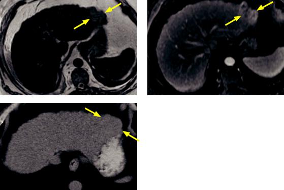
Hemangioma
Early arterial post-contrast T1-weighted image |
Portal venous post-contrast T1-weighted image |
Delayed post-contrast T1-weighted image |
T2-weighted image |
Hemangioma: Dynamic contrast-enhanced T1-weighted MRI (early arterial top left image to delayed bottom left image) shows a large mass (yellow arrows) in hepatic segment 7 demonstrating peripheral discontinuous nodular enhancement. There is progressively increasing centripetal enhancement towards the center of the lesion on delayed images. The signal intensity of the peripheral enhancement is similar to that of the aorta. On delayed images, there are a few central foci of nonenhancement (red arrows), consistent with cystic degeneration.
The hemangioma is hyperintense on the T2-weighted image (bottom right image) with subtle central areas of even higher signal centrally (red arrows), corresponding to the areas of cystic degeneration.
Case courtesy of Cheryl Sadow, MD, Brigham and Women’s Hospital.
•A hepatic hemangioma is a benign mass composed of disorganized endotheliallined pockets of blood vessels, supplied by a branch of the hepatic artery at the periphery.
•Hemangioma is more common in females and uncommon in cirrhosis. When a known hemangioma is sequentially followed in a patient with early cirrhosis, the hemangioma involutes as the liver becomes more cirrhotic.
•Hemangiomas may range in size from <1 cm to >10 cm. Giant hemangiomas tend to have a nonenhancing central area representing cystic degeneration.
•A virtually pathognomonic imaging feature is peripheral, discontinuous, progressive, nodular enhancement. The attenuation (or signal intensity on MR) of the enhancement is identical to the aorta and features gradual centripetal fill-in on later phases.
•The unenhanced CT appearance of a hemangioma is a nonspecific hypoattenuating liver mass.
97

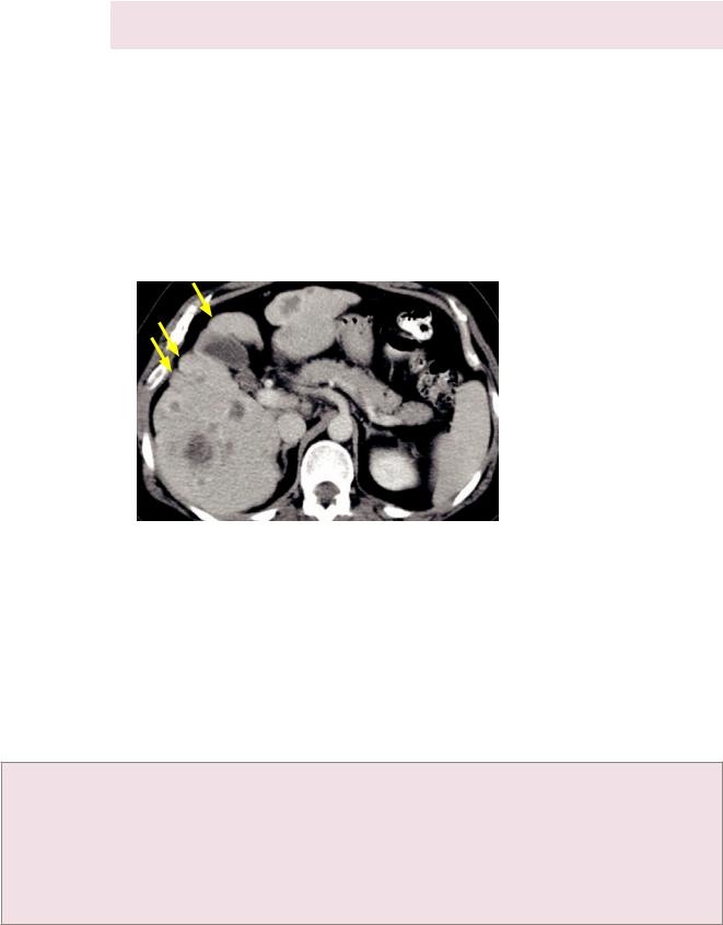
Hepatic adenoma
Hepatic adenoma: Coronal portal venous phase contrast-enhanced CT demonstrates a heterogeneously enhancing hypoattenuating mass (arrows) in the liver, which is a nonspecific appearance.
In- (left image) and out-of-phase (right image) MRI shows a mass in the right lobe of the liver (arrows) that demonstrates signal loss on out-of-phase images, consistent with a lesion containing intracellular lipid.
Case courtesy Cheryl Sadow, MD, Brigham and Women's Hospital.
•Hepatic adenoma is a benign hepatic neoplasm containing hepatocytes, scattered Kupffer cells, and no bile ducts.
The absence of bile ducts makes a nuclear medicine HIDA scan a useful test to distinguish between focal nodular hyperplasia (which contains bile ducts and would be positive on HIDA) and a hepatic adenoma, which does not contain bile ducts.
•Adenomas are much more common in females, especially with prolonged oral contraceptive use. When seen in males, adenoma may be associated with anabolic steroids.
•Adenomas have a relatively high risk of hemorrhage, which is often the presenting symptom. For this reason, incidentally discovered adenomas are usually resected.
•Multiple hepatic adenomas are seen in von Gierke disease (type I glycogen storage disease).
•A pseudocapsule may be present, which tends to enhance late.
•Adenomaslackportalvenousdrainageandthusarehypervascularonarterialphase.The presenceofmicroscopicfat,whenpresent,isbestseenonin-andout-of-phaseMRI. IntralesionalhemorrhagemaycauseT1hyperintensity.Adenomasmaybedifficult to differentiate fromotherhypervascularliverlesionsintheabsenceoffatorhemorrhage.
98

Vascular liver disease
Budd-Chiari
•Budd–Chiariishepaticvenousoutflowobstruction,whichcanbethromboticornon- thrombotic.Budd–Chiarimaybeduetohypercoagulativestatesincludinghematological disorders,pregnancy,oralcontraceptives,malignancy,infection,andtrauma.Itisveryrare tohaveprimaryBudd–Chiariduetocongenitalhepaticveinanomaly,aspicturedbelow.
•Acute Budd–Chiari presents with a clinical triad of hepatomegaly, ascites, and abdominal pain.
• Direct vascular findings include lack of flow within hepatic veins, thrombus in the hepatic veins/IVC, and the formation of
collateral vessels.
•Acute intraparenchymal findings include an edematous peripheral liver with sparing of the caudate lobe. The caudate is spared as it drains directly into the IVC.
•Progressive liver failure may result in chronic disease, producing caudate lobe hypertrophy and atrophy of peripheral liver with prominent regenerative nodules.
Veno-occlusive disease
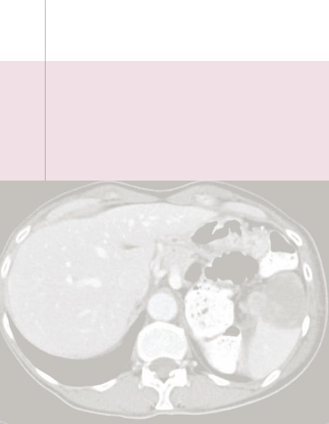
Congenital Budd–Chiari: Axial contrast-enhanced CT shows massive enlargement of the caudate lobe (yellow arrows) and the right posterior segment. There is atrophy of the left lobe and right anterior segments of the liver. Collateral venous drainage in the periphery of the liver is seen as geographic peripheral hyperenhancement (red arrows). The causative congenital hepatic vein anomaly is not visualized at this level.
Case courtesy Cheryl Sadow, MD, Brigham and Women's Hospital.
•Veno-occlusivedisease(VOD)isdestructionofpost-sinusoidalvenules,withpatenthepatic veins.VODisseeninbonemarrowtransplantpatients,possiblyduetochemotherapy.
•Imaging findings are nonspecific. Periportal edema, narrowing of the hepatic veins, hepatomegaly, and heterogeneous hepatic enhancement have been reported. In contrast to Budd–Chiari, the caudate lobe is not spared.
Cardiac hepatopathy
•Cardiac hepatopathy is passive hepatic congestion from heart failure, constrictive pericarditis, or right-sided valvular disease, which ultimately may lead to cirrhosis.
•Imaging clues are enlarged hepatic veins and IVC, with reflux of intravenous contrast from the right atrium into the IVC and hepatic veins. The liver is typically enlarged and demonstrates mottled enhancement. Ascites is usually present.
Congenital cystic liver disease
Biliary hamartomas (von Meyenburg complexes)
•Biliaryhamartomasareincidentalsmallcystic hepatic lesionsthatdonotcommunicate withthebiliarytree,causedbyembryologicfailureofnormalbileductformation.
•Biliary hamartomas tend to be smaller and more irregularly shaped than simple cysts.
Autosomal dominant polycystic liver disease (ADPLD)
•40%ofpatientswithautosomaldominantpolycystickidneydisease(ADPKD)haveasimilar diseaseprocessintheliver,calledADPLD.Eveninseveredisease,hepaticfailureisrare.
•On imaging, there are innumerable nonenhancing simple cysts throughout the liver.
99
Liver trauma
Overview of liver trauma
•Theliveristhesecondmostcommonlyinjuredsolidorganduetoblunttrauma,second tothespleen.TheCTdescriptionandgradingofliverinjuryissimilartosplenicinjury.
•The American Association for the Surgery of Trauma (AAST) classification describes hepatic injury based on findings at laparotomy.
•The MDCT hepatic injury grading scale is based on CT findings and is more commonly used by radiologists. It is similar to the MDCT grading scale for splenic trauma.
MDCT grading of hepatic injury
MDCT hepatictrauma |
• Grade I: Superficial laceration or subcapsular hematoma <1 cm in size. |
|
• Grade II: Laceration or subcapsular/intraparenchymal hematoma >1 and <3 cm in size. |
||
• Grade III: Laceration or subcapsular/intraparenchymal hematoma >3 cm in diameter. |
||
• |
Grade V: Destruction or devascularization of both hepatic lobes. |
|
|
• |
Grade IV: Massive hematoma >10 cm, or destruction/devascularization of one hepatic lobe. |
|
|
|
Biliary imaging
Introduction to MRCP
Magnetic resonance cholangiopancreatography (MRCP) overview
•Magnetic resonance cholangiopancreatography (MRCP) is an abdominal MRI acquired with heavily T2-weighted sequences that increase the contrast between T2 hyperintense stationary fluid in the biliary tract and surrounding structures.
•Fast spin echo sequences are most commonly used for MRCP acquisition. Various techniques can be employed to optimize imaging including breath-hold sequences and respiratory-triggered sequences.
•Heavily T2-weighted sequences primarily image the biliary tree.
•Sequences with intermediate T2 (TE 80–100 ms) are best suited for visualization of the biliary ductal system and surrounding tissue, in particular to evaluate extraluminal structures.
•Advantages of MRCP over ERCP include:
MRCP has the ability to see extra-luminal findings.
MRCP can visualize excluded (obstructed) ducts.
MRCP is non-invasive.
•Disadvantages of MRCP compared to ERCP include:
MRCP does not allow for concurrent therapeutic intervention.
MRCP does not actively distend the biliary ductal system with contrast.
MRCP has worse spatial resolution compared to ERCP.
•Contrast-enhanced MRCP can also be performed with fat-saturated T1-weighted imaging after injection of gadolinium contrast agents that have biliary excretion, such as gadoxetic acid disodium (Eovist, Bayer Healthcare, Germany) and gadobenate dimeglumine (Multihance, Bracco Diagnostics). These agents shorten T1 relaxation, resulting in T1 hyperintense biliary fluid, but require a 20–45 minute delay prior to imaging to allow time for biliary excretion.
100
Choledochal cysts
Overview and Todani classification of choledochal cysts
GB, gallbladder
CHD, common hepatic duct CBD, common bile duct RHD, right hepatic duct
sphincter LHD, left hepatic duct of Oddi 
Type I choledochal cyst:
Fusiform common bile duct dilation
LHD
RHD
GB CHD
CD
CBD
Most common, makes up ~50% of choledochal cysts
Type II choledochal cyst: |
Type III choledochal cyst: |
||
Extrahepatic saccular dilation |
Dilation of intraduodenal bile duct |
||
|
LHD |
|
LHD |
RHD |
|
RHD |
|
GB |
CHD |
GB |
CHD |
|
CD |
|
CD |
|
|
|
CBD |
Type IV choledochal cyst: |
Type V choledochal cyst: |
||
Multiple segments dilated |
Intrahepatic dilation = Caroli disease |
||
|
LHD |
|
LHD |
RHD |
|
RHD |
|
GB |
CHD |
GB |
CHD |
|
CD |
|
CD |
|
CBD |
|
CBD |
Type IVA: Intra and extrahepatic dilation (pictured)
Type IVB: Extrahepatic dilation only
101
•Choledochal cysts are thought to represent a heterogeneous group of diseases with a common end pathway of intrahepatic or extrahepatic biliary ductal dilation.
•The Todani system divides the cysts into types I–V based on their number, distribution, and morphology.
•Most choledochal cysts are diagnosed in childhood, but less commonly may be a new diagnosis for an adult. Clinically, choledochal cysts can present with nonspecific abdominal pain or may be found incidentally.
•Choledochal cysts are often resected due to increased cholangiocarcinoma risk, which can be as high as 25%.
•Incontrasttobiliaryhamartomas,choledochalcystsdo communicatewiththebiliarytree.
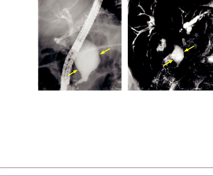
Type I choledochal cyst
Type I choledochal cyst: ERCP (left image) and thick-slab coronal MRCP heavily T2-weighted sequence (right image) shows a fusiform dilation of common bile duct (arrows).
Case courtesy Cheryl Sadow, MD, Brigham and Women's Hospital.
•A type I choledochal cyst, representing extrahepatic dilation of the common bile duct, is the most common type of extrahepatic cyst.
Caroli disease (Type V choledochal cysts)
•Caroli disease represents saccular dilation of the intrahepatic bile ducts, which may be segmental or diffuse. Caroli disease may be associated with polycystic kidneys.
•Caroli syndrome is Caroli disease plus hepatic fibrosis.
•The central-dot sign describes the small branches of the portal vein and hepatic artery bridging the dilated bile ducts, which look like a central dot on contrast-enhanced CT.
Biliary anatomical variants
Low insertion of cystic duct
•With a low insertion of the cystic duct, the surgeon may misidentify the common duct as the cystic duct if the patient undergoes cholecystectomy, possibly leading to inadvertent common duct ligation.
Aberrant right posterior duct
•Anaberrantrightposteriorductisonlyimportantifthepatientisarighthepaticlobeliver donor,asthetworighthepaticductsneedtobeanastomosedseparatelyintherecipient.
102

Gallbladder infection and inflammation
•Gallbladder pathology is further discussed in the ultrasound section.
Acute cholecystitis
•Cholecystitis is inflammation and localized infection secondary to obstruction of the gallbladder neck or cystic duct.
•Calculous cholecystitis is caused by a gallstone which blocks the cystic duct.
•Acalculous cholecystitis is a functional obstruction of the cystic duct without a culprit stone. It is typically seen in ICU patients.
•Acute cholecystitis is typically diagnosed by ultrasound, but similar criteria can be applied to CT:
Gallbladder wall thickening >3 mm (a nonspecific finding).
Pericholecystic fluid or inflammatory changes in the pericholecystic fat.
Gallbladder hyperemia.
Gallbladder calculi (although not all gallstones are radiopaque; ultrasound is more sensitive).
•Complications of acute cholecystitis include gangrenous cholecystitis, gallbladder perforation, and emphysematous cholecystitis.
•Gangrenous cholecystitis is due to increased intraluminal pressure, leading to gallbladder wall ischemia. On imaging, the gallbladder wall thickening may be notably asymmetric and intraluminal membranes may be present. Due to the increased risk of perforation, treatment is emergent cholecystectomy or cholecystostomy.
•Acute gallbladder perforation has a very high mortality due to generalized bile peritonitis. Subacute perforation may lead to a pericholecystic abscess and chronic perforation may cause a cholecystoenteric fistula.
•Emphysematous cholecystitis is a severe complication of acute cholecystitis caused by gas-forming bacteria. Gas may be present either within the lumen or the wall of the gallbladder. The typical patient susceptible to emphysematous cholecystitis is an elderly diabetic. Treatment of emphysematous cholecystitis is most often emergent cholecystectomy or cholecystostomy, although treatment can be conservative in patients with a very high surgical risk.
Porcelain gallbladder
•Porcelain gallbladder describes a peripherally calcified gallbladder wall, thought to be a sequela of chronic cholecystitis.
•Porcelain gallbladder is associated with a (somewhat controversial) increased risk of gallbladder carcinoma. Typically, a porcelain gallbladder is an indication for non-emergent cholecystectomy.
103

Bile duct infection and inflammation
Ascending cholangitis
•Obstruction of the biliary tree, most commonly due to choledocholithiasis, may cause ascending cholangitis, which presents with the clinical triad of fever, abdominal pain, and jaundice (Charcot’s triad).
•On imaging, the key finding is hyperenhancement and thickening of the walls of the bile ducts, often with a common bile duct stone present. On ultrasound, debris within the biliary system may be apparent.
•Initial treatment is antibiotics and fluid resuscitation. Endoscopic biliary intervention may be necessary if the patient does not respond to conservative management.
Primary sclerosing cholangitis (PSC)
Primary sclerosing cholangitis: ERCP (left image) and thick-slab coronal MRCP heavily T2-weighted sequence (right image) show a beaded, irregular appearance to the intrahepatic bile ducts.
Case courtesy Cheryl Sadow, MD, Brigham and Women's Hospital.
•Primary sclerosing cholangitis (PSC) is idiopathic inflammation and destruction of bile ducts.
•PSC is associated with ulcerative colitis (UC) and is more common in males.
Most (75%) patients with PSC have UC, while only a few (4–5% ) of patients with UC have PSC.
•Biliary imaging shows a characteristic beaded, irregular appearance of the common bile duct and intrahepatic bile ducts.
•PSC appears similar to HIV-cholangiopathy, although cholangitis in HIV patients is more commonly associated with papillary stenosis.
•Long-term complications of PSC include cirrhosis, cholangiocarcinoma, and recurrent biliary infections. Cross-sectional imaging is better at evaluating for these complications compared to ERCP.
Primary biliary cirrhosis (PBC)
•Primary biliary cirrhosis (PBC) is inflammation and destruction of smaller bile ducts compared to PSC. PBC affects middle-aged women and often initially presents with pruritus.
•Similar to PSC, chronic PBC can lead to hepatic cirrhosis.
104
AIDS cholangitis (AIDS cholangiopathy)
•Patients with acquired immunodeficiency syndrome are susceptible to biliary infection with Cryptosporidium and CMV, which clinically present with right upper quadrant pain, fever, and elevated LFTs.
•TheimagingofAIDScholangitisappearsnearlyidenticaltoprimarysclerosingcholangitis, withmultiplestricturesandabeadedappearanceofthebileducts.Adistinguishing featureofAIDScholangitisispapillarystenosis,whichisnottypicallyseeninPSC.
Recurrent pyogenic cholangitis (oriental cholangiohepatitis)
•Recurrent pyogenic cholangitis, also known as oriental cholangiohepatitis, is thought to be caused by the parasite Clonorchis sinensis, which leads to pigment stone formation, biliary stasis, and cholangitis. Nutritional deficiency may also play a role. The disease typically affects patients indigenous to Southeast Asia. Clinically, patients present with recurrent jaundice and fevers.
•Recurrent pyogenic cholangitis features an imaging triad of:
1)Pneumobilia.
2)Lamellated bile duct filling defects.
3)Intrahepatic and extrahepatic bile duct dilation and strictures.
•Patients with recurrent pyogenic cholangitis have an increased risk of cholangiocarcinoma.
Biliary neoplasia
Biliary cystadenoma
Biliary cystadenoma: T1-weighted post-contrast (left image) and T2-weighted (right image) MRI shows a large multiloculated cystic mass (arrows) in the right lobe of the liver, with enhancing septations. There are no enhancing nodules. There is also a small simple-appearing cyst in the left liver (segment 4; red arrow).
Case courtesy Cheryl Sadow, MD, Brigham and Women's Hospital.
•Biliarycystadenomaisabenigncysticneoplasm,occurringpredominantlyinmiddle-aged women.Biliarycystadenomamaybequitelargeatpresentationandcausenonspecific symptomssuchasabdominalpain,nausea,vomiting,andobstructivejaundice.
•Biliary cystadenoma does not communicate with the biliary system.
•On imaging, biliary cystadenoma appears as a large, multiloculated, cystic mass. The presence of septations distinguishes cystadenoma from a simple cyst. The septations may mimic an echinococcal cyst. In contrast to hepatic abscess or necrotic metastasis, a thick enhancing wall is not a feature of cystadenoma.
•Although benign, cystadenoma may uncommonly recur after resection.
•Malignant degeneration to biliary cystadenocarcinoma has been reported but is rare. The presence of a large solid component or thick calcification should raise concern for cystadenocarcinoma.
105

Cholangiocarcinoma
•Cholangiocarcinoma is a highly malignant tumor of the biliary ductal epithelium.
•A hilar tumor (at the confluence of the right and left intrahepatic biliary ducts), known as a Klatskin tumor, is the most common form of cholangiocarcinoma. In contrast, peripheral cholangiocarcinoma is rare.
•Cholangiocarcinoma tends to obstruct bile ducts and cause intrahepatic ductal dilation. Eventually, the obstruction may lead to lobar atrophy.
•Risk factors for development of cholangiocarcinoma include:
Choledochal cyst(s).
Primary sclerosing cholangitis.
Familial adenomatous polyposis syndrome.
Clonorchis sinensis infection.
Thorium dioxide (alpha-emitter contrast agent), not used since the 1950s. Thorium dioxide is also associated with angiosarcoma and HCC.
•On cross-sectional imaging, cholangiocarcinoma typically presents as an intrahepatic mass at the confluence of the central bile ducts (Klatskin tumor), with resultant bile duct dilation and capsular retraction. Tumor fingers often extend into the bile ducts.
Gallbladder carcinoma
•Gallbladder carcinoma is rare and is usually due to chronic gallbladder inflammation.
•Gallstones and concomitant chronic cholecystitis are typically present. Porcelain gallbladder, a result of chronic cholecystitis, is thought to be a risk factor for gallbladder cancer, although this is controversial.
•Gallbladder carcinoma most commonly presents as a scirrhous infiltrating mass that invades through the gallbladder wall into the liver. Less commonly, gallbladder carcinoma may appear as a polypoid mass. Very rarely it can present as mural thickening.
•Tumor spread is via direct extension into the liver, although lymphatic and hematogenous metastases are also common.
•Prognosis is generally poor, although small polypoid lesions may undergo curative resection.
Gallbladder metastasis
•Melanoma has a propensity to metastasize to the gallbladder.
106

Pancreas
Overview of pancreatic neoplasms
Pancreatic neoplasms
Solid epithelial neoplasm
Cystic epithelial neoplasm
Endocrine
neoplasm
 Ductal adenocarcinoma
Acinar cell carcinoma
Serous cystic
Mucinous cystic
Solid and papillary epithelial neoplasm
Intraductal papillary mucinous neoplasm
Insulinoma
Gastrinoma
Glucagonoma
80−90% of pancreatic tumors
rare, aggressive, can cause fat necrosis
benign, many small cysts, elderly women
malignant potential, surgical lesion,
single or few large cysts, middle-aged women
young women, heterogeneous, prone to hemorrhage
malignant potential, elderly males
most are benign and small
causes Zollinger−Ellison syndrome
VIPoma
Somatostatinoma
107
Solid pancreatic epithelial neoplasms
Adenocarcinoma (ductal adenocarcinoma)
CBD
PD
PD
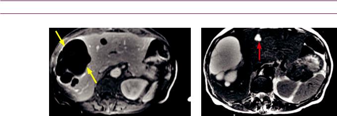
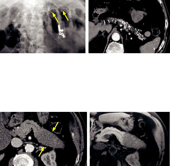
Pancreatic adenocarcinoma causing the double duct sign: Two coronal images from a contrastenhanced CT show marked dilation of the common bile duct (CBD), moderate dilation of the pancreatic duct (PD), and an ill-defined hypoattenuating mass in the pancreatic head (red arrows).
Case courtesy Cheryl Sadow, MD, Brigham and Women's Hospital.
•Pancreatic ductal adenocarcinoma makes up 80–90% of all pancreatic tumors. It is typically seen in patients over age 60, with a slight male predominance. Risk factors include smoking, alcohol, and chronic pancreatitis.
•A pancreatic-mass CT includes unenhanced, late arterial phase, and portal venous phase images. The late arterial phase (pancreatic parenchymal phase) has the greatest conspicuity for detecting the hypoenhancing tumor against the background enhancing pancreas.
•The most common location of ductal adenocarcinoma is the pancreatic head.
•Theclassicappearanceisahypodense(CT),T1hypointense(MR),ill-defined,hypovascular masscausingductalobstructionandatrophyofthepancreatictail.The double duct sign describesdilationofboththepancreaticductandthecommonbileduct.
•Since pancreatic adenocarcinoma is almost always associated with a dilated pancreatic duct, an alternative diagnosis should be strongly considered if there is a pancreatic mass with no ductal dilation, such as:
Autoimmune pancreatitis. |
Duodenal gastrointestinal stromal tumor (GIST). |
Groove pancreatitis. |
Peripancreatic lymph node. |
Cystic pancreatic tumor. |
Pancreatic metastasis (e.g., renal cell, thyroid, or melanoma). |
Neuroendocrine tumor. |
Lymphoma. |
|
|
•Conversely, if the double duct sign is present but no mass is visible, one should still be suspicious for pancreatic adenocarcinoma. Approximately 10% of cases will be isoattenuating relative to pancreas in the pancreatic parenchymal (late arterial) phase and thus extremely difficult to directly detect.
•Most cancers present at an advanced, unresectable stage. Unresectable tumors show encasement (>180° circumference) of the SMA, extensive venous invasion, or evidence of metastasis.
•For lower-stage tumors, complete surgical resection is the only chance for cure. A resectable tumor features no evidence of celiac, SMA, or portal venous invasion. Limited extension to the duodenum, distal stomach, or CBD does not preclude resection, as these structures are resected during the Whipple procedure. Limited
venous extension may be resectable.
108
Acinar cell carcinoma
•Acinar cell carcinoma is a rare, aggressive variant of pancreatic adenocarcinoma, exclusively seen in elderly males.
•The malignant cells produce a large amount of lipase to cause the clinical triad of lipase hypersecretion syndrome: Subcutaneous fat necrosis; bone infarcts causing polyarthralgias; and eosinophilia.
Cystic pancreatic epithelial neoplasms
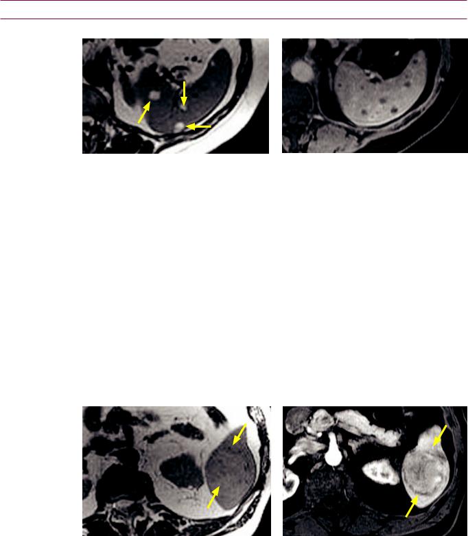
Serous cystadenoma
•Serous cystadenoma is a benign tumor that occurs in elderly women and has been nicknamed the grandmother tumor.
•Itconsistsofmanysmallcysts(>6cysts thatare<2cm)thatmayhaveasolid appearanceonCTduetoappositionof manycystwalls.MRIisusefultoshow thecysticnatureofthelesion.
•Serouscystadenomaishypervascular, uniqueamongcysticpancreatictumors.
•Unlikeadenocarcinoma,serous cystadenomadoesnotcause pancreaticductdilationortailatrophy.
•A classic imaging feature is central stellate calcification.
Mucinous cystic neoplasm
Serous cystadenoma: Axial oral-contrast-only CT shows a large multicystic pancreatic mass containing central stellate calcification (arrows).
Case courtesy Cheryl Sadow, MD, Brigham and Women's Hospital.
•Mucinous cystic neoplasm affects middle-aged women and has therefore been nicknamed the mother tumor.
•Mucinous cystic neoplasm is benign, but does have malignant potential. Treatment is typically resection due to malignant potential.
•The tumor consists of a single or a few large cysts (<6 cysts that are >2 cm) and typically occurs in the pancreatic body and tail.
•Mucinous cystic neoplasm has a capsule. The only other pancreatic tumor with a capsule is SPEN (below).
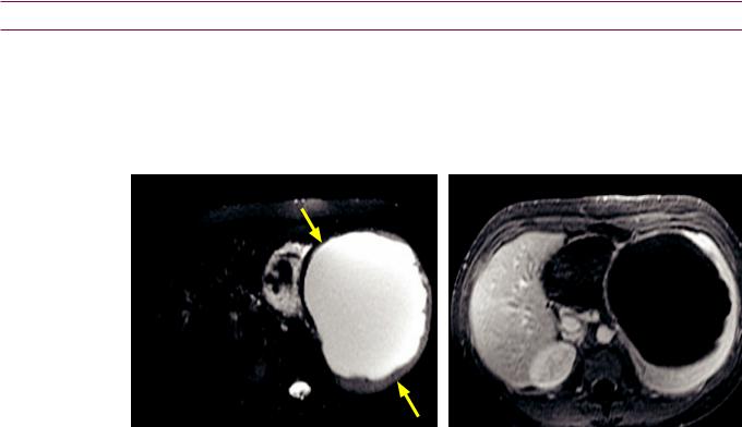
Solid and papillary epithelial neoplasm (SPEN)
Mucinous cystic neoplasm: Axial noncontrast CT shows a cystic, peripherally calcified mass in the tail of the pancreas (arrow). Pathology at resection showed borderline malignancy.
Case courtesy Cheryl Sadow, MD, Brigham and Women's Hospital.
•Solid and papillary epithelial neoplasm (SPEN) occurs in young women and children and is nicknamed the daughter tumor. It may be a rare cause of abdominal pain.
•It has a low malignant potential and is typically resected.
•On imaging, SPEN appears as a large mass with heterogeneous solid and cystic areas. Hemorrhage is typical. SPEN features a capsule, as does mucinous cystic neoplasm.
109
Intraductal papillary mucinous neoplasm (IPMN)
•Intraductal papillary mucinous neoplasm (IPMN) occurs most commonly in elderly males and is nicknamed the grandfather tumor, although these tumors exhibit the greatest age and sex variability of the cystic pancreatic neoplasms.
•IPMN features a spectrum of biological behavior from benign to indolent to aggressive carcinoma. IPMNs may arise from the main pancreatic duct or a sidebranch. The main duct IPMNs have greater malignant potential.
•Theclassicappearanceonendoscopyisafish-mouth papillapouringoutmucin.Crosssectional imagingshowsacystic intrapancreatic lesionincontiguity withthe ductor sidebranch.Anynodularorenhancingcomponentshouldraise concernformalignancy.
•The recommended imaging follow-up and criteria for resectability are controversial. Current guidelines published in 2006 recommend following simple pancreatic cysts <1 cm annually by imaging (typically MR). However, up to 40% of elderly males have a pancreatic cyst, suggesting that they may be an acquired condition of aging rather than a premalignancy.
•In general, a suspected IPMN is resected if it is >3 cm in size, if there is a mural nodule, or if there is associated dilation of the pancreatic duct to >10 mm.
Pancreatic endocrine neoplasms
Overview
•Pancreatic neuroendocrine tumors may be hyperfunctioning or non-hyperfunctioning.
•Hyperfunctioningtumorscometoclinicalattention duetosymptomsofendocrineexcess.
•Non-hyperfunctioningtumorstendtobelargeratdiagnosis.Thesetumorsmay undergocysticchangeandshouldbeconsideredinthedifferential ofacysticpancreatic neoplasm.Thereisoftencentralnecrosisandcalcification intheselargetumorsaswell.
•Pancreatic endocrine tumors tend to be hypervascular and are best seen in the late arterial phase. Most are solid unless large. A hypervascular liver mass with an
associated pancreatic mass is most likely a metastatic pancreatic endocrine neoplasm.
Insulinoma
•Insulinoma is the most common pancreatic endocrine tumor. Due to symptoms of hypoglycemia, insulinomas tend to present early and have the best prognosis of all neuroendocrine tumors, with only 10% demonstrating malignant behavior.
•TheWhippletriaddescribestheclinicalsymptomsofinsulinoma:Hypoglycemia,clinical symptomsofhypoglycemia,andalleviationofsymptomsafteradministrationofglucose.
Gastrinoma
•GastrinomacauseshypersecretionofgastricacidresultinginZollinger–Ellisonsyndrome. Gastrinomaisthesecondmostcommonpancreaticendocrinetumor.Gastrinomais associatedwithmultipleendocrineneoplasia(MEN)type1.WhenassociatedwithMEN-1, gastrinomastendtobemultipleandlocatedintheduodenumratherthanthepancreas.
•The gastrinoma triangle describes the typical location of gastrinomas, in an area bounded by the junction of the cystic duct and CBD, the duodenum inferiorly, and the neck/body of the pancreas medially.
•High gastrin levels may cause formation of carcinoid tumors in the stomach, which may regress after the gastrinoma is resected.
Other pancreatic endocrine tumors
•Glucagonoma is the third most common pancreatic endocrine tumor. Prognosis is
poor. VIPoma and somatostatinoma are very rare and also have poor prognoses.
110
Congenital pancreatic anomalies
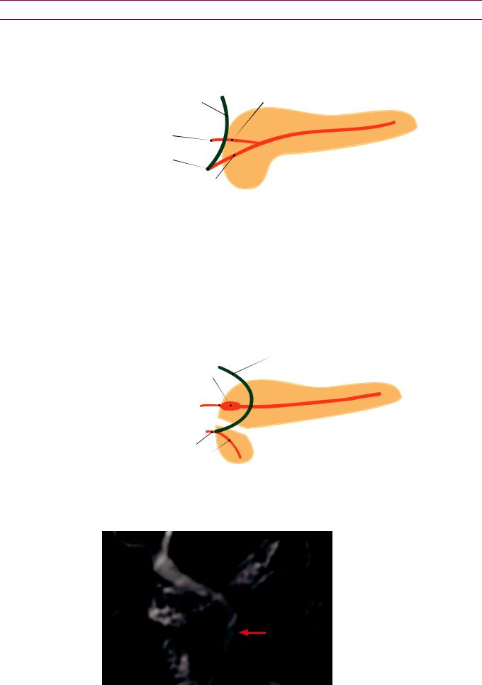
Normal ductal anatomy
Normally |
the main pancreatic duct drains to the major papilla (the ampulla of Vater) |
|||||
through the duct of Wirsung |
while the duct of Santorin drains to the minor papilla. |
|||||
The sphincter of Oddi s circular band of muscle encircling the ampulla of Vater |
||||||
|
|
duct of Santorini |
|
|
||
|
common bile duct |
(drains to minor papilla) |
|
|
||
|
|
|
|
|
|
|
meets the duct of Wirsung to |
|
|
|
|
|
|
drain into the major papilla |
|
|
main pancreatic duct |
|
|
|
minor papilla |
|
|
|
|
|
|
major papilla |
|
|
|
|
|
|
(ampulla of Vater) |
|
|
|
|
|
|
|
duct of Wirsung |
|
|
|
|
|
|
(drains to major papilla) |
|
|
|
|
|
Mnemonic for norma anatomy Santorini is uperior and drains to |
|
mal (minor) papilla. |
||||
|
|
|
|
|
||
The following anatomy s always constant regardless of whether |
|
anomaly s present |
||||
1) The |
bile duct always drains to the major papilla where t meets the duct of Wirsung |
|||||
2)The main pancreatic duct always drains the pancreatic tail.
3)The duct of Santorin always drains to the minor papilla.
Pancreas divisum
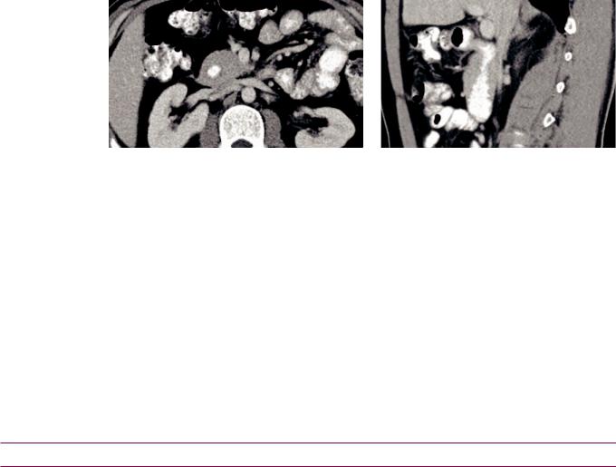
Pancreas divisum s the most |
congenita pancreatic anomaly t s caused by |
failure offusion of ventra and dorsa |
pancreatic ducts The ventra duct (Wirsung) |
only drains portion of the |
while the majority of the pancreatic exocrine |
gland output s drained through the smaller duct of Santorin nto the minor papilla.
Santorinicele
minor papilla
common bile duct
meets the ventral pancreatic duct (Wirsung) to drain into the major papilla
dorsal |
(main) |
|
major papilla |
crossing sign: CBD crosses the dorsal (main) |
|
pancreatic duct as it courses to join the ventral duct |
||
ventral (Wirsung) pancreatic duct |
dorsal and ventral pancreas do not fuse |
|
|
|
|
Pancreas divisum |
be |
of pancreatitis due to obstruction at the minor papilla |
from Santorinicele. A Santorinicele s foca dilation of the termina duct of Santorini
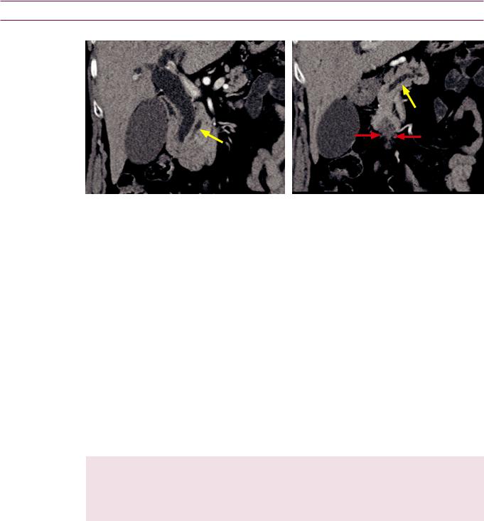
The crossing sign describes the CBD crossing Wirsung
MPD
CBD
VPD
the main duct to join the duct of
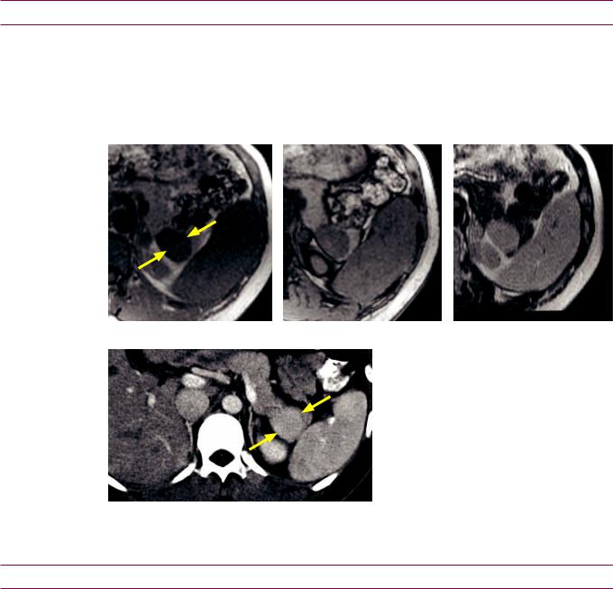
Crossing sign of |
divisum: |
|
Thick-slab corona |
MRCP heavily |
|
T2-weighted |
shows the |
|
|
bile duct (CBD) crossing |
|
the main pancreatic duct (MPD) |
||
at the |
The CBD |
|
towards the ventra pancreatic duct (VPD) to empty nto the major papilla. The main/dorsal pancreatic duct drains separately nto the minor papilla.
Case courtesy Cheryl Sadow, MD, Brigham and Women's Hospital
111
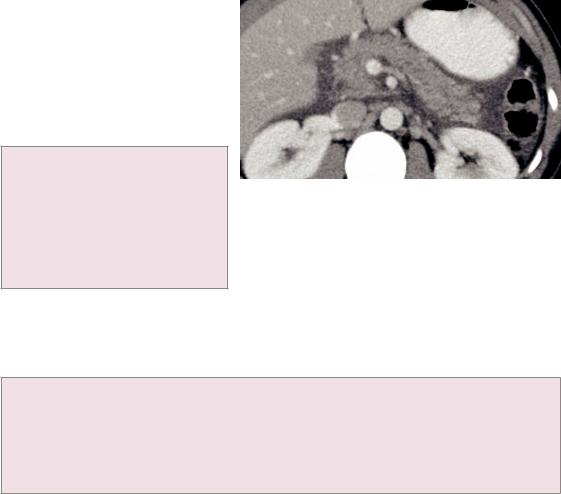
Annular pancreas
•Annular pancreas is a rare congenital anomaly where a portion of the pancreas wraps completely around the duodenum, secondary to incomplete rotation of the ventral pancreatic bud.
Panc
D
PancPanc
D
Annularpancreas:Axial(leftimage)andsagittal(rightimage)contrast-enhancedCTshowscircumferential encirclingofthepancreas(Panc)aroundtheduodenum(D),whichisfilledwithoralcontrast.
Case courtesy Cheryl Sadow, MD, Brigham and Women's Hospital.
•In an adult, annular pancreas can cause pancreatitis, peptic ulcer disease, and duodenal obstruction. In a neonate, it can cause duodenal obstruction and is in the differential for the double bubble sign.
Common channel syndrome/pancreaticobiliary maljunction
•Normally the common bile duct and duct of Wirsung both drain to the major papilla, where there is usually a thin septum separating these two systems.
•In common channel syndrome, also known as pancreaticobiliary maljunction, the distal CBD and pancreatic duct are missing the septum, allowing reflux between the two systems.
•Commonchannelsyndromemaybeinthespectrumofcholedochalcystdiseasewiththe commonchannelrepresentingaverymildformofcholedochocele.Commonchannel syndromemaypredisposetocholangiocarcinoma,butthisisrareandcontroversial.
Systemic diseases that affect the pancreas
Pancreatic manifestations of von Hippel–Lindau
•von Hippel–Lindau is an inherited multisystemic disease with increased risk of multiple malignancies and formation of cysts in various organs including the pancreas.
•Pancreatic neoplasms seen in von Hippel–Lindau include serous cystadenoma and pancreatic neuroendocrine tumors.
Cystic fibrosis (CF)
•Cystic fibrosis (CF) is the most common cause of childhood pancreatic atrophy.
•CF can cause either fatty atrophy of the pancreas or pancreatic cystosis (diffuse replacement of the pancreas with innumerable cysts).
Schwachman–Diamond
•Schwachman–Diamond is a rare inherited disorder characterized by diffuse fatty replacement of the pancreas, resultant pancreatic exocrine insufficiency, neutropenia, and bone dysplasia.
•Schwachman–Diamond is the second-most common cause of childhood pancreatic atrophy.
Obesity and exogenous steroid use
•Both obesity and steroids can cause fatty atrophy of the pancreas.
112
Miscellaneous pancreatic lesions
Intrapancreatic accessory spleen
•Intrapancreatic accessory spleen is a benign mimic of a hypervascular pancreatic neoplasm.
•On imaging, an intrapancreatic spleen typically is a small (1–3 cm), well-defined mass usually found in the pancreatic tail. It follows the density, signal intensity, and enhancement of the spleen on all CT and MRI sequences.
In-phase MRI |
Out-of-phase MRI |
T2-weighted MRI |
Intrapancreatic spleen: MRI images show a mass in the tail of the pancreas (arrows) that completely follows splenic signal intensity on all sequences. CT of the same areas (bottom left image) shows identical enhancement pattern of this mass compared to the spleen.
Case courtesy of Cheryl Sadow, MD, Brigham and Women’s Hospital
Contrast-enhanced CT
•MRI is usually diagnostic. Either technetium-99m sulfur colloid or technetium-99m RBC scintigraphy can confirm the diagnosis in ambiguous cases.
Pancreatitis
•Pancreatitis is inflammation of the pancreas, which may be due to a wide variety of etiologies that share a final common pathway of premature activation of pancreatic enzymes and resultant autodigestion of pancreatic parenchyma.
•Pancreatitis may range in severity from mild self-limited disease to necrotizing pancreatitis resulting in multi-organ failure and death.
CT protocol and role of imaging
•Imaging of pancreatitis is optimally performed in the pancreatic parenchymal phase (late arterial; ~40 seconds after contrast injection), which is the most sensitive timing to detect subtle areas of decreased enhancement suggestive of necrosis.
•CT is key for pancreatitis imaging. In addition to often identifying an etiology of the pancreatitis, CT can grade severity, detect complications, and guide possible percutaneous interventions.
•CT imaging is not indicated in patients with clinical diagnosis of mild acute pancreatitis, especially if they are improving. CT imaging may be negative or show a mildly edematous pancreas in these cases.
113
Acute pancreatitis
•Acute pancreatitis is most commonly caused by alcohol or an obstructing gallstone.
•Acute pancreatitis can be classified either with the Balthazar grading system or by the CT severity index.
•Balthazar grading system:
A:Normal-appearing pancreas
B:Focal or diffuse pancreatic enlargement
C:Mild peripancreatic inflammatory changes
D:Single fluid collection
E:Two or more fluid collections
Acute pancreatitis: Contrast-enhanced axial CT demonstrates diffuse pancreatic enlargement and peripancreatic edema. The pancreatic parenchyma enhances uniformly, without evidence for necrosis. This would be a Balthazar grade B, with 0 points added for necrosis, for a CT severity index of 2.
0% mortality, 4% morbidity for grades A, B, and C.
14% mortality, 54% morbidity for grades D and E (a fluid collection is a poor prognostic indicator).
•CT severity index (CTSI) integrates the Balthazar grading system with the degree of necrosis:
Assigns 0–4 points for Balthazar A–E, with 0 points for Balthazar A and 4 points for Balthazar E.
Adds 0–6 points for necrosis to create a total score from 0-10.
0 points: 0% necrosis
2 points: <30% necrosis
4 points: 30–50% necrosis
6 points: >50% necrosis
CTSI 0–3: 3% mortality, 8% morbidity
CTSI 7–10: 17% mortality, 92% morbidity
•Pancreatic and peripancreatic complications:
Pancreaticnecrosis:Onimaging,pancreaticnecrosisappearsasafocalordiffuseareaofnonenhancing pancreaticparenchyma.Evaluationofnecrosisisbestperformed48–72hoursafteronsetofacute pancreatitis. Latearterialphaseimaginghasthehighestsensitivityfordetectingpancreaticnecrosis. Patientswithpancreaticnecrosisareatincreasedriskforinfectionandseveremorbidity.
Fluid collections: Peripancreatic fluid may resolve or may evolve either into peripancreatic abscess or pseudocyst.
Pseudocyst: A pancreatic pseudocyst is a collection of pancreatic enzymes and fluid enclosed by a fibrous wall lacking an epithelial lining. The fibrous wall usually takes about 4–6 weeks to mature.
Pancreatic abscess: Pancreatic abscess is a purulent collection featuring thicker, more irregular walls compared to a pseudocyst. Gas locules may be present within the abscess.
•Extrapancreatic complications:
Extrapancreatic pseudocyst may occur nearly anywhere below the diaphragm and should always be considered in the differential of a cystic structure in a patient with history of pancreatitis. In particular, an intrasplenic pseudocyst may lead to intrasplenic hemorrhage.
Perihilar renal inflammation, which may lead to venous compression or thrombosis. Bowel involvement, especially of the transverse colon.
•Secondary inflammation of adjacent vessels can cause vascular complications:
Arterial bleeding, most commonly due to erosion into the splenic artery.
Pseudoaneurysm, most commonly of the splenic artery.
Venous thrombosis,mostcommonlysplenicveinthrombosis,whichmayleadtoportalhypertension.
114
Chronic pancreatitis
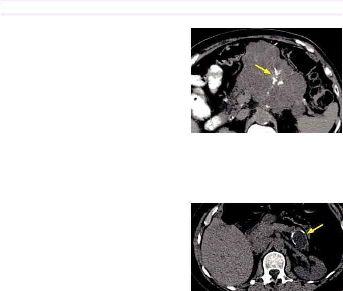
Chronic pancreatitis: Abdominal radiograph (left image) and contrast-enhanced axial CT (right image) show numerous coarse calcifications in the pancreas (arrows).
Case courtesy Cheryl Sadow, MD, Brigham and Women's Hospital.
•Chronic pancreatitis, most commonly from long-term alcohol abuse, causes irreversible pancreatic damage.
A much less common cause of chronic pancreatitis is pancreas divisum.
•Calcifications in the distribution of the pancreatic duct are pathognomonic for chronic pancreatitis.
Autoimmune pancreatitis
Segmental autoimmune pancreatitis: Contrast-enhanced axial CT (left image) shows a segmental region of low attenuation enlargement of the pancreatic tail and body (arrows), with loss of the normal ductal architecture.
T1-weighted unenhanced MRI (right image) shows a corresponding segmental loss of the normal T1hyperintense pancreatic signal, with effacement of the pancreatic duct in the affected body and tail.
The differential diagnosis for this appearance would include pancreatic lymphoma, less likely pancreatic adenocarcinoma as there is no ductal dilation.
Case courtesy Cheryl Sadow, MD, Brigham and Women's Hospital.
•Autoimmune pancreatitis is caused by an inflammatory lymphoplasmacytic infiltrate. It is associated with Sjögren syndrome and causes elevated serum IgG-4 levels.
•The typical imaging appearance of autoimmune pancreatitis is diffuse “sausageshaped” enlargement of the entire pancreas; however, a focal or segmental form may mimic a pancreatic mass.
•Treatment is with steroids, which can lead to a complete resolution.
115

Groove pancreatitis
Diagram demonstrates inflammation within
the |
between the head ofthe |
|
duodenum, and |
bile duct. |
|
Groove pancreatitis s the head of the usually affects
|
form offoca |
pancreatitis of the |
between |
duodenum, and |
bile duct Groove pancreatitis |
||
who |
heavy drinkers |
|
|
The histopathologic hallmark s fibrosis |
n the pancreaticoduodena |
Chronic |
nflammation of the duodenum |
varying evels of duodena stenosis cystic |
|
chang of the duodena wall. maging reflects these findings, with duodena thickening and cystic change often apparent The cystic change s best appreciated MR
The main differentia consideration s adenocarcinoma of the head of the
Spleen
Congenital splenic variations and anomalies
Splenule (accessory spleen) |
|
|
|
|
|
|
|
|
|
|
|
|
|
|
|
|
Also called |
|
|
|
spleen, splenule s |
focus of norma |
splenic tissue separate |
||||||||||
fr |
the main body of the spleen, due to embryologic failure offusion of the splenic |
|||||||||||||||
anlag |
The most |
|
|
ocation |
s the splenic hilum. |
|
|
|
|
|||||||
Although usually |
|
incidenta |
finding, the |
|
|
of |
splenule does have |
|
||||||||
signific |
n certain clinica |
settings |
For |
nstance |
splenectomy for consumptive |
|||||||||||
thrombocytopenia |
|
|
not be curative if there s sufficient unresected |
|
|
|||||||||||
splenic tissue present |
A splenule |
|
be mistaken for |
ymph node |
|
when |
||||||||||
n |
unusua ocation |
As previously discussed, |
ntrapancreatic splenule |
be |
||||||||||||
mistaken for hypervascular pancreatic |
|
|
|
|
|
|
|
|||||||||
A splenule should follow splenic tissue |
al |
MR |
|
f |
n doubt, |
Tc-99m |
||||||||||
sulfur colloid |
|
|
heat-damaged Tc-99m RBC |
|
be confirmatory. |
|
||||||||||
Polysplenia syndrome |
|
|
|
|
|
|
|
|
|
|
|
|
|
|
|
|
Polysplenia |
yndrome |
s |
ctrum of |
tomic disorders characterized b |
|
de |
||||||||||
of viscera heterotaxia |
n addition to multiple discrete foci of splenic tissue. Multiple |
|||||||||||||||
sple |
|
be |
the right |
|
eft, but |
|
always |
the |
side the stomach. |
|||||||
Polysplenia |
s usually associated with |
|
|
congenita cardiac anomalies. Most |
||||||||||||
tients die |
n early childhood, but |
few |
|
have only minor cardiac defects and |
||||||||||||
be incidentally discovered |
adults. |
|
|
|
|
|
|
|
||||||||
Polysplenia |
s associated with |
|
|
anomalies including nterruption of the |
VC with |
|||||||||||
|
hemiazygos continuation. A ess |
|
association s |
preduodena porta vein |
||||||||||||
Wandering spleen |
|
|
|
|
|
|
|
|
|
|
|
|
|
|
|
|
A wandering spleen |
s |
norma spleen with abnorma axity |
absence of |
ts fixed |
||||||||||||
igamentous attachments. |
|
|
|
|
|
|
|
|
|
|
|
|||||
Wandering spleen |
|
|
present clinically |
|
abdomina |
|
|
acute |
||||||||
abdomina pain secondary to torsion |
|
|
|
|
|
|
|
|
|
|||||||
116
Benign non-cystic splenic lesions
Hemangioma
Multiple splenic hemangiomas: T2-weighted (left image) and post-contrast T1-weighted (right image) MRI shows multiple T2 hyperintense splenic lesions (arrows) that demonstrate subtle peripheral enhancement.
Case courtesy Cheryl Sadow, MD, Brigham and Women's Hospital.
•Hemangioma is the most common benign splenic neoplasm. Hemangioma may be solitary or multiple, and lesions tend to be small.
•Splenic hemangiomas are associated with Kasabach–Merritt syndrome (anemia, thrombocytopenia, and consumptive coagulopathy) and Klippel–Trenaunay–Weber syndrome (cutaneous hemangiomas, varicose veins, and extremity hypertrophy). These visceral hemangiomatosis syndromes are usually associated with phleboliths.
•On CT, hemangiomas are typically isoor hypoattenuating pre-contrast and hyperenhancing. On MR, hemangiomas are typically hyperintense on T2-weighted images and may enhance peripherally or homogeneously. However, the classic pattern of discontinuous nodular enhancement seen in hepatic hemangiomas is uncommon.
•Nuclear medicine scintigraphy with Tc-99m labeled red blood cells would show increased activity within the lesion on delayed images. In contrast, Tc-99m sulfur colloid scanning may show either increased or decreased activity.
Hamartoma
Splenic hamartoma: T2-weighted (left image) and arterial-phase enhanced T1-weighted (right image)
MRI shows a vague T2 isointense splenic mass (arrows) that enhances heterogeneously.
Case courtesy Cheryl Sadow, MD, Brigham and Women's Hospital.
•Splenic hamartoma is a rare, benign lesion composed of malformed red pulp elements. It may be associated with tuberous sclerosis.
•Splenichamartomaistypicallyawell-circumscribed,iso-orhypoattenuating mass onunenhancedCTthatenhancesheterogeneouslyafter contrastadministration. On MR,ahamartomaisiso-toslightlyhyperintense onT2-weightedimages,featuring
heterogeneousearlyenhancementandrelativelyhomogeneousdelayedenhancement.
117
Benign cystic splenic lesions
Congenital true (epithelial) cyst
•A congenital true cyst is defined as having an epithelial lining. Interestingly, a splenic epithelial cyst may cause elevation of tumor markers including CA19-9, CA125, and CEA, despite its completely benign nature.
•Unlike a post-traumatic pseudocyst, a true cyst may have septations, but mural calcification is uncommon.
Splenic epithelial cyst: T2-weighted MRI with fat saturation (left image) and enhanced T1-weighted MRI (right image) shows a large T2 hyperintense, T1 hypointense, nonenhancing structure (arrows) replacing nearly the entire splenic parenchyma.
Case courtesy Cheryl Sadow, MD, Brigham and Women's Hospital.
Post-traumatic pseudocyst
•A post-traumatic pseudocyst is the end result of evolution of a splenic hematoma.
•Unlike a true (epithelial) splenic cyst, the periphery of a pseudocyst is not cellular but made of fibrotic tissue.
•On imaging, a post-traumatic pseudocyst appears as a well-circumscribed, fluiddensity lesion, with no peripheral enhancement.
•In contrast to a true cyst, septations are uncommon but there may be mural calcification.
Intrasplenic pancreatic pseudocyst
•A post-pancreatitis pseudocyst involving the tail of the pancreas may extend into the spleen. There is almost always a history of pancreatitis.
•Unlike a true congenital cyst, an epithelial lining is lacking and histology more closely resembles a post-traumatic pseudocyst.
•Splenic rupture has been reported in some cases of intrasplenic post-pancreatitis pseudocysts.
Lymphangioma
•Splenic lymphangioma is a rare, benign neoplasm usually diagnosed in childhood, which may be solitary or multiple.
•Lymphangioma features a classic imaging appearance of a multilocular cystic structure with thin septations. Post-contrast images may show septal enhancement.
118

Inflammatory splenic lesions
Sarcoidosis
•Sarcoidosis is a systemic disease of unknown etiology characterized histologically by multiple nodules composed of noncaseating granulomas.
•When sarcoidosis involves the spleen, splenomegaly is the most common presentation, often associated with hepatomegaly and lymphadenopathy.
•Lesscommonly,sarcoidosismayinvolvethe spleeninamultinodular pattern with numeroushypoattenuating 1–3cmlesionsdemonstrating essentially noenhancement.
ThesenodulesareformedbycoalescentsarcoidgranulomasandhavelowsignalonallMRIsequences.
Sarcoid nodules are most conspicuous on T2-weighted images and early-phase post-contrast T1weighted images. On the post-contrast images the nonenhancing nodules will stand out against the avidly enhancing splenic parenchyma.
•Imaging appearance is generally indistinguishable from splenic lymphoma.
Inflammatory pseudotumor
•Splenic inflammatory pseudotumor is a rare focal collection of immune cells and associated inflammatory exudate, of unclear etiology. Patients often have constitutional symptoms including fever and malaise.
•Inflammatory pseudotumor has a variable and nonspecific imaging appearance, but a typical presentation is of a well-circumscribed, heterogeneously enhancing mass.
Splenic infection
Pyogenic abscess
•Splenic bacterial abscesses are uncommon and usually seen in immunocompromised patients. A solitary abscess is much more likely to be bacterial. In contrast, multifocal small abscesses are more likely to be fungal.
•On CT, a bacterial abscess usually has an irregular, enhancing wall. Gas is not usually seen, but is highly specific for a bacterial abscess when present.
•A characteristic ultrasound finding is the wheel within a wheel or bull’s-eye appearance, which describes concentric hyperechoic and hypoechoic rings surrounding the abscess.
•TreatmentisCT-orultrasound-guidedpercutaneousdrainageinaddition toantibiotics.
Fungal abscess
•Splenic fungal abscesses are typically multiple and small, usually <1 cm in size. Almost all patients with splenic fungal abscesses are immunocompromised.
•The most common causative agents include Candida, Aspergillus, and Cryptococcus, which all appear as multiple tiny hypoattenuating foci on CT.
•Splenic Pneumocystis jiroveci (formerly known as Pneumocystis carinii) infection is rare, almost always seen in advanced AIDS, and has a classic appearance of multiple calcified splenic lesions.
Echinococcal cyst
•Splenic involvement of Echinococcus granulosus infection is unusual, seen in only 1 to 3% of echinococcal infections, and is almost always associated with infection of other organs as well.
•An echinococcal cyst is a true cyst with a cellular lining. As with echinococcal abscess elsewhere, characteristic imaging findings are a cystic lesion with internal undulating membrane and daughter cysts.
119
Malignant splenic lesions
Splenic lymphoma
•Splenic lymphoma is the most common splenic malignancy.
•Primary splenic lymphoma is rare, accounting for less than 1% of all cases of lymphoma, and usually presents as a solitary hypovascular mass. In contrast to splenic hamartoma or metastatic disease, primary splenic lymphoma may extend beyond the splenic capsule and involve adjacent organs.
•Secondary splenic involvement of systemic lymphoma is much more common. Four imaging presentations have been described, depending on the size of the lymphomatous masses:
Miliary masses, in which discrete tiny masses may be difficult to see.
Multiple small to moderate sized masses.
One large mass.
Splenomegaly without discrete mass.
•Conspicuity is highest on post-contrast T1-weighted images, where the involved portion of the spleen tends to be hypoenhancing.
•On ultrasound, lymphoma may appear cystic but color Doppler shows internal flow.
Splenic metastasis
Splenic metastasis from lung cancer: Contrast-enhanced axial CT shows a hypoattenuating, irregular mass in the inferior anterior aspect of the spleen (arrows) demonstrating wispy, heterogeneous enhancement.
Case courtesy Cheryl Sadow, MD, Brigham and Women’s Hospital.
•Splenic metastases may be solitary or multiple, cystic or solid. However, metastases to the spleen are rare, occurring in 2–9% of cancer patients. Most patients with splenic metastases already have known widespread disease.
Isolatedsplenicmetastasesareseeninonly5%ofpatientswithmetastaticinvolvementofthespleen.
•Several theories for the low rate of splenic metastases have been proposed, which is thought to be due to the antineoplastic properties of lymphoid-rich splenic tissue and lack of afferent lymphatics to bring tumor cells into the spleen.
•The most common primary tumors known to metastasize to the spleen include breast, lung, ovarian, and melanoma. Ovarian cancer and melanoma typically cause cystic metastasis.
•Calcification is rare unless the primary is a mucinous adenocarcinoma.
Angiosarcoma
•Angiosarcoma is a rare, extremely aggressive malignancy, with 20% 6-month survival.
•Unlike hepatic angiosarcoma, the association with Thorotrast, vinyl chloride, and arsenic has not been well established.
•Angiosarcoma usually presents as enlarged, heterogeneous mass that may completely
replace the normal spleen. Enhancement is variable and heterogeneous.
120
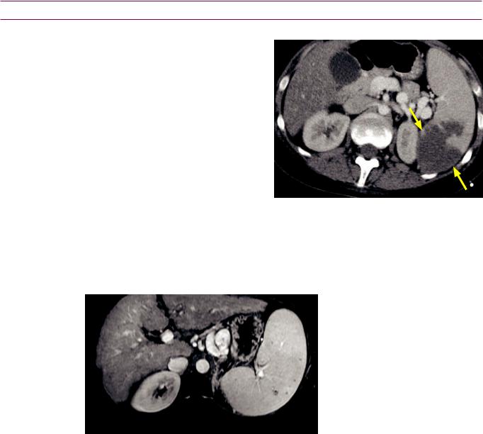
Miscellaneous splenic lesions
Splenic infarct
•Splenic infarcts are most commonly due to emboli (in older patients) and thrombosis (in younger patients with hematologic disease).
•Splenic infarct classically manifests as a wedge-shaped peripheral region of nonenhancement, but a more heterogeneous, mass-like appearance can also be seen.
•On MR, the affected regions may be T1 hyperintense if acute and
hemorrhagic, while chronic infarcts are T1 hypointense and T2 hyperintense.
•Lack of enhancement of the entire spleen should raise the concern for complete infarction, possibly due to a
wandering spleen with torsion.
Gamna-Gandy bodies
Splenic infarct: Axial contrast-enhanced CT shows a posterior peripheral region of splenic
nonenhancement (arrows).
Case courtesy Cheryl Sadow, MD, Brigham and Women’s Hospital.
Gamna–Gandy bodies: Contrastenhanced T1-weighted MRI shows numerous hypointense foci scattered throughout the spleen in a patient with evidence of cirrhosis (nodular liver contour, splenomegaly, and varices).
Case courtesy Cheryl Sadow, MD, Brigham and Women’s Hospital.
•Gamna–Gandy bodies are multiple tiny foci of hemosiderin deposition resulting from portal hypertension.
•Usually, other signs of portal hypertension and cirrhosis are apparent on imaging, such as splenomegaly, varices, recanalized umbilical vein, ascites, and nodular liver contour.
•The hemosiderin deposits demonstrate low signal on all sequences. On inand out- of-phase gradient-echo sequences, the longer TE of the in-phase images demonstrate blooming (relatively decreased signal on in-phase images), due to longer dephasing time and exaggeration of T2* effect.
Gaucher disease
•Gaucher disease is an autosomal recessive deficiency of glucocerebrosidase, leading to accumulation of glucocerebrosides in the reticuloendothelial system.
•Splenic manifestations of Gaucher disease include splenomegaly (almost always) and multiple splenic nodules (seen in one third of Gaucher patients).
•Associated bony findings seen in Gaucher disease include the characteristic Erlenmeyer flask deformity of the distal femurs, femoral head avascular necrosis, and H-shaped vertebral bodies from endplate avascular necrosis.
121

Splenic trauma
Overview of splenic trauma
•The spleen is the most commonly injured abdominal organ from blunt trauma.
•Splenectomy dramatically increases the risk of subsequent sepsis, which has driven the trend towards splenic preservation and conservative management of splenic trauma.
•The spleen must be evaluated for injury in the portal venous phase, as physiologic heterogeneous enhancement in the arterial phase can both mask and mimic injury.
•A splenic hematoma is a focal collection of blood (hypoattenuating relative to enhanced spleen and hyperattenuating relative to unenhanced spleen), most commonly subcapsular. Less commonly, a hematoma may be intraparenchymal, where it may assume an irregular shape.
•A splenic laceration can only be well seen on a contrast-enhanced study, where it appears as a linear or branching area of decreased attenuation.
•Active contrast extravasation due to vessel injury appears as an area of increased attenuation (initially iso-enhancing to the arterial blood pool), which further increases in size on delayed scanning due to ongoing bleeding.
•In contrast to active extravasation, pseudoaneurysm and arteriovenous fistula are contained vascular injuries that also initially appear as a well-circumscribed area of increased attenuation, but do not increase on delayed scanning.
A pseudoaneurysm is caused by injury to the intima and media of the arterial wall, and is essentially a rupture contained only by the adventitia. There is a high chance of rupture without treatment.
A traumatic arteriovenous fistula is indistinguishable from pseudoaneurysm by CT and is due to injury of an artery and the adjacent vein. Splenic arteriography is the only way to differentiate pseudoaneurysm from arteriovenous fistula.
American Association for the Surgery of Trauma (AAST) grading scale
•The American Association for the Surgery of Trauma (AAST) splenic injury grading scale is based on the extent of injury seen at laparotomy, and is therefore limited in practicality as many splenic injuries are now managed conservatively.
•An additional limitation of the AAST grading scale is the exclusion of vascular injury.
MDCT-based splenic injury grading system.
MDCT grading of splenic trauma
•The most widely used CT grading system is the MDCT-based splenic injury grading system, which is similar to the AAST grading scale but incorporates vascular injury.
•MDCT grade I: Small (<1 cm) subcapsular hematoma, laceration, or parenchymal hematoma.
•MDCT grade II: Medium (>1 and <3 cm) subcapsular hematoma, laceration, or parenchymal hematoma.
•MDCT grade III: Splenic capsular disruption; or large (>3 cm) laceration or parenchymal hematomas.
•MDCT grade IVA: Active extravasation, vascular injury (pseudoaneurysm or arteriovenous fistula), or
shattered spleen.
• MDCT grade IVB: Active intraperitoneal bleeding.
122

Esophagus
Anatomy
Pharynx
•Nasopharynx: Extends from the base of the skull to the soft palate.
•Oropharynx:Locatedbehindthemouthandextendsfromthe uvulatothehyoidbone.
•Hypopharynx: Extends from the hyoid bone to the cricopharyngeus muscle, which is located at the lower end of the cricoid cartilage.
Esophagus
•Thecricopharyngeusmuscle,locatedatC5–6,istheupperesophagealsphincterand demarcatesthetransition betweenthepharynxsuperiorlyandthe cervicalesophagus.
•The esophagus extends from the neck to the gastroesophageal junction. The distal esophagus passes through the diaphragmatic hiatus at approximately T10.
•The three anatomic rings of the distal esophagus are the A (muscular), B (mucosal), and C (diaphragmatic impression) rings.
Esophageal rings and webs
Esophageal web
•Anesophagealwebisathinanteriorinfolding/indentationoftheupperesophagus, whichisusuallyasymptomaticbutmaybeacauseofdysphagia.Thereisacontroversial associationwithanemia(Plummer–Vinsonsyndrome)andupperesophagealcarcinoma.
Schatzki ring
•A Schatzki ring is a focal narrowing of the B (mucosal) ring of the distal esophagus, causing intermittent dysphagia.
•A true Schatzki ring requires clinical symptoms of dysphagia in addition to esophageal narrowing seen on imaging.
Asymptomatic narrowing of the B ring is referred to as a lower esophageal ring.
•An upper GI study is more sensitive than endoscopy. The key imaging feature is focal circumferential constriction near the gastroesophageal junction, almost always associated with a hiatal hernia.
On an upper GI study, most symptomatic rings do not allow passage of a 12 mm tablet.
•The differential of circumferential esophageal constriction includes:
Focal stricture.
Muscular esophageal ring above the GE junction (also known as an A ring).
Esophageal cancer.
Esophageal web (rarely circumferential, usually in cervical esophagus).
Schatzki ring: Double-contrast barium swallow in a patient with dysphagia shows a circumferential narrowing (arrows) at the gastroesophageal junction, which is slightly superiorly displaced in a hiatal hernia.
Case courtesy of Cheryl Sadow, MD, Brigham and Women's Hospital.
123
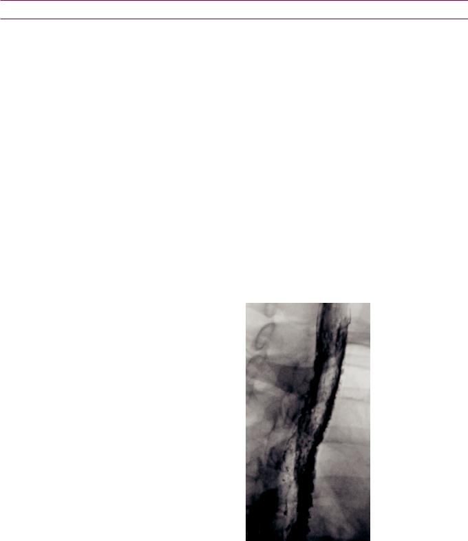
Esophagitis
Reflux (peptic) esophagitis
•Reflux (peptic) esophagitis is caused by exposure of the esophageal mucosa to acidic gastric secretions, which leads to distal ulcerations and eventual stricture.
•Pepticesophagitisismostcommonlycausedbygastroesophagealreflux,butisalsoseenin:
Zollinger–Ellison, due to increased acid production.
Scleroderma, due to gastroesophageal sphincter fibrosis and resultant incompetence.
•Reflux esophagitis appears as thickened distal esophageal folds.
•Chronic esophagitis and scarring develops after prolonged exposure to acid, which causes a smoothly tapered stricture above the GE junction.
Barrett esophagus
•An important long-term sequela of peptic esophagitis is Barrett esophagus, which is metaplasia of normal squamous epithelium to gastric-type adenomatous mucosa. Barrett esophagus is a precursor lesion to esophageal carcinoma.
•Nearly10%ofpatientswithrefluxesophagitismayhavesomeadenomatousmetaplasia.
•On imaging, Barrett demonstrates a featureless distal esophagus, with signs of active reflux esophagitis (mucosal granularity and superficial erosions) more proximally.
•Barrett esophagus is often associated with esophageal stricture, which is abnormally high in location compared to a peptic stricture.
Infectious esophagitis
•Although radiographic distinction between types of infections has been described, endoscopy and biopsy are typically performed in clinical practice.
•Esophageal candidiasis can present as a spectrum from scattered plaque-like lesions in mild disease to very shaggy esophagus in severe cases.
•Herpes esophagitis typically causes discrete small ulcerations scattered randomly throughout esophagus.
•CMV/HIV esophagitis characteristically causes a large, flat, ovoid ulcer.
Medication esophagitis
Candida esophagitis:
Spot image from double-contrast esophagram shows a diffuse, shaggy appearance to the entire visualized esophagus, consistent with severe candida esophagitis.
Case courtesy of Cheryl Sadow, MD, Brigham and Women's Hospital.
•Medication-induced esophagitis typically causes an ulcer at the level of the aortic arch or distal esophagus, which are areas of relative narrowing that may predispose to temporary hold-ups in passage.
Crohn esophagitis
•Crohn esophagitis is very rare and is usually seen in the setting of severe disease in the small bowel and colon.
•Aphthous ulcers (discrete ulcers surrounded by mounds of edema) may become confluent.
124
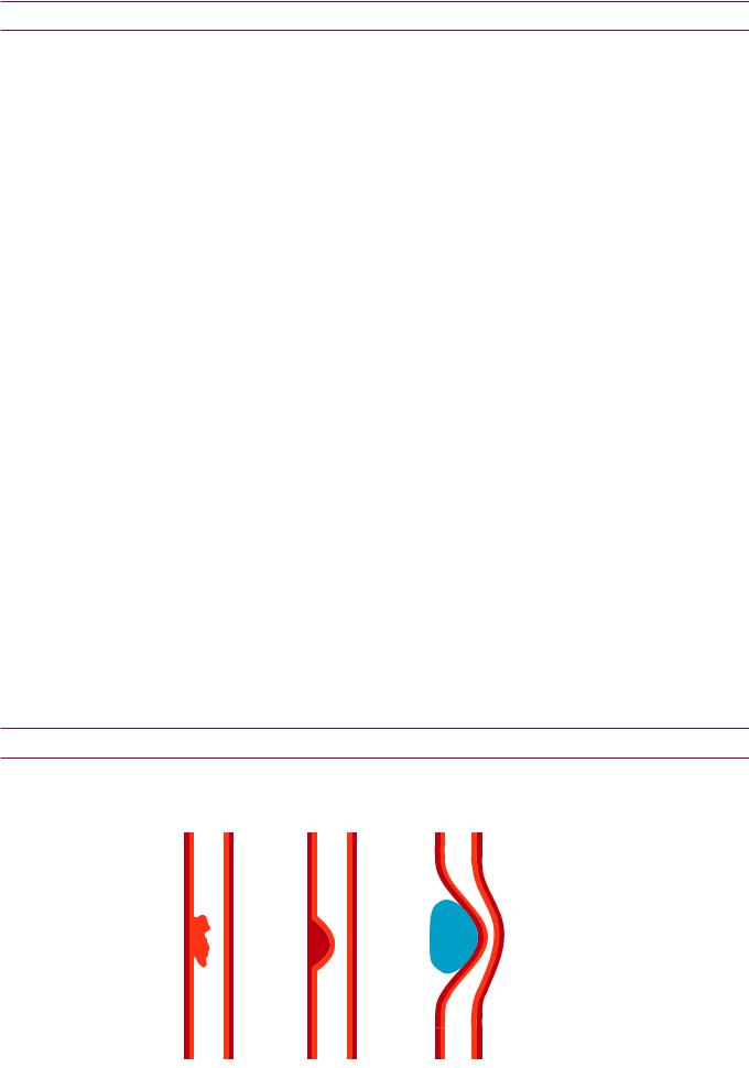
Esophageal strictures
Peptic stricture
•As previously discussed, a peptic stricture is secondary to chronic reflux.
•Peptic strictures are located distally, usually just above the GE junction.
•A peptic stricture may be focal or involve a longer segment of esophagus.
•Fibrosis can cause esophageal shortening, leading to a hiatal hernia as the stomach is pulled into the thorax.
Barrett esophagus stricture
•A Barrett stricture typically occurs in the mid-esophagus, above the metaplastic adenomatous transition. Barrett strictures occur higher than peptic strictures because adenomatous tissue is acid-resistant and therefore unaffected by gastric secretions.
Malignant stricture (due to esophageal carcinoma)
•Key imaging finding is shouldered margins, which suggests circumferential luminal narrowing by a mass.
Caustic stricture/nasogastric (NG) tube stricture
•Both caustic strictures and strictures secondary to nasogastric tube placement are typically long, smooth, and narrow.
•Strictures develop 1–3 months after the caustic ingestion or NG tube placement.
•Caustic strictures are associated with an increased risk of cancer, with a long lag time of up to 20 years after the initial insult. Caustic strictures are usually longer than peptic strictures.
Radiation stricture
•Radiation strictures are long, smooth and narrow, similar to caustic strictures. However, in contrast to strictures from an NG tube, caustic ingestion, and reflux, radiation strictures usually spare the GE junction.
•It generally requires more than 50 Gy of radiation to cause an esophageal stricture.
•Acute radiation esophagitis occurs 1–4 weeks after radiation therapy. Radiation strictures develop later, occurring 4–8 months after radiation.
Extrinsic compression from mediastinal adenopathy
•Cross-sectional imaging would best evaluate if extrinsic compression is suspected.
Evaluation of esophageal masses
•Masses arising from the mucosa, submucosa, and extrinsic to the esophagus produce
characteristic effects on the esophagus, which are usually able to be seen on imaging.
mucosal |
submucosal |
extrinsic compression |
125

Benign esophageal masses
Mesenchymal tumor
•Benign mesenchymal tumors are the most common submucosal tumors and include gastrointestinal stromal tumor (GIST), leiomyoma, lipoma, hemangioma, and others. The classification varies in the literature, with both GIST and leiomyoma described as the most common. On a barium swallow, a mesenchymal tumor typically appears as smooth, round, submucosal filling defect.
Adenoma
•An esophageal adenoma is a benign mucosal lesion with malignant potential, usually arising within Barrett esophagus. Most are <1.5 cm in size and resected at endoscopy.
Inflammatory polyp
•An inflammatory polyp is a non-neoplastic, enlarged gastric fold that protrudes up into the lower esophagus. Inflammatory polyps are almost always associated with reflux and always contiguous with a gastric fold. They are mucosal in location.
Fibrovascular polyp
•A fibrovascular polyp is a pedunculated mass composed of mesenchymal elements with a significant fatty component. In contrast to an esophageal adenoma, there is no malignant potential. Fibrovascular polyps usually occur in the cervical esophagus. The clinical presentation can be dramatic, with regurgitation of a fleshy mass.
•CT is usually diagnostic, demonstrating intra-lesional fatty component.
Varices
•Esophageal varices are most commonly due to portal hypertension. Varices can usually be distinguished from a solid mass since varices change in size and shape with peristalsis. However, thrombosed varices may mimic a tumor.
•Uphill varices, due to portal hypertension, affect the distal esophagus.
Blood flows “uphill” from the portal vein left gastric (coronary vein) periesophageal venous plexus azygos/hemiazygos collaterals SVC.
•Downhill varices are much less common, are caused by superior vena cava obstruction, and usually affect the proximal esophagus.
Enlarged collateral vessels include the supreme intercostal veins (drain the first intercostal space), bronchial veins, and inferior thyroidal veins.
Foregut duplication cysts
•Esophagealduplicationcystislinedwithsquamousepithelium,hasasmoothmusclewall, andisusuallyintheposteriormediastinum.Itmaybeeitherextrinsictotheesophagusor submucosal;thelatterisimpossibletodifferentiate fromaleiomyomabyesophagram.
•Bronchogenic cyst is lined by respiratory epithelium. It is generally indistinguishable from an esophageal duplication cyst on esophagram and CT.
•Neurenteric cyst is associated with vertebral body anomalies.
Esophageal foreign body
•AradiopaqueesophagealforeignbodyisbestvisualizedwithalateralradiographorCT.
•Bony foreign objects usually get stuck in the cervical esophagus.
•Meat impaction usually occurs at the gastroesophageal junction. There is a risk of esophageal perforation from transmural ischemia if the food bolus is impacted for >24 hours. Most cases of food bolus impaction are treated with endoscopic removal of the impacted food. Historically, meat impaction was treated with effervescent granules and meat tenderizer, but this technique is no longer commonly performed.
126
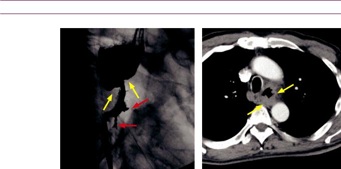
Malignant esophageal masses
Esophageal carcinoma
Esophageal carcinoma: Oblique photospot image from a barium swallow (left image) shows an irregular stricture in the proximal esophagus (yellow arrows) with proximal dilation of the esophagus. Two linear ulcerations (red arrows) project into the mural mass. Contrast-enhanced axial CT (right image) shows irregular thickening of the esophagus (arrows).
Case courtesy of Cheryl Sadow, MD, Brigham and Women's Hospital.
•Esophageal carcinoma has a broad range of appearances. Early esophageal cancer may be apparent on barium swallow as a plaque-like lesion, polypoid lesion, or focal irregularity of the esophageal wall. A classic appearance of advanced esophageal carcinoma is a mass causing a stricture with a “shouldered” edge and irregular contour.The uncommon varicoid appearance can be initially confused with varices, but the tumor does not change shape with peristaltic waves as varices typically do.
•Esophageal carcinoma may be squamous cell carcinoma (SCC) or adenocarcinoma, which cannot be reliably differentiated on barium studies. SCC tends to involve the upper or mid-esophagus and adenocarcinoma typically involves the distal esophagus and may extend into the stomach.
•Squamous cell carcinoma (SCC) is most commonly due to smoking and alcohol. Less common risk factors include celiac disease, Plummer–Vinson, achalasia, and human papilloma virus (which more commonly causes laryngeal squamous cell carcinoma).
•Adenocarcinoma is due to chronic reflux, arising from Barrett esophagus distally. Its incidence has been rising in recent years.
Metastasis
•Direct invasion of the esophagus is most commonly from gastric, lung, or breast primaries. Hematogenous spread is very rare.
•Most often, mediastinal lymph node metastases will be prominent. The midesophagus is most commonly affected due to its proximity to mediastinal lymph nodes.
Lymphoma
•Esophageal lymphoma is often indistinguishable from primary esophageal cancer.
Malignant GIST
•Malignant GIST tends to be bulkier and more irregular than the benign variant.
127
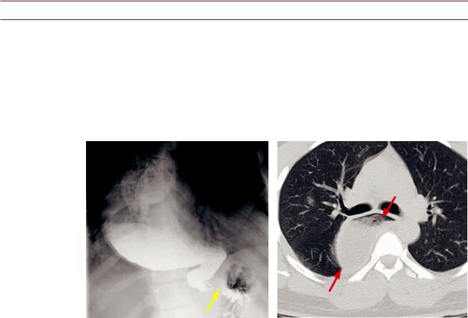
Esophageal motility disorders
Contraction waves
•A primary contraction wave is a normal, physiologic wave initiated by a swallow.
•A secondary contraction wave is a normal, physiologic wave initiated by a bolus in the esophagus.
•A tertiary wave is a nonpropulsive contraction that does not result in esophageal clearing. Tertiary contractions are seen more commonly in the elderly. They are not normal, but are also not thought to be clinically significant when seen.
Achalasia
Achalasia: Photospot image from an upper GI series (left image) shows a markedly dilated esophagus terminating in a bird’s beak (arrow) at the GE junction, due to failure of the distal esophageal sphincter to relax. Axial non-contrast CT confirms the markedly dilated, debris-filled esophagus (red arrows).
•Achalasia is a motility disorder of the distal esophagus, which is unable to relax due to an abnormality of myenteric ganglia in the Auerbach plexus. Vigorous achalasia is a less severe form of achalasia consisting of repetitive nonpropulsive contractions.
•Chagas disease causes a secondary achalasia that is indistinguishable radiographically from primary achalasia.
•Potential complications of chronic achalasia include esophageal cancer, which has a lag period of at least 20 years, and candidal infection from stasis.
•The classic imaging appearance of achalasia is a massively dilated esophagus with a bird’s beak stricture near the gastroesophageal junction.
•Surgical treatment of achalasia is the Heller myotomy, which is an incision of the lower esophageal muscle fibers.
•Pseudoachalasia is caused by an obstructing gastroesophageal junction cancer. In achalasia, there is transient relaxation of the stricture when the patient stands. In pseudoachalasia, however, the fixed obstruction does not relax with standing.
Diffuse esophageal spasm (corkscrew esophagus; shish kebab esophagus)
•Diffuse esophageal spasm is a clinical syndrome of chest pain or dysphagia caused by repetitive, nonpropulsive esophageal contractions. The nonpropulsive contractions have a characteristic appearance on barium swallow, leading to the descriptive names of corkscrew esophagus and shish kebab esophagus.
•Nutcrackeresophagus isarelateddisordercharacterizedbyhigh-amplitudecontractions onmanometryinconjunctionwithchestpain,withnormalradiologicalfindings.
128

Esophageal diverticula
Types of diverticula
•Pulsion diverticula are caused by increased esophageal pressure and comprise nearly all diverticula seen in the USA.
•Traction diverticula are caused by traction of adjacent structures, typically resulting from tuberculous mediastinal adenopathy. They are rarely seen.
Zenker diverticulum
•Zenkerdiverticulumisanesophagealdiverticulumcausedbyfailureofthe cricopharyngeusmuscletorelax,leadingtoelevatedhypopharyngealpressure.Symptoms ofaZenkerdiverticulumincludehalitosis,aspiration,andregurgitationofundigestedfood.
•A Zenker diverticulum is posteriorly protruding. As a secondary finding, the cricopharyngeus muscle is usually hypertrophied.
•Treatment is with cricopharyngeal myotomy and diverticulopexy or diverticulectomy.
•A pseudo-Zenker diverticulum is barium trapped in a pharyngeal contraction wave.
Killian–Jamieson (KJ) diverticulum
•A Killian–Jamieson (KJ) diverticulum is located at the Killian–Jamieson space, which is an area of weakness below the attachment of the cricopharyngeus muscle.
•In contrast to Zenker diverticulum, KJ diverticula are more often bilateral.
•KJ diverticula protrude anteriorly, best seen on the lateral view.
Pseudodiverticulosis
•Pseudodiverticulosisistheimagingfindingofmultiple tinyoutpouchingsintothe esophageallumencausedbydilatedsubmucosalglandsfromchronicrefluxesophagitis.
•These submucosal glands are analogous to the Rokitansky–Aschoff sinuses of the gallbladder.
•Pseudodiverticulosis is often associated with a smooth stricture in mid/upper esophagus, which may cause symptoms.
•Candida is frequently cultured, but infection is not believed to be a causal factor.
Miscellaneous esophageal disorders
Feline esophagus
•Feline esophagus is thought to be a normal variant characterized by multiple transverse esophageal folds.
•There is a controversial association with esophagitis, where the incidence of esophagitis may be increased in the presence of feline esophagus.
Feline esophagus: Spot image from double-contrast esophagram shows characteristic multiple transverse folds in the lower esophagus.
Case courtesy Cheryl Sadow, MD, Brigham and Women's Hospital.
129
Aberrant right subclavian artery
RV
aRS
LS
RCC LCC * LV
Aberrant right subclavian artery: Oblique single-contrast barium esophagram (left image) demonstrates a smooth posterior indentation of the proximal thoracic esophagus (arrow). Aortogram in the same patient (right image) demonstrates the aberrant right subclavian artery (aRS, * at origin) crossing the midline before heading towards the right arm. This patient has an additional aortic branching anomaly, with the left vertebral artery (LV) arising directly from the aorta. The right vertebral artery (RV), arising from the aberrant right subclavian, is hypoplastic. The right and left common carotid arteries (RCC and LCC), and left subclavian (LS) are normal.
Case courtesy Cheryl Sadow, MD, Brigham and Women's Hospital.
•Aberrant right subclavian artery (with a normal left arch) is seen in approximately 1% of patients and is almost always asymptomatic. The aberrant right subclavian artery travels posterior to the esophagus, where it may rarely produce dysphagia.
•OnanupperGIstudy,theresultantposterioresophagealindentation isalwayssmooth.
Scleroderma
•Sclerodermaisasystemicdiseaseinvolvingexcesscollagendepositioninmultiple tissues.
•The esophagus is involved in 80% of patients with scleroderma, producing lack of peristalsis of the distal 2/3 of the esophagus due to smooth muscle atrophy and fibrosis, which leads to marked esophageal dilation.
•Secondary candidiasis or aspiration pneumonia can result from prolonged esophageal stasis.
•The esophageal dilation is often apparent before the typical skin changes of scleroderma become evident.
Esophageal hernias
Hiatal hernia (HH)
•Ahiatalhernia(HH)ispresentwhengastricfoldsareseenabovethediaphragm.Ahiatal herniamaybesliding(mostcommon)orshort(secondarytochronicrefluxesophagitis).
Paraesophageal hernia
•Withaparaesophagealhernia,theGEjunctionislocatednormallybelowthediaphragm, butaportionofthestomachherniatesintothethoraxthroughtheesophagealhiatus.
•ParaesophagealherniaismorepronetostrangulationthanHH.Mostaresurgicallyrepaired.
130

Stomach
Thickened gastric folds
•Thickened gastric folds are most commonly due to inflammatory gastritis, which characteristically produces smooth fold thickening.
•Nodular fold thickening is suggestive of neoplasm, such as gastric lymphoma or submucosal carcinoma.
Helicobacter pylori gastritis
•Helicobacter pylori is a major cause of gastritis, gastric ulcers and duodenal ulcers.
Zollinger–Ellison (ZE)
•Zollinger–Ellison (ZE) is gastrin over-production from a gastrinoma, which is a pancreatic islet cell tumor that has a 50% rate of malignancy.
ZE features elevated gastrin level and a paradoxical increase in gastrin after secretin administration.
•25% of patients with gastrinoma have multiple endocrine neoplasia (MEN) type 1.
MEN-1 consists of parathyroid adenoma, pituitary adenoma, and pancreatic islet cell tumors.
Eosinophilic gastritis
•Eosinophilic gastritis is characterized by thickened folds in the stomach and small bowel in a patient with a history of allergy.
Menetrier disease
•Menetrierdiseaseisaprotein-losingenteropathythatisoften adiagnosisofexclusion. Itusuallyaffects theproximalstomachandispathologicallycharacterizedby replacementofparietalcellsbyhyperplastic epithelialcells, leadingtoachlorhydria.
•Menetrier disease has a controversial association with gastric carcinoma.
Crohn disease
•Gastric Crohn disease is almost always associated with small bowel disease. Usually the distal half of the stomach is affected.
•The earliest pathologic change is the formation of aphthous ulcers.
Other causes of thickened gastric folds
•Gastric varices (from portal hypertension), gastric lymphoma, and submucosal carcinoma are non-inflammatory causes of thickened gastric folds.
Gastric polyps
Hyperplastic polyp (inflammatory polyp)
•A hyperplastic polyp, also known as an inflammatory polyp, is cystic dilation of a gastric gland that develops in response to chronic inflammation. Hyperplastic polyps are almost always benign, with very rare cases of malignant transformation having been reported.
•Fundic gland polyposis syndrome is a variant of familial adenomatous polyposis syndrome that also involves the stomach. In the stomach, most polyps are hyperplastic, but elsewhere in the GI tract the polyps are adenomatous.
Adenomatous polyp
•An adenomatous polyp is a neoplastic polyp with malignant potential. There is an elevated risk of malignant transforation to adenocarcinoma if >2 cm in size.
•Adenomatous polyps are usually treated with endoscopic biopsy and polypectomy.
131
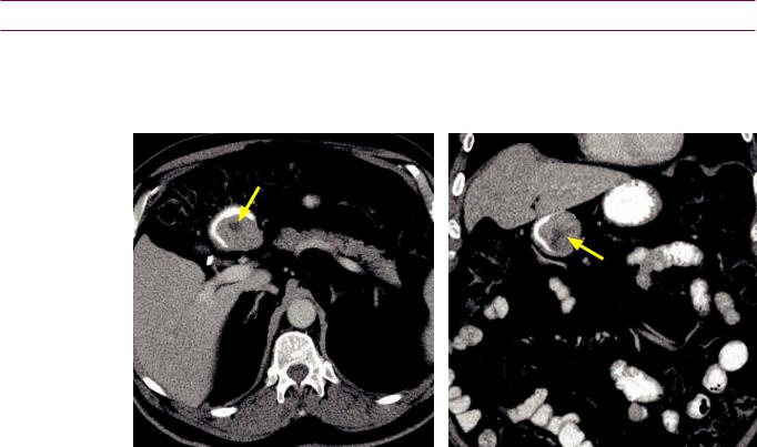
Hamartomatous polyp
•Hamartomatous polyps are benign polyps usually associated with syndromes such as Peutz–Jeghers, juvenile polyposis, and Cronkhite–Canada syndromes.
Benign gastric masses
Lipoma (benign)
•Alipomaisabenign,submucosal,mesenchymalneoplasm.Atfluoroscopy, agastric lipomaisindistinguishablefromaGIST.Fattyattenuation onCTisdiagnosticofalipoma.
Gastrointestinal stromal tumor (GIST)
SubmucosalGIST:Axial(leftimage)andcoronal(rightimage)contrast-enhancedCTshowsawell- circumscribed,relativelyhomogeneousmasswithacentralfocusofnecrosis(arrows),locatedinthe submucosalposteriorwallofthegastricantrum.IncidentalcholecystectomyclipsareseenontheaxialCT.
Case courtesy Cheryl Sadow, MD, Brigham and Women's Hospital.
•Gastrointestinal stromal tumor (GIST) is the most common submucosal gastric tumor. The tumor arises from the interstitial cells of Cajal, which are pacemaker cells that drive peristalsis. GIST may occur anywhere in the gastrointestinal tract.
•GISTmaybebenignormalignant,withriskformalignancydeterminedbysizeand numberofmitoses.Regardlessofsizeandnumberofmitoses,gastricGISTislesslikely tobemalignantcomparedtosimilar-sizedGISTsintheduodenum,jejunum/ileum,or rectum.Gastrictumors≤2cminsizeareessentiallyalwaysbenign.Largertumorscarrya riskofmalignancyashighas86%foragastricGIST>10cmwithanelevatedmitoticrate.
•Small gastric GISTs are usually asymptomatic, but may be a cause of melena.
•On imaging, a smooth endoluminal surface is characteristic due to its submucosal location. Larger tumors have a tendency to become exophytic, or less commonly to invade the lumen.
•The differential diagnosis of a submucosal gastric mass includes mesenchymal tumors (GIST, fibroma, lipoma, neurofibromas, etc.), carcinoid, and ectopic pancreatic rest.
Ectopic pancreatic rest
•Anectopicpancreaticrestisafocusofheterotopicpancreasinthegastricsubmucosa. Theectopicpancreatictissueissusceptibletopancreaticdiseases,includingpancreatitis andcarcinoma.Onimaging,theclassicappearanceisanumbilicatedsubmucosalnodule, withtheumbilicationrepresentingafocusofnormalepithelium.Theulcerationisnot alwaysseen,inwhichcasetheimagingisofanonspecificsubmucosalgastricmass.
132

Malignant gastric masses
Gastric cancer
•Gastric adenocarcinoma may present either as a mass or as a gastric ulcer.
•Gastric cancer is generally caused by chronic inflammation, with specific risk factors including:
Ingestion of polycyclic hydrocarbons and |
Pernicious anemia. |
|
nitrosamines (from processed meats). |
Post-subtotal gastrectomy. |
|
Atrophic gastritis. |
||
|
•Gastric carcinoma may spread locally from the mucosal surface to the serosa, in which case 90% of patients will have omental involvement from trans-serosal spread.
•Lymphatic spread is along lesser curvature gastrohepatic ligament and greater curvature.
•A Krukenberg tumor is classically described as metastatic spread of gastric carcinoma to the ovary; however, the term has also been used to describe any mucinous metastasis to the ovary.
GIST (malignant)
•Malignant GIST tends to be larger than benign GIST, often reaching sizes of greater than 10 cm, with central necrosis. Although the tumor begins in the submucosa, it can be difficult to determine the site of origin of large tumors.
Lymphoma
•Gastriclymphomacanhaveawidevarietyofpresentations.Ifsolitary,lymphomacan mimicgastriccarcinoma.Todifferentiate betweenlymphomaandgastriccarcinoma,the patternofadenopathycanbehelpful.Ingastriccancer,adenopathyatorbelowthelevel oftherenalhilaisunusual,butoccursmorecommonlyinpatientswithlymphoma.
•The stomach is a common extranodal site for non-Hodgkin lymphoma.
Metastases
•Metastaticdiseasetothestomachisrare.Breast,lung,andmelanomaaremostcommon.
Gastric ulcers
Benign gastric ulcer
•Althoughlesscommonlyencounteredinthemoderneraofprotonpumpinhibitorsand Helicobacter pylori treatment,benigngastriculcerstendtohavetypicalimagingfindings:
Radiating gastric folds are smooth and symmetric.
Ulcer extends beyond the normal contour of the gastric lumen.
The Hampton line represents nonulcerated acid-resistant mucosa surrounding the ulcer crater.
Mostbenignulcersoccuralongthelessercurvatureofthestomach,althoughbenignulcersassociated withaspiriningestioncanoccurinthegreatercurvatureandantrum,whicharedependentlocations.
Gastric carcinoma
•Gastric carcinoma may present with malignant ulceration, which can usually be distinguished from a benign ulcer by the following features:
Asymmetric ulcer crater, with surrounding nodular tissue.
Abrupt transition between normal gastric wall and surrounding tissue. Ulcer crater does not project beyond the expected location of gastric wall.
The Carman meniscus sign is considered pathognomonic for tumor. It describes the splaying open of a large, flat malignant ulcer when compression is applied.
133
Overview of gastric bypass surgery
Postoperative anatomy of Roux-en-Y gastric bypass (RYGB)
distal  esophagus
blind-ending  limb
limb
gastrojejunostomy
gastric pouch
excluded |
Roux |
stomach |
limb |
ligament  of Treitz
a erent limb |
|
(pancreaticobiliary limb) |
jejunojejunostomy |
|
n order to evaluate for and accurately describe complications of Roux-en-Y gastric b (RYGB) t s mportant to be familiar with the procedure and norma
tsurgica anatomy
A smal gastric pouch s created with |
volume of approximately 15 to 30 |
by |
xcluding the dista stomach from the path offood. |
|
|
The Roux imb s created by transecting the jejunum approximately 35–45 |
dista to |
|
the igament of Treitz then bringing t |
to be anastomosed to the gastric pouch via |
|
gastrojejunostomy stoma |
|
|
The current favored approach for placement of the Roux imb s antecolic (in front of the transverse colon). The Roux imb used to be placed retrocolic, which required
the creation of |
surgica defect through the transverse mesocolon (mesentery of the |
|||
tr |
colon). A retrocolic Roux imb has higher risk of transmesocolic hernia |
|||
due to the defect |
n the transverse mesocolon. |
|
||
Although the antecolic approach s |
commonly performed, there |
patients who |
||
ha |
previously undergone retrocolic approach. |
|
||
A dista side-to-side jejunojejunostomy s created to connect the pancreaticobiliary imb to the jejunum
The RYGB eads to weight oss both from early satiety (due to smal size of the gastric pouch) and malabsorption (due to surgica bypass of the proxima jejunum).
134

Complications of Roux-en-Y surgery
Postoperative leak
•Postoperative leak is usually diagnosed by 10 days after surgery.
•AnupperGIstudywithwater-solublecontrastisthestudyofchoiceifaleakissuspected.
•Leaks may arise from the distal esophagus, gastric pouch, or blind-ending jejunal limb. It is rare for a leak to arise from the distal jejunojejunostomy.
Gastrogastric fistula
•A gastrogastric fistula is a communication between the gastric pouch and the excluded stomach, which may be an early or late complication of RYGB.
•A gastrogastric fistula may be a cause of inadequate weight loss or recurrent weight gain.
Small bowel obstruction (SBO)
•Small bowel obstruction (SBO) in the acute postoperative period is most often due to edema or hematoma at the gastrojejunostomy or jejunojejunostomy.
With a retrocolic Roux limb, edema at the transverse mesocolon defect may also cause obstruction.
Treatment is usually conservative, with most cases resolving as the edema and/or hematoma resolves.
•A late presentation of small bowel obstruction may be due to internal hernia (more common with laparoscopic surgery) or adhesions (more common with open surgery).
Internal hernia
•Laparoscopic Roux-en-Y procedures are associated with a higher rate of internal hernias (seen in 2.5% of laparoscopic procedures) compared to open procedures (0.5%). Internal hernias can be difficult to diagnose, both clinically and by imaging.
•Internal hernias usually present within 2 years of bypass and are the most common cause of SBO after a laparoscopic Roux-en-Y.
•Most RYGB-associated internal hernias occur in three characteristic locations.
•The surgically created defect in the mesentery of the transverse colon is the most common site (the transmesocolic hernia), associated with a retrocolic Roux limb.
•Less common sites of internal hernia include Peterson’s space (located between the mesentery of the Roux limb and the transverse mesocolon) and the mesenteric defect created by the jejunojejunostomy.
•Imaging features of internal hernia include swirling of the mesentery, a mushroom shape of the mesentery, and/or the presence of small bowel loops posterior to the superior mesenteric artery.
Stomal stenosis
•Narrowing of the gastrojejunostomy stoma may occur in up to 10% of patients, leading to dilation of the pouch and distal esophagus. Stomal stenosis is usually treated with endoscopic dilation.
•Narrowingofthedistaljejunojejunostomyismuchmorerareandusuallyrequiressurgery.
Marginal ulcers
•The jejunal mucosa adjacent to the gastrojejunal anastomosis is susceptible to gastric secretions, which can cause marginal ulcers in up to 3% of patients.
•A marginal ulcer is diagnosed by upper GI as a thickening and small outpouching of a gastric fold.
•Treatment is conservative.
135

Small bowel
Small bowel anatomy
•The wall of the small intestine is made of four layers, from outside in:
Serosa. |
Submucosa. |
Muscularis (thin longitudinal and thick |
Mucosa (consists of intestinal villi, circular |
circumferential smooth muscle). |
folds, glands, and lymphoid tissue). |
•Valvulae conniventes create the characteristic small bowel fold pattern.
•Thesuperiormesentericartery(SMA)suppliesboththejejunumandileum.Acommon smallbowelmesenteryanchorsthejejunumandileumtotheposteriorabdominalwall. Thejejunumfeatureslarger,morefeature-fullfoldsandlargervillicomparedtotheileum.
Small bowel obstruction (SBO)
•Small bowel obstruction (SBO) is common and most often due to adhesions from prior surgery or hernia. Neoplasm, stricture, and intussusception are less common causes.
Radiographic evaluation of small bowel obstruction
•Anabdominalradiographisoftentheinitialimagingevaluationforsuspectedobstruction.
•Radiographic findings of SBO include small bowel distention and multiple air–fluid levels at different heights seen on the upright view. In addition, the lack of gas in the colon is especially suggestive of obstruction.
An upright or decubitus view is generally necessary to confidently diagnose obstruction.
•Potential false positives for diagnosing SBO on plain radiographs include:
Ileus with prior colectomy: Would not see gas in the colon.
Ileus with ascites: Ascites often compresses the ascending and descending colon and rectum as these structures are not on a mesentery. However, gas in the transverse colon and sigmoid colon is still apparent.
CT imaging of small bowel obstruction
•CTishighlysensitiveandspecificfordiagnosisofSBO.Smallboweldistention≥3cm withatransitionpointtocollapsedbowelishighlyspecificforasmallbowelobstruction.
•Inadditiontodiagnosingobstruction,CTcanshowthetransitionpoint,thecauseof obstruction,andpotentialcomplicationsofobstructionsuchasischemiaorstrangulation.
•It is important to approach the interpretation of an obstruction in a systematic way.
•First, look for the transition point to decompressed bowel to determine the cause.
•Second, always determine if the obstruction is simple or closed-loop. A closed-loop obstruction is a never miss lesion as there is very high risk for bowel ischemia and severe morbidity and mortality.
•Third, evaluate for signs of ischemia or impending ischemia, which include (in rough order of severity):
Engorged mesenteric vessels.
Ascites surrounding the bowel, due to increased capillary permeability. Wall thickening, due to submucosal edema.
Lackofbowelwallenhancement,duetovasoconstrictionorunder-perfusion.Notethatthepresence orabsenceofbowelwallenhancementcanonlybeassessedifpositiveoralcontrastwasnotgiven.
Pneumatosis intestinalis, which is gas in the bowel wall due to necrosis. Pneumatosis produces multiple small locules of gas seen circumferentially in the bowel wall.
136
•Inadditiontosmallboweldistention >3cmandatransitionpointto decompressedbowel,anadditional helpfulCTfindingofSBOisthesmall bowel feces sign, whichdescribes particulatefeculentmaterialmixedwith gasbubblesinthesmallbowelthat resemblestheCTappearanceofstool.
The small bowel feces sign is often seen just proximal to the transition point and is helpful to localize the site of transition.
Thesmallbowelfecessignmaybeespecially helpfulinsubacuteorpartialobstruction, whichcanotherwisebedifficult todiagnose.
The small bowel feces sign is thought to be due to bacterial overgrowth and
Closed loop obstructionundigested food.
Small bowel feces sign: Axial CT shows an obstruction. A loop of small bowel in the right lower quadrant (arrow) demonstrates numerous gas locules and particulate material in the small bowel.
•Closed loop obstruction is a surgical emergency that may lead to bowel ischemia. Closed loop obstruction represents obstruction of both the efferent and afferent segments of a single loop of bowel.
•Closed loop obstruction may be secondary to adhesions or hernia. The formation of a narrow pedicle can lead to volvulus, which predisposes to ischemia.
•CT imaging features include a U-shaped distribution of the bowel loop with radially oriented vessels. If volvulus is present, the whirl sign may be seen, due to twisting of mesenteric vessels.
Obstruction due to adhesions
Small bowel obstruction due to adhesions: Coronal (left image) and sagittal CT (right image) shows multiple dilated, fluid-filled loops of small bowel. A transition point is located in the midline pelvis (arrows on sagittal image), with no obstructing mass or evidence of hernia.
•Adhesions from prior surgery or intra-peritoneal inflammatory process are the most common cause of small bowel obstruction.
•Adhesions are an imaging diagnosis of exclusion. On CT, a transition point is seen, but no obvious cause for the transition (e.g., no mass or hernia, etc.) is identified.
•ThevastmajorityofpatientswithSBOduetoadhesionshavehadpriorabdominalsurgery.
137

Obstruction due to external hernia
•Protrusion of bowel through the abdominal wall is the second most common cause of small bowel obstruction. Approximately 75% of external hernias occur in the groin, with the majority being inguinal hernias.
•An inguinal hernia may be either indirect or direct, depending on the relation of the hernia to the inferior epigastric vessels.
Indirect: Indirect inguinal hernia is the most common type and is more common in males. The neck of the hernia is lateral to the inferior epigastric vessels. Hernia contents travel with the spermatic cord, often into the scrotum. Indirect inguinal hernias are considered a congenital lesion due to a patent processus vaginalis.
Direct:Theneckofanindirectinguinalherniaismedialtotheinferiorepigastricvessels,protruding throughaweakareaintheanteriorabdominalwall.Theherniacontentsdo not go into scrotum.
•In an obturator hernia, bowel herniates through the obturator canal. Obturator hernias are almost always seen in elderly women due to pelvic floor laxity.
The key imaging finding is bowel located between the pectineus and obturator muscles.
It is important to correctly diagnose an obturator hernia preoperatively. An obturator hernia
requires a very different surgery from inguinal hernia, and has an especially high morbidity and
mortality if incarcerated.
•Ventral hernia is often due to prior laparotomy.
Obstruction due to internal hernia
•Protrusion of bowel through the peritoneum or mesentery into a compartment in the abdominal cavity is a relatively uncommon cause of small bowel obstruction.
•Transmesenteric hernia is a broad category of bowel herniation through defects in any of the three true mesenteries (small bowel mesentery, transverse mesocolon, and sigmoid mesentery). The most common type of transmesenteric hernia is the transmesocolic hernia, due to a defect in the transverse mesocolon (mesentery of the transverse colon). Transmesocolic hernia is seen most commonly post Roux-en-Y gastric bypass or biliary-enteric anastomosis from liver transplant.
The lack of confining hernia sac and variable imaging appearance make diagnosis difficult. A clue on imaging may be posterior displacement of the colon, with small bowel located anterior to the colon. The SMA and SMV may be displaced and engorged.
Internal hernias carry a high rate of volvulus. If volvulus is present, the whirl sign may be visible.
Transmesenteric hernias are also the most common type of hernia in children, not due to surgery but secondary to a congenital mesenteric defect thought to be from prenatal intestinal ischemia. In children, the mesenteric defect has a variable position.
•Paraduodenal hernia was previously the most common internal hernia (older literature states 53% of internal hernias were paraduodenal), prior to the rise in gastric bypass surgery. Paraduodenal hernias are congenital anomalies, due to embryologic failure of mesenteric fusion and resultant mesenteric defect. They more commonly occur on the left.
Paraduodenal hernia is associated with abnormal rotation of the intestine.
A common clinical complaint described by patients with paraduodenal hernia is chronic postprandial pain often relieved by massaging, which reduces the hernia.
In the more common left paraduodenal hernia, the bowel can herniate through a mesenteric defect named Landzert’s fossa, located behind the ascending (fourth) duodenum. The key imaging finding is a cluster of small bowel loops between the pancreas and stomach.
•Foramen of Winslow hernia: The foramen of Winslow is the communication between the lesser sac and the greater peritoneal cavity.
The key imaging features of a foramen of Winslow hernia are dilated loops of bowel in the upper abdomen and presence of mesentery between the IVC and main portal vein.
138

Obstruction due to neoplasm
•Amassintrinsictothebowelorexternalcompressionfromanextrinsicmassmaycause smallbowelobstruction.AnextrinsicmassisusuallystraightforwardtodiagnosebyCT.
•Although the presence of an intraluminal mass may be more difficult to detect on CT, clues to the presence of an intrinsic mass include irregular bowel wall thickening and/ or regional lymphadenopathy.
•Primarysmallbowelneoplasmcausingintrinsicbowelobstructionmaybedueto adenocarcinoma,GIST,andcarcinoid.Metastaticcausesofintrinsicbowelneoplasminclude melanoma,ovarian,andlungcancer.Melanomaisknowntocauseintussusception.
•Lymphomaisgenerallya“soft”tumorandrarelycausesobstruction.Aneurysmal expansion ofthesmallbowelwallisaclassicappearance,butpresentationishighlyvariable.
Obstruction due to intussusception
Intussusception causing small bowel obstruction: Coronal contrastenhanced CT demonstrates a segmental jejunojejunal intussusception (arrows), causing an early or partial proximal small bowel obstruction. This was
a case of metastatic melanoma (metastatic lesion not visualized on this image).
•While transient intussusceptions are a common incidental finding, an intussusception causing obstruction should raise suspicion for an underlying lesion and prompt surgery.
Obstruction due to Crohn disease
Obstruction due to Crohn ileitis: Coronal (left image) and axial contrast-enhanced CT shows dilated loops of proximal small bowel. The terminal ileum (yellow arrows) and several loops of distal ileum (red arrows) are thickened, reflecting enteritis.
•Stricture or active enteritis is an important cause of bowel obstruction in Crohn disease, especially the fibrostenotic subtype. Crohn disease is discussed on the following page.
Obstruction due to gallstone
•Gallstone ileus is due to a gallstone that has eroded through into the small bowel, causing the classic Rigler’s triad of pneumobilia (from cholecystoduodenal fistula), small bowel obstruction, and ectopic gallstone within the small bowel.
139
Enteritis
•Enteritis is inflammation of the small bowel. The most common CT manifestation of enteritis is bowel wall thickening. Mesenteric stranding or free fluid may also be present.
Crohn disease
•Crohn disease is a chronic granulomatous inflammatory condition that may affect any part of the gastrointestinal tract from the mouth to the anus. Involvement is discontinuous, with characteristic skip lesions of intervening normal GI tract. The most common site of involvement is the small bowel, especially the terminal ileum.
•The earliest histologic changes occur in the submucosa, seen on imaging as aphthous ulcers due to lymphoid hyperplasia and lymphedema.
•Endoscopy and barium fluoroscopy (small bowel follow-through, enteroclysis, and barium enema) have historically been the modalities to evaluate Crohn disease. More recently, however, CT and MR enterography are emerging as the exams of choice.
The advantages of CT and MRI are the ability to visualize beyond the bowel lumen to evaluate the bowel wall, presence of extraintestinal complications, and the vasculature.
The disadvantages of CT and MRI compared to fluoroscopy and endoscopy are reduced spatial resolution and limited sensitivity for detecting subtle early signs of disease.
•The most common imaging finding on all modalities is wall thickening of the terminal ileum.
•Fluoroscopic findings include thickened, nodular folds in the affected regions of small bowel, luminal narrowing, mucosal ulceration, and separation of bowel loops. The typical cobblestone appearance seen on endoscopy and fluoroscopy is a result of crisscrossing deep ulcerations.
Crohn disease (terminal ileitis): Small bowel follow-through (left image) shows terminal ileum nodular fold thickening, mucosal ulceration, and separation of the terminal ileum (arrows) from adjacent loops of small bowel. Right lower quadrant color Doppler ultrasound (right image) in the same patient demonstrates the nodular fold thickening (arrows) and hyperemic wall.
Case courtesy Michael Callahan, MD, Boston Children’s Hospital.
140

•The fibrostenotic subtype of Crohn disease may clinically present with bowel obstruction. Asymmetric bowel fibrosis from ulcerations of the mesenteric side of the bowel produces pseudosacculations on the antimesenteric side. The fibrosis can lead to a segmental stricture, called the string sign.
Crohn disease (fibrostenotic subtype): Small bowel follow-through (left image) shows the string sign of Crohn disease (yellow arrows) in a loop of distal ileum, with a few small antimesenteric pseudosacculations (red arrows). Contrast-enhanced CT (right image) shows bowel wall thickening and fibrofatty mesenteric changes (blue arrows), known by pathologists and surgeons as creeping fat.
Case courtesy Michael Callahan, MD, Boston Children’s Hospital.
•Complications of Crohn disease include bowel strictures, fistulae, and abscesses.
Perirectal abscess and enterocutaneous fistula secondary to Crohn disease: Contrast-enhanced CT (left image) shows a peripherally enhancing fluid collection to the right of the rectum (arrows). T2-weighted MRI (right image) shows the distal portion of an enterocutaneous fistula extending to the skin surface (red arrow). There is marked subcutaneous edema (blue arrows) extending into the subcutaneous tissues of the right buttock.
Case courtesy Michael Callahan, MD, Boston Children’s Hospital.
141

Scleroderma
•Scleroderma is a systemic disease characterized by the deposition of collagen into multiple internal organs and the skin.
•The primary insult to the gastrointestinal tract in scleroderma is impaired motility due to replacement of the muscular layers with collagen, which leads to slowed transit and subsequent bacterial overgrowth, progressive dilation, and pseudo-obstruction.
•Radiographic findings are sacculations on the antimesenteric border (side opposite where the mesentery attaches) and a hidebound bowel due to thin, straight bowel folds stacked together.
•Treatment is with antibiotics for bacterial overgrowth and prokinetic drugs such as erythromycin or octreotide for bowel motility.
Celiac disease (sprue, gluten-sensitive enteropathy)
•Celiac disease, also known as sprue and gluten-sensitive enteropathy, is an autoimmune, proximal enteritis caused by a T-cell-mediated immune response triggered by antigens in ingested gluten.
•The primary sites of involvement are the duodenum and jejunum.
•Themostcharacteristicimagingfindingofceliacdiseaseisreversalofjejunalandilealfold patterns.Normally,thejejunumhasmorefoldsthantheileum.However,inceliacdisease, thelossofjejunalfoldscausesacompensatoryincreaseinthenumberofilealfolds.
•Villous atrophy causes the loss of jejunal folds and hypersecretion of intraluminal fluid that creates flocculations of barium due to lack of contrast adhesion to the bowel wall. The moulage (french for casting) sign is seen on a barium study and refers to a castlike appearance of the featureless jejunum.
•The CT findings of celiac disease include dilated, fluid-filled bowels, often with intra-luminal flocculations of enteric contrast. Contrast can be seen both insinuated between the small bowel folds and centrally within the bowel, with a peripheral layer of low-attenuation secretions. Other CT findings of celiac disease include mesenteric adenopathy and engorgement of mesenteric vessels.
•Unlikeothercausesofenteritis,diffusebowelwallthickeningandascitesarelesscommon.
•An important complication of celiac disease is small bowel T-cell lymphoma, which may manifest as an exophytic mass, circumferential bowel wall thickening, or enlarged mesenteric lymph nodes.
•Other complications of celiac disease include:
Intussusception, thought to be due to uncoordinated peristalsis, without a lead-point mass.
Pneumatosis intestinalis,thoughttobeduetodissection ofintraluminalgasthroughthe inflamed bowelwall.Pneumatosisinthesetting ofceliacdisease isnotthoughttoreflect bowelischemia.
Splenic atrophy.
Increased risk of venous thromboembolism.
Lab abnormalities include anemia (secondary to malabsorption), leukopenia, and immunoglobulin deficiency. Skin abnormalities include the characteristic dermatitis herpetiformis rash.
Cavitating mesenteric lymph node syndrome (CMLNS) is a very rare complication of celiac disease, with only 36 reported cases in the literature. The central portion of affected lymph nodes shows low attenuation due to liquid necrosis. CMLNS is thought to be highly specific for celiac disease when seen in combination with villous atrophy and splenic atrophy. The differential diagnosis of low attenuation mesenteric lymph nodes includes tuberculosis, Whipple disease, treated lymphoma, and CMLNS.
142
Infectious enteritis
•Several bacterial, viral, and fungal organisms may cause enteritis.
•Yersinia and tuberculosis have a propensity to affect the terminal ileum, mimicking Crohn disease.
•Salmonella is the most common cause of food-borne gastroenteritis and causes segmental distal small bowel thickening on CT and segmental nodular thickened folds on fluoroscopy.
Radiation enteritis
•Long-term effects of radiation to the pelvis include adhesive and fibrotic changes to the mesentery and small bowel.
•Clues to the diagnosis of radiation enteritis include a history of radiation therapy and regional involvement of bowel loops not confined to a vascular territory.
•Imagingfindings includemuralthickeningandmucosalhyperenhancementwith narrowingofthelumen.Radiation enteritis maybe acauseofsmallbowelobstruction.
Whipple disease
•Whipple disease is due to infection by Tropheryma whippelii, which manifests in the GI tract as malabsorption and abdominal pain. Whipple disease may cause arthralgias and increased skin pigmentation.
•Whippledisease characteristically causeslowattenuation adenopathythatmayappear similartothecavitating mesentericlymphnodesyndromeseeninceliacdisease.
•Radiographically,Whipplediseasecausesthickeningandnodularityofduodenaland proximalsmallbowelfolds.Incontrasttoceliacdisease,thereistypicallynohypersecretion.
Graft versus host disease (GVHD)
GVHD: Axial (left image) and coronal contrast-enhanced CT shows marked thickening of the wall of the distal ileum (arrows). This finding is not specific and may also represent Crohn disease; however, this patient has a history of stem cell transplant for a congenital immunodeficiency.
•Graft versus host disease is a complication of bone marrow transplantation. The skin, liver, and gastrointestinal tract are most commonly affected.
•Imaging findings of GVHD include nonspecific wall thickening and effacement of the normal small bowel fold pattern. While the classic barium finding is the ribbon bowel, this is not often seen.
143
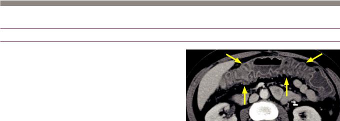
Large bowel
Colitis
Overview of colitis
•Colitis is inflammation of the colon that may be caused by several unrelated etiologies, often with overlapping imaging findings.
•The primary imaging feature of colitis is bowel wall thickening. Generally, a full clinical evaluation, stool studies, and sometimes colonic biopsy are required for a definitive diagnosis.
•Incidental colonic wall thickening is
Ischemic colitis found in as many as 10% of CT scans.
Pancolitis: Contrast enhanced CT shows severe mural thickening of the entire visualized colon (arrows) with mucosal hyperenhancement. Although nonspecific, this was a case of pseudomembranous colitis.
•Colonic ischemia can be caused by acute arterial thrombus, chronic arterial stenosis, low-flow states (e.g., congestive heart failure), and venous thrombosis.
•The splenic flexure is the watershed region between the superior and inferior mesenteric arteries and is especially susceptible to ischemia in low-flow states.
•Therectumissuppliedbyadualbloodsupplyandisalmostneveraffectedbyischemia. Thesuperiorrectalartery(terminalbranchoftheIMA)andtheinferiorandmiddlerectal arteries(arisingfromtheinternaliliacarteryanteriordivision)formperirectalcollaterals.
•A suggestive CT finding of ischemic colitis is segmental, continuous thickening of the affected colon in a vascular distribution, with sparing of the rectum.
If arterial thromboembolic disease is suspected, one should evaluate for the presence of aortic atherosclerotic disease or a left atrial thrombus in the setting of atrial fibrillation.
Ifchronicarterialstenosisissuspected,oneshouldevaluateforatherosclerosisofthemesentericvessels.
Infectious colitis
•Infectious colitis can be bacterial, tubercular, viral, or amoebic. There is a large overlap in the clinical presentation and imaging findings of the various pathogens.
•In general, infectious colitis features pericolonic stranding and ascites in addition to the colonic wall thickening seen in all forms of colitis.
•Yersinia,Salmonella,andcolonictubercolosisaffecttherightcolon.Tuberculosisisknownto involvetheileocecalvalve,resultinginadesmoplasticreactionthatmimicsCrohndisease.
•E. coli, CMV, and C. difficile colitis (discussed below) most commonly cause pancolitis.
Pseudomembranous colitis
•Pseudomembranous colitis is an especially prevalent form of infectious colitis caused by overgrowth of Clostridium difficile, most commonly due to alteration in colonic bacterial flora after antibiotic use. Pseudomembranous colitis may also occur without a history of antibiotics, especially in hospitalized or nursing home patients.
•A key imaging finding is marked thickening of the colonic wall, typically with involvement of the entire colon (pancolitis). The accordion sign describes severe colonic wall thickening combined with undulation of enhancing inner mucosa. It signifies severe colonic edema but is not specific to C. difficile. Thumbprinting is a fluoroscopic finding of thickened haustra and is also due to edema.
144
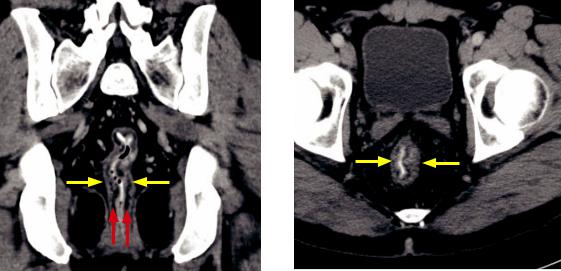
Ulcerative colitis (UC)
Chronic ulcerative colitis: Coronal (left image) and axial contrast-enhanced CT shows continuous thickening of the rectal wall (yellow arrows) with fat attenuation of the submucosa (red arrows).
•Ulcerative colitis (UC) is an idiopathic inflammatory bowel disease that begins distally in the rectum and spreads proximally in a continual manner (unlike Crohn disease, which features skip areas).
Of note, it is possible for the rectum to appear normal with more proximal colonic involvement present if the patient has been treated with corticosteroid enemas.
•Patients with UC have an increased risk of primary sclerosing cholangitis, colon cancer, and cholangiocarcinoma.
•Extra-abdominal manifestations of UC include sacroiliitis, iritis, erythema nodosum (tender red subcutaneous nodules), and pyoderma gangrenosum (cutaneous ulcers).
•UC does not extend more proximally than the cecum; however, a backwash ileitis caused by reflux of inflammatory debris into the ileum may mimic Crohn disease.
•Imaging of ulcerative colitis features circumferential wall thickening with a granular mucosal pattern that is best seen on barium enema. Pseudopolyps may be present during acute inflammation, representing islands of normal mucosa surrounded by inflamed mucosa. A collar-button ulcer is nonspecific but represents mucosal ulceration undermined by submucosal extension.
•Chronic changes of ulcerative colitis include a featureless and foreshortened lead pipe colon. Similar to Crohn disease, fat-attenuation of the colonic wall suggests chronic disease, as seen in the case above.
•Toxic megacolon is a severe complication of ulcerative colitis (and less commonly, Crohn disease) caused by inflammation extending through the muscular layer. Imaging of toxic megacolon shows dilation of the colon to greater than 6 cm in association with an adynamic ileus. Colonic perforation may occur and colonoscopy is contraindicated in suspected toxic megacolon.
Typhlitis (neutropenic enterocolitis)
•Typhlitis is a right-sided colitis seen in immunocompromised patients.
•Treatment is with broad-spectrum antibiotics and antifungals.
145

Polyposis syndromes affecting the bowel
Familial adenomatous polyposis (FAP)
•Familial adenomatous polyposis (FAP) is an autosomal-dominant syndrome featuring innumerable premalignant adenomatous polyps in the colon and to a lesser extent the small bowel. Prophylactic colectomy is the standard of care to prevent colon cancer.
•Gastric polyps are also present, although the gastric polyps are hyperplastic and are not premalignant.
•Gardner syndrome is a variant of FAP. In addition to colon polyps, patients also have:
Desmoid tumors. |
Papillary thyroid cancer. |
Osteomas. |
Epidermoid cysts. |
Mnemonic: DOPE Gardner
•Turcot syndrome is another variant of FAP. In addition to colon polyps, patients also have CNS tumors (gliomas and medulloblastomas).
Hereditary nonpolyposis colon cancer syndrome (HNPCC) = Lynch syndrome
•Hereditary nonpolyposis colon cancer (HNPCC) syndrome (also called Lynch syndrome) is an autosomal dominant polyposis syndrome caused by DNA mismatch repair, leading to colon cancer from microsatellite instability on a molecular level.
•Similar to FAP, the colon polyps of HNPCC are adenomatous.
•HNPCC is associated with other cancers, including endometrial, stomach, small bowel, liver, and biliary malignancies.
Peutz–Jeghers
•Peutz–Jeghers is an autosomal dominant syndrome that features multiple hamartomatous pedunculated polyps, usually in the small bowel. These polyps may act as lead points and cause intussusception.
•Characteristic skin manifestations include perioral mucocutaneous blue/brown pigmented spots on the lips and gums.
•Peutz–Jeghers is associated with gynecologic neoplasms as well as gastric, duodenal, and colonic malignancies.
Cowden syndrome
•Cowden syndrome is an autosomal dominant syndrome of multiple hamartomatous polyps most commonly found in the skin and external mucous membranes, but also in the gastrointestinal tract.
•Cowden syndrome is associated with an increased risk of thyroid cancer (usually follicular), as well as skin, oral, breast, and uterine malignancies.
Cronkhite–Canada
•Cronkhite–Canada is a non-inherited disorder (the only polyposis syndrome in this list that is not autosomal dominant) consisting of hamartomatous polyps throughout the gastrointestinal tract.
•Cutaneous manifestations include abnormal skin pigmentation, alopecia, and onychodystrophy (malformation of the nails).
146
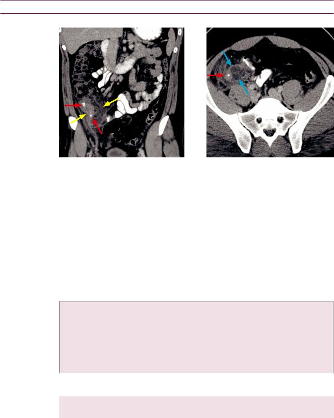
Acute bowel
Appendicitis
Appendicitis: Coronal (left image) and axial contrast-enhanced CT shows a large focus of inflammatory stranding centered around the appendix in the right lower quadrant (yellow arrows). The margins of the appendix itself are indistinct and there is the suggestion of an early fluid collection (blue arrows). Note the two appendicoliths within the appendix (red arrows).
•Appendicitis is the most common surgical cause of acute abdomen. Acute inflammation of the appendix is thought to be due to obstruction of the appendiceal lumen, leading to venous congestion, mural ischemia, and bacterial translocation.
•Appendicitis represents a spectrum of severity ranging from tip appendicitis (inflammation isolated to the distal appendix) to gangrenous appendicitis with abscess if the disease is not diagnosed until late.
•Greater than 97% of patients undergo a preoperative CT prior to appendectomy, with resultant decrease in negative appendectomy rate from 23% in 1990 to 1.7% in 2007.
•Imaging of appendicitis relies on direct and indirect imaging findings.
•Direct findings of appendicitis are due to abnormalities of the appendix itself:
Distended, fluid-filled appendix: 6 mm is used as cutoff for normal diameter of the appendix, although there is wide normal variability and 6 mm is from the ultrasound literature using compression. A normal appendix distended with air can measure >6 mm; therefore, some authors advocate using caution with a numeric cutoff in an otherwise normal-appearing appendix filled with air or enteric contrast.
Appendiceal wall-thickening.
Appendicolith, which may be a cause of luminal obstruction; however, appendicoliths are commonly seen without associated appendicitis.
•Indirect findings of appendicitis are due to the spread of inflammation to adjacent sites:
Periappendiceal fat stranding. |
Hydroureter. |
Cecal wall thickening. |
Small bowel ileus. |
•Appendicitis can also be evaluated by ultrasound, with the key sonographic finding a tubular, blind-ending, non-compressible right lower quadrant structure measuring >6 mm in diameter. Is is generally necessary to use graded compression to evaluate for compressibility.
Secondary findings of appendicitis can be evaluated by ultrasound, including free fluid and periappendicular abscess.
147

Diverticulitis
Complicateddiverticulitis: AxialCTshowsalarge diverticulumarisingfromthesigmoidcolon containingentericcontrast(yellowarrow),with surroundingmesentericfatstranding.Afewadjacent loculesofextraluminalgas(redarrow)arepresent.
Uncomplicated diverticulitis: Axial CT in a different patient shows fat stranding surrounding a diverticulum at the hepatic flexure (arrow).
•Diverticulitis is microperforation and acute inflammation of a colonic diverticulum. CT is the primary modality for diagnosis, triage, and evaluation of severity and complications.
•The left colon is affected far more commonly than the right.
•Itisoftenimpossibletodistinguishacutediverticulitis frommicroperforatedcoloncancer. Manyauthorsrecommendfollow-upcolonoscopyaftertheacuteepisodehasresolved, althoughthisrecommendationissomewhatcontroversialandvariesbyinstitution.
•Uncomplicated diverticulitis does not have any imaging evidence of bowel perforation (even though histopathologically all diverticulitis is associated with bacterial translocation across the bowel wall). CT findings of uncomplicated diverticulitis include bowel wall thickening and pericolonic fat stranding, usually centered around a culprit diverticulum.
•Complicated diverticulitis impliesthepresenceofanadditionalcomplication, including:
Pericolonic or hepatic abscess.
Extraluminal air.
Bowel obstruction.
Bowel fistula (colovesical fistula most common, apparent on imaging as gas in the bladder not explained by Foley catheter placement).
Mesenteric venous thrombosis.
•Uncomplicated diverticulitis is typically treated conservatively.
•Abscesses can usually be drained percutaneously.
•Indications for surgery include the presence of a fistula or recurrent diverticulitis, with two prior episodes of diverticulitis treated conservatively.
Epiploic appendagitis
•Epiploic appendagitis is a benign, clinical mimic of diverticulitis caused by torsion of a normal fatty tag (appendage) hanging from the colon.
•Epiploic appendagitis has a pathognomonic imaging appearance of an oval fatattenuation lesion abutting a normal colonic wall, with mild associated fat stranding. A central hyperdense dot in cross-section represents the thrombosed central vein of the epiploic appendage.
•Treatment is with anti-inflammatories, not antibiotics or surgery.
148

Mesentery and peritoneum
Anatomy
lesser omentum 
transverse colon
liver
stomach
T12 |
|
|
mesentery |
|
|
||
|
|
|
omentum |
|
|
|
|
|
|
|
|
L1
 transverse mesocolon

 pancreas (retroperitoneal)

 duodenum (retroperitoneal)
greater omentum 
small bowel loops
|
small bowel mesentery |
|
peritoneum |
sigmoid |
sigmoid mesentery |
uterus
|
|
|
|
|
|
|
|
|
|
|
|
|
|
|
|
|
|
|
|
|
|
|
|
|
|
|
|
|
|
|
|
|
|
|
|
|
|
|
|
|
|
|
|
|
|
|
|
|
|
|
|
|
|
|
|
|
pouch of Douglas |
|
|
|
|
|
|
|
|
|
|
|
||||||||||||||||||
|
|
|
|
|
|
|
|
|
|
|
|
|
|
|
|
|
|
|
|
|
|
|
|
|
|
|
|
|
|
|
|
|
|
|
|
|
|
|
|
|
rectum |
|
|
|
|
|
|
|
|
|
|
|
|
|||||||||||||||||||||||||||||||||
|
|
|
|
|
|
|
|
|
|
|
|
|
|
|
|
|
|
|
|
|
|
|
|
|
|
|
|
|
|
|
|
|
|
|
|
|
|
|
|
|
|
|
|
|
|
|
|
|
|
|
|
|
|
|
|
|
|
|
|
|
|
|
|
|
|
|
|
|
|
|
|
|||||||||||||||
Peritoneum |
|
|
|
|
|
|
|
|
|
|
|
|
|
|
|
|
|
|
|
|
|
|
|
|
|
|
|
|
|
|
|
|
|
|
|
|
|
|
|
|
|
|
|
|
|
|
|
|
|
|
|
|
|
|
|
|
|
|
|
|
|
|
|
|
|
|
|
|
|
|
|
|
|
|
|
|
|
|
|
|
||||||
|
|
|
|
|
|
|
|
|
|
|
|
|
|
|
|
|
|
|
|
|
|
|
|
|
|
|
|
|
|
|
|
|
|
|
|
|
|
|
|
|
|
|
|
|
|
|
|
|
|
|
|
|
|
|
|
|
|
|
|
|
|
|
|
|
|
|
|
|
|
|
|
|
|
|
|
|
|
|
|
|||||||
|
|
|
|
|
|
|
|
|
|
|
|
|
|
|
|
|
|
|
|
|
|
|
|
|
|
|
|
|
|
|
|
|
|
|
|
|
|
|
|
|
|
|
|
|
|
|
|
|
|
|
|
|
|
|
|
|
|
|
|
|
|
|
|
|
|
|
|
|
|
|
|
|
|
|
|
|
|
|
|
|||||||
|
The peritoneum s |
|
|
thin membrane consisting of |
single ayer of mesothelia cells |
|||||||||||||||||||||||||||||||||||||||||||||||||||||||||||||||||||||||||||||||||
|
that |
supported by subserosa |
|
|
fat cells, ymphatic cells, and white blood cells. |
|||||||||||||||||||||||||||||||||||||||||||||||||||||||||||||||||||||||||||||||||
|
The viscera peritoneum |
|
|
ines the surface of al intraperitonea |
while the |
|||||||||||||||||||||||||||||||||||||||||||||||||||||||||||||||||||||||||||||||||
|
parieta |
peritoneum ines the outer walls of the peritonea cavity |
|
|
|
|
|
|
|
|
|
|
|
|||||||||||||||||||||||||||||||||||||||||||||||||||||||||||||||||||||||||
|
The most dependent portion of the peritonea cavity (both supine and upright) s the |
|||||||||||||||||||||||||||||||||||||||||||||||||||||||||||||||||||||||||||||||||||||
|
pouch of Douglas |
n |
|
|
|
|
and the retrovesica |
|
|
|
|
n |
|
|
|
|
|
|
|
|
|
|
|
|||||||||||||||||||||||||||||||||||||||||||||||||||||||||||||||
Mesentery |
|
|
|
|
|
|
|
|
|
|
|
|
|
|
|
|
|
|
|
|
|
|
|
|
|
|
|
|
|
|
|
|
|
|
|
|
|
|
|
|
|
|
|
|
|
|
|
|
|
|
|
|
|
|
|
|
|
|
|
|
|
|
|
|
|
|
|
|
|
|
|
|
|
|
|
|
|
|
|
|
||||||
|
There |
|
three true mesenteries which each supply portion of the bowe and |
|||||||||||||||||||||||||||||||||||||||||||||||||||||||||||||||||||||||||||||||||||
|
onnect to the posterior abdomina wall. Each mesentery consists of |
network of |
||||||||||||||||||||||||||||||||||||||||||||||||||||||||||||||||||||||||||||||||||||
|
blood vessels and |
ymphatics, sandwiched between the peritonea ayers. The three |
||||||||||||||||||||||||||||||||||||||||||||||||||||||||||||||||||||||||||||||||||||
|
true mesenteries |
|
|
|
|
|
|
|
|
|
|
|
|
|
|
|
|
|
|
|
|
|
|
|
|
|
|
|
|
|
|
|
|
|
|
|
|
|
|
|
|
|
|
|
|
|
|
|
|
|
|
|
|
|
|
|
|
|
|
|
|
|
|
|
|
|
|
|
|
|
|
|||||||||||||||
|
|
|
|
|
|
|
|
|
|
|
|
|
|
|
|
|
|
|
|
|
|
|
|
|
|
|
|
|
|
|
|
|
|
|
|
|
|
|
|
|
|
|
|
|
|
|
|
|
|
|
|
|
|
|
|
|
|
|
|
|
|
|
|
|
|
|
|
|
|
|
|
|
|
|
|
|
|
|
|
|
|
|||||
|
|
|
|
|
|
|
|
|
|
|
|
|
|
|
|
|
|
|
|
|
|
|
|
|
|
|
|
|
|
|
|
|
|
|
|
|
|
|
|
|
|
|
|
|
|
|
|
|
|
|
|
|
|
|
|
|
|
|
|
|
|
|
|
|
|
|
|
|
|
|
|
|
|
|
|
|
|
|
|
|
|
|||||
|
|
|
|
|
|
|
|
|
|
|
|
|
|
|
|
|
|
|
|
|
|
|
|
|
|
|
|
|
|
|
|
|
|
|
|
|
|
|
|
|
|
|
|
|
|
|
|
|
|
|
|
|
|
|
|
|
|
|
|
|
|
|
|
|
|
|
|
|
|
|
|
|
|
|
|
|
|
from |
||||||||
|
|
Small bowel mesentery |
|
|
|
Supplies |
|
both |
|
the |
|
jejunum |
|
and |
ileum. |
Oriented |
obliquely |
the |
||||||||||||||||||||||||||||||||||||||||||||||||||||||||||||||||||||
|
|
|
||||||||||||||||||||||||||||||||||||||||||||||||||||||||||||||||||||||||||||||||||||
|
|
|
|
|
|
|
|
|
|
|
|
|
|
|
|
|
|
|
|
|
|
|
|
|
|
|
|
|
|
|
|
|
|
|
|
|
|
|
|
|
|
|
|
|
|
|
|
|
|
|
|
|
|
|
|
|
|
|
|
|
|
|
|
|
|
|
||||||||||||||||||||
|
|
|
|
|
|
|
|
|
of |
eft |
||||||||||||||||||||||||||||||||||||||||||||||||||||||||||||||||||||||||||||
|
|
ig |
the |
the |
ileoceca |
the |
right |
ower |
||||||||||||||||||||||||||||||||||||||||||||||||||||||||||||||||||||||||||||||
|
|
|
|
|
|
|
|
|
|
|
|
|
|
|
|
|
|
|
|
|
|
|
quadrant |
quadrant |
||||||||||||||||||||||||||||||||||||||||||||||||||||||||||||||
|
|
|
|
|
|
|
|
Treitz |
n |
junction |
n |
|||||||||||||||||||||||||||||||||||||||||||||||||||||||||||||||||||||||||||
|
|
ament |
|
|
|
|
|
|
|
|
|
|
|
|
|
to |
||||||||||||||||||||||||||||||||||||||||||||||||||||||||||||||||||||||
|
|
|
|
|
|
|
|
|
|
|
|
|
|
|
|
|
|
to |
|
|
|
transverse |
|
|
|
|
|
|
|
|
transverse |
|
||||||||||||||||||||||||||||||||||||||||||||||||||||||
|
|
|
|
ransverse mesocolon |
the |
colon, |
the |
|||||||||||||||||||||||||||||||||||||||||||||||||||||||||||||||||||||||||||||||
|
|
T |
|
|
Mesentery |
|
|
|
|
connecting |
posterior |
|||||||||||||||||||||||||||||||||||||||||||||||||||||||||||||||||||||||||||
|
|
|
|
|
|
|
|
|
|
|
|
|
|
|
|
|
|
|
|
|
|
|
|
|
|
|
|
|
|
|
|
|
|
|
|
|
|
|
|
|
|
|
|
|
|
|
||||||||||||||||||||||||||||||||||||||||
|
|
|
olon |
the |
abdomina |
wal |
|
|
|
|
|
|
|
|
|
|
|
|
|
|
|
|
|
|
|
|
|
|
|
|
|
|
|
|
|
|
|
|||||||||||||||||||||||||||||||||||||||||||||||||
|
|
|
|
|
to |
|
|
posterior |
|
|
|
|
|
|
|
|
|
|
|
|
|
|
|
|
|
|
|
|
|
|
|
|
|
|
|
|
|
|
|
|||||||||||||||||||||||||||||||||||||||||||||||
|
|
|
|
|
|
|
|
|
|
|
|
|
|
|
|
|
|
|
|
|
|
|
|
|
|
|
|
|
|
|
|
|
|
|||||||||||||||||||||||||||||||||||||||||||||||||||||
|
|
|
|
|
|
|
|
|
|
|
|
|
|
|
|
|
|
|
|
|
|
|
|
|
|
|
|
|
|
|
|
|
|
|
|
|
|
|
|
|
|
|
|
|
|
|
|
|
|
|
|
|
|
|
|
|
|
|
|
|
|
|
|
|
|
|
|
|
|
|
|
|
|
|
|
|
|
|
|
|
|
|
|
|
|
|
Sigmoid mesentery Mesentery to the sigmoid colon.
The greater and esser omentum specialized mesenteries that attach to the tomach. The greater and esser omentum do not connect to the posterior abdomina all.
Greater omentum Large drape-like mesentery n the anterior abdomen, which connects the
 tomach to the anterior aspect ofthe transverse colon.
Lesser omentum Connects stomach to iver.
149
Flow of peritoneal fluid
•Peritoneal fluid is constantly produced, circulated, and finally resorbed around the diaphragm, where it eventually drains into the thoracic duct.
“Misty” mesentery
Overview of the “misty” mesentery
•As previously discussed, the abdominal mesenteries are fatty folds through which the arterial supply and venous and lymphatic drainage of the bowel run.
•The mesenteries themselves are not seen on CT because they are made primarily of fat and blend in with intra-abdominal fat. However, the vessels which course through the mesentery are normally seen.
•Infiltration of the mesentery by fluid, inflammatory cells, tumor, or fibrosis may increase the attenuation of the mesentery and cause the mesenteric vasculature to appear indistinct. These findings are often the first clue to certain pathologies.
Mesenteric edema
•Edema of the mesentery may be secondary to either systemic or intra-abdominal etiologies.
•Systemic causes of edema include congestive heart failure, low protein states, and third-spacing, all of which can lead to diffuse mesenteric edema.
•Focal mesenteric edema may be secondary to an intra-abdominal vascular cause, such as mesenteric vessel thrombosis, Budd–Chiari syndrome, or IVC obstruction. Abdominal vascular insults may cause bowel ischemia, which manifest on imaging as bowel wall thickening, pneumatosis, or mesenteric venous gas.
Mesenteric inflammation
•The most common cause of mesenteric inflammation in the upper abdomen is acute pancreatitis. However, any focal inflammatory process such as appendicitis, inflammatory bowel disease, and diverticulitis may cause local mesenteric inflammation leading to the “misty” mesentery appearance.
•Mesenteric panniculitis is an idiopathic inflammatory condition, which may cause a diffuse “misty” mesentery.
Intra-abdominal hemorrhage
•Intra-abdominal hemorrhage tends to be localized, surrounding the culprit bleeding vessel unless large. Hemorrhage may be secondary to trauma, post-procedural, or due to anticoagulation.
Neoplastic infiltration
•Neoplastic infiltration of the mesentery may cause the “misty” mesentery. The most common tumor involving the mesentery is non-Hodgkin lymphoma, which typically also causes bulky adenopathy.
•Mesenteric involvement may be especially apparent after treatment, where the “misty” mesentery is limited to the portion of the mesentery that contained the treated lymph nodes.
•Other tumors that may involve the mesentery include pancreatic, colon, breast, gastrointestinal stromal tumor, and mesothelioma.
150
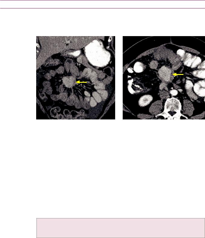
Mesenteric masses
Overview of mesenteric masses
•Primary mesenteric tumors are rare, although the mesentery is a relatively common site of metastasis.
Carcinoid
Carcinoid metastatic to the mesentery: Coronal (left image) and axial contrast-enhanced CT shows a hyperenhancing mesenteric mass (arrows) that contains a few tiny foci of calcification peripherally. There are numerous linear soft tissue strands radiating from the mass.
Case courtesy Cheryl Sadow, MD, Brigham and Women’s Hospital.
•Gastrointestinal carcinoid is a relatively rare tumor compared to other gastrointestinal malignancies, but is the most common small bowel tumor. It typically occurs in the distal ileum.
•Carcinoid usually arises as an intraluminal mass and may secondarily spread to the mesentery either by direct extension or lymphatic spread. Up to 80% of carcinoids spread to the mesentery.
•A classic imaging appearance of carcinoid affecting the mesentery is an enhancing soft-tissue mass with radiating linear bands extending into the mesenteric fat. Calcification is common.
The radiating linear bands do not represent infiltrative tumor but are the result of an intense desmoplastic reaction caused by the release of serotonin by the tumor.
•The differential diagnosis of a sclerosing mesenteric mass includes:
Carcinoid.
Desmoid tumor.
Sclerosing mesenteritis.
Desmoid tumor
•Desmoid tumor is a benign, locally aggressive mass composed of proliferating fibrous tissue.
•Desmoid may be sporadic, but mesenteric desmoid tumors are more common in patients with Gardner syndrome (a variant of familial adenomatous polyposis).
•On CT, most desmoids are isoattenuating to muscle, but large tumors may show central necrosis. A characteristic imaging feature is strands of tissue radiating into the adjacent mesenteric fat, similar to mesenteric carcinoid and sclerosing mesenteritis.
151

Sclerosing mesenteritis
•Sclerosing mesenteritis is a rare inflammatory condition that leads to fatty necrosis and fibrosis of the mesenteric root.
•Imaging of sclerosing mesenteritis shows mesenteric masses with striations of soft tissue extending into the adjacent fat. Calcification may be present.
•Mesenteric panniculitis is a variant where inflammation predominates and presents as acute abdominal pain. On CT, there is a “misty” mesentery, sometimes with linear bands of soft tissue representing early fibrosis.
Mesenteric metastases and lymphoma
•Gastric, ovarian, breast, lung, pancreatic, biliary, colon cancer, and melanoma can metastasize to mesenteric lymph nodes.
•Mesenteric lymphoma can produce the sandwich sign, where the mesenteric fat and vessels (the sandwich filling) are engulfed on two sides by bulky lymphomatous masses (the bread).
Diffuse peritoneal disease
Peritoneal carcinomatosis
Peritoneal carcinomatosis due to gastric cancer, with Krukenberg tumors of the ovaries: Coronal contrast-enhanced CT (left image) shows marked thickening of the gastric wall (yellow arrow). There are bilateral adnexal masses (red arrows), representing ovarian metastases. Moderate ascites is present. Axial CT (right image) demonstrates peritoneal nodularity and thickening, especially in the left anterior abdomen (blue arrows), consistent with peritoneal carcinomatosis.
Case courtesy Cheryl Sadow, MD, Brigham and Women’s Hospital.
•Peritoneal carcinomatosis represents disseminated metastases to the peritoneal surface.
•The term omental caking describes the replacement of omental fat by tumor and fibrosis.
•Mucinous adenocarcinoma is the most common tumor type to cause peritoneal carcinomatosis. peritoneal carcinomatosis due to mucinous adenocarcinoma should not be confused with pseudomyxoma peritonei, discussed on the following page.
152
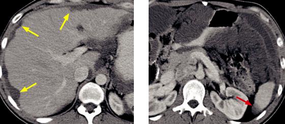
Pseudomyxoma peritonei
Pseudomyxoma peritonei: Axial contrast-enhanced CT through the liver (left image) shows scalloping of the hepatic capsule (arrows). A lower image through the kidneys shows the lobulated, mucinous ascites exerting mass effect on adjacent bowel loops. A splenic implant is also visible (red arrow).
Case courtesy Cheryl Sadow, MD, Brigham and Women’s Hospital.
•Pseudomyxoma peritonei is a low-grade malignancy characterized by copious mucus in the peritoneal cavity.
•In general, pseudomyxoma peritonei is thought to be produced by a mucin-producing adenoma or adenocarcinoma of the appendix; however, there is some controversy as to whether the ovary or colon can be a primary site as well.
Pseudomyxoma peritonei is often associated with an ovarian mass (up to 30% of female patients), but it is thought that these are most often metastatic deposits.
•Pseudomyxoma peritonei was previously thought to be produced by a benign appendiceal mucocele, which is now believed to occur much less commonly than originally thought.
20% of all appendiceal adenomas or adenocarcinomas will cause pseudomyxoma peritonei.
Only 2% of all appendiceal mucoceles (which occur slightly less commonly than appendiceal adenomatous lesions) will cause pseudomyxoma peritonei.
•Tumor deposits tend to be spread throughout the entire peritoneal cavity due to intraperitoneal fluid currents.
•Clinically, pseudomyxoma peritonei presents with recurrent mucinous ascites. The surgeons refer to the mucinous ascites as a “jelly belly.”
•CT shows lobular ascites that is typically of slightly higher attenuation (5–20 Hounsfield units) compared to fluid ascites. Occasionally, mucus can be seen in the region of the appendix, but the flow of peritoneal contents tends to spread the mucinous ascites diffusely throughout the peritoneum.
•Advanced disease shows pathognomonic scalloping of the hepatic margin.
•Treatment continues to evolve, but the best outcomes are primarily with surgical treatment and hyperthermic intraperitoneal chemotherapy lavage.
153

References, resources, and further reading
General references:
Johnson, D.C. & Schmit, G.D. Mayo Clinic Gastrointestinal Imaging Review. Mayo Clinic Scientific Press. (2005).
Ros, P.R., Mortele K.J. CT and MRI of the Abdomen and Pelvis: A Teaching File. Lippincott Williams & Wilkins. (2006).
Roth, C.G. Fundamentals of Body MRI (1st ed.). Elsevier/Saunders. (2012).
Liver:
Alústiza, J.M. et al. Iron overload in the liver diagnostic and quantification. European journal of radiology 61, 499-506(2007).
Blachar, A., Federle, M.P. & Sosna, J. Liver Lesions With Hepatic Capsular Retraction. Seminars in Ultrasound, CT, and MRI 30, 426-35(2009).
Brancatelli, G. et al. Budd-Chiari Syndrome: Spectrum of Imaging Findings. American Journal of Roentgenology 188, 168-76(2007).doi:10.2214/AJR.05.0168
Chung, Y.E. et al. Varying appearances of cholangiocarcinoma: radiologic-pathologic correlation. Radiographics 29, 683-700(2009).
Elsayes, K.M. et al. Focal hepatic lesions: diagnostic value of enhancement pattern approach with contrast-enhanced 3D gradient-echo MR imaging. Radiographics 25, 1299-320(2005).
Hanna, R. et al. Cirrhosis-associated Hepatocellular Nodules: Correlation of Histopathologic and MR imaging features. Radiographics 28, 747-69(2008).
Kamaya, A. et al. Hypervascular Liver Lesions. Seminars in Ultrasound, CT, and MRI 30, 387-407(2009).
Kim, T., Hori, M. & Onishi, H. Liver Masses With Central or Eccentric Scar. Seminars in Ultrasound, CT, and MRI 30, 418-25(2009).
Martin, D. & Semelka, R. Magnetic Resonance Imaging of the Liver: Review of Techniques and Approach to Common Diseases. Seminars in Ultrasound, CT, and MRI 26, 116-31(2005).
Martin, D.R., Danrad, R. & Hussain, S.M. MR imaging of the liver. Radiologic clinics of North America 43, 861-86, viii(2005).
Mortele, K.J. & Ros, P.R. Cystic Focal Liver Lesions in the Adult: Differential CT and MR Imaging Features. Radiographics 21, 895-910(2001).
Pancreaticobiliary:
Acar, M., Tatli, S. Cystic tumors of the pancreas: a radiological perspective. Diagnostic and interventional radiology (Ankara, Turkey), 17(2), 143-49(2011).
Blachar, a., Federle, M.P. & Brancatelli, G. Primary biliary cirrhosis: clinical, pathologic, and helical CT findings in 53 patients. Radiology 220, 329-36(2001).
Blasbalg, R. et al. MRI features of groove pancreatitis. AJR. American journal of roentgenology 189, 73-80(2007).
Fasanella, K.E. & McGrath, K. Cystic lesions and intraductal neoplasms of the pancreas. Best practice & research. Clinical gastroenterology 23, 35-48(2009).
Kim, S.H. et al. Intrapancreatic accessory spleen: findings on MR Imaging, CT, US and scintigraphy, and the pathologic analysis. Korean journal of radiology 9, 162-74(2008).
Koo, B.C., Chinogureyi, a. & Shaw, a.S. Imaging acute pancreatitis. The British journal of radiology 83, 104-12(2010).
Levy, A.D. et al. From the Archives of the AFIP: Benign Tumors and Tumorlike Lesions of the Gallbladder and Extrahepatic Bile Ducts: Radiologic-Pathologic Correlation. Radiographics 22, 387-413(2002).
Mortele, K.J. et al. Multimodality Imaging of Pancreatic and Biliary Congenital Anomalies. Radiographics 26, 715-31(2006).
Nakajima, Y., Yamada, T. & Sho, M. Malignant potential of intraductal papillary mucinous neoplasms of the pancreas. Surgery today 40, 816-24(2010).
Nishimori, I., Onishi, S. & Otsuki, M. Review of diagnostic criteria for autoimmune pancreatitis; for establishment of international criteria. Clinical Journal of Gastroenterology 1, 7-17(2008).
Pedrosa, I. & Boparai, D. Imaging considerations in intraductal papillary mucinous neoplasms of the pancreas. World journal of gastrointestinal surgery 2, 324-30(2010).
154

Saokar, A., Rabinowitz, C.B. & Sahani, D.V. Cross-sectional imaging in acute pancreatitis. Radiologic clinics of North America 45, 447-60, viii(2007).
Sawai, Y. et al. Development of pancreatic cancers during long-term follow-up of side-branch intraductal papillary mucinous neoplasms. Endoscopy 43, 79(2011).
Scaglione, M. et al. Imaging Assessment of Acute Pancreatitis: A Review. Seminars in Ultrasound, CT, and MRI 29, 322-40(2008).
Sidden, C.R., Mortele, K.J. Cystic Tumors of the Pancreas: Ultrasound, Computed Tomography, and Magnetic Resonance Imaging Features. Seminars in Ultrasound, CT, and MRI 28(5), 339-56(2007).
Spencer, L.a., Spizarny, D.L. & Williams, T.R. Imaging features of intrapancreatic accessory spleen. The British journal of radiology 83, 668-73(2010).
Triantopoulou, C. et al. Groove pancreatitis: a diagnostic challenge. European radiology 19, 1736-43(2009).
Trout, A.T. et al. Imaging of acute pancreatitis: prognostic value of computed tomographic findings. Journal of computer assisted tomography 34, 485-95(2010).
Vikram, R. et al. Pancreas: peritoneal reflections, ligamentous connections, and pathways of disease spread. Radiographics 29, e34(2011).
Spleen:
Boscak, A. & Shanmuganathan, K. Splenic trauma: what is new? Radiologic clinics of North America, 50(1), 105-22(2012).
Elsayes, K.M. et al. MR imaging of the spleen: spectrum of abnormalities. Radiographics 25, 967-82(2005).
Gayer, G. et al. Congenital Anomalies of the Spleen. Seminars in Ultrasound, CT, and MRI 27, 358-369(2006).
Kamaya, a., Weinstein, S. & Desser, T. Multiple Lesions of the Spleen: Differential Diagnosis of Cystic and Solid Lesions.
Seminars in Ultrasound, CT, and MRI 27, 389-403(2006).
Warshauer, D. & Hall, H. Solitary Splenic Lesions. Seminars in Ultrasound, CT, and MRI 27, 370-88(2006).
Esophagus and Stomach:
Carucci, L.R. & Turner, M.A. Imaging after bariatric surgery for morbid obesity: Roux-en-Y gastric bypass and laparoscopic adjustable gastric banding. Seminars in roentgenology 44, 283-96(2009).
Blachar, a. et al. Radiographic manifestations of normal postoperative anatomy and gastrointestinal complications of bariatric surgery, with emphasis on CT imaging findings. Seminars in Ultrasound, CT, and MRI 25, 239-51(2004).
Oh, J.Y. et al. Benign submucosal lesions of the stomach and duodenum: imaging characteristics with endoscopic and pathologic correlation. European journal of radiology, 67(1), 112–24(2008).
Paroz, a., Calmes, J.M., Giusti, V. & Suter, M. Internal hernia after laparoscopic Roux-en-Y gastric bypass for morbid obesity: a continuous challenge in bariatric surgery. Obesity surgery, 16(11), 1482-7(2006).
Patel, R.Y. et al. Internal hernia complications of gastric bypass surgery in the acute setting: spectrum of imaging findings. Emergency radiology 16, 283-9(2009).
Sunnapwar, A. et al. Taxonomy and imaging spectrum of small bowel obstruction after Roux-en-Y gastric bypass surgery. AJR. American journal of roentgenology, 194(1), 120-8(2010).
Trenkner, S.W. Imaging of morbid obesity procedures and their complications. Abdominal imaging, 34(3), 335-44(2009).
Small Bowel:
Dilauro, S. & Crum-Cianflone, N.F. Ileitis: when it is not Crohnʼs disease. Current gastroenterology reports 12, 249-58(2010). Furukawa, A. et al. Cross-sectional Imaging in Crohn Disease 1. Radiographics 24, 689(2004).
Khurana, B. Signs in Imaging. The Whirl Sign. Radiology 1, 69-70(2003).
Khurana, B. et al. Bowel obstruction revealed by multidetector CT. AJR. American journal of roentgenology 178, 1139-44(2002).
Martin, L.C., Merkle, E.M. & Thompson, W.M. Review of internal hernias: radiographic and clinical findings. AJR. American journal of roentgenology 186, 703-17(2006).
Scholz F, Afnan J. CT Findings in Adult Celiac Disease. Radiographics 31, 977-93(2011).
Silva, A.C., Pimenta, M. & Guimarães, L.S. Small bowel obstruction: what to look for. Radiographics 29, 423-39(2009).
155

Soyer, P. et al. Celiac disease in adults: evaluation with MDCT enteroclysis. AJR. American journal of roentgenology 191, 1483-92(2008).
Takeyama, N. et al. CT of Internal Hernias. Radiographics 25, 997-1015(2005).
Colon:
Buckley, O. et al. Computed tomography in the imaging of colonic diverticulitis. Clinical radiology 59, 977-83(2004). Chintapalli, K.N. et al. Diverticulitis versus colon cancer: differentiation with helical CT findings. Radiology 210, 429-35(1999). Cloutier, R.L. Neutropenic enterocolitis. Emergency medicine clinics of North America 27, 415-22(2009).
Kawamoto, S., Horton, K.M. & Fishman, E.K. Pseudomembranous colitis: spectrum of imaging findings with clinical and pathologic correlation. Radiographics 19, 887-97(1999).
Macari, M. & Balthazar, E.J. CT of bowel wall thickening: significance and pitfalls of interpretation. AJR. American journal of roentgenology 176, 1105-16(2001).
Macari, M., Balthazar, E.J. & Megibow, a.J. The accordion sign at CT: a nonspecific finding in patients with colonic edema. Radiology 211, 743-6(1999).
Moraitis, D. et al. Colonic wall thickening on computed tomography scan and clinical correlation. Does it suggest the presence of an underlying neoplasia? The American surgeon 72, 269-71(2006).
Sarma, D. & Longo, W.E. Diagnostic imaging for diverticulitis. Journal of clinical gastroenterology 42, 1139-41(2008). Taourel, P. et al. Imaging of ischemic colitis. Radiologic clinics of North America 46, 909-24, vi(2008).
Touzios, J.G. & Dozois, E.J. Diverticulosis and acute diverticulitis. Gastroenterology clinics of North America 38, 513-25(2009).
Peritoneum and Mesentery:
Levy, A. & Shaw, J. Secondary Tumors and Tumorlike Lesions of the Peritoneal Cavity: Imaging Features with Pathologic Correlation 1. Radiographics 29, 4799(2009).
Lucey, B.C., Stuhlfaut, J.W. & Soto, J.A. Mesenteric Lymph Nodes Seen at Imaging: Causes and Significance. Radiographics 25, 351(2005).
Mindelzun, R.E. et al. The misty mesentery on CT: differential diagnosis. American Journal of Roentgenology 167, 61(1996).
Sheth, S. et al. Mesenteric Neoplasms: CT Appearances of Primary and Secondary Tumors and Differential Diagnosis. Radiographics 23, 457-73(2003).
Smeenk, R.M., Bruin, S.C., van Velthuysen, M.-L.F., & Verwaal, V.J. Pseudomyxoma peritonei. Current problems in surgery, 45(8), 527-75(2008).
156