


198 4.2 Benign Nontraumatic Abnormalities
4.2Benign Nontraumatic Abnormalities
#94 Cholecystolithiasis,
Classical Appearance
Related topic: #72 (stones in the common bile duct)
KEY FACTS: DISEASE
●Incidence: depends on age and sex (25% in women over 50 years)
●Ratio of women to men: 3 : 1
●Types:
–Cholesterol stones (± 80% in Western countries): pure cholesterol (10%); cholesterol plus calcium (70%)
–Pigment stones (calcium bilirubinate)
●Predisposing factors for cholesterol stones:
–Estrogens (female sex, pregnancy)
–Obesity
–High age
–Hypertriglyceridemia
–Clofibrate therapy
●Predisposing factors for pigment stones:
–Hemolytic disease (e.g., spherocytosis, prosthetic cardiac valves)
–Cirrhosis
–High age
–Biliary disease (e.g., stricture)
●Symptoms and complications:
–Biliary colic
–Pancreatitis
–Acute and chronic cholecystitis
–Fistula
–Cancer of gallbladder (incidence doubled)
KEY FACTS: MRI
●Signal intensity:
–Most commonly hypointense on T1and T2-weighted images
–May be partially or (more rarely) entirely hyperintense on T1 (Fig. 94a, b)
●Comments:
–Cross-sectional T2-weighted images are more sensitive than projective and T1-weighted images
–Ultrasonography is the primary modality for detection of stones in the gallbladder
References
Baron RL, Shuman WP, Lee SP et al. (1989) MRI appearance of gallstones in vitro at 1.5 T: correlation with chemical composition. AJR Am J Roentgenol 153 : 497–502
Bret P, Reinhold C (1997) MRI of the gallbladder. In: Rossi P (ed) Biliary tract radiology. Springer, Berlin Heidelberg New York, pp 59–69
Calvo MM,Bujanda L,Heras I,et al. (2002) Magnetic resonance cholangiography versus ultrasound in the evaluation of the gallbladder. J Clin Gastroenterol 34 : 233–236
4 Gallbladder and Cystic Duct 199
a |
b |
c |
d |
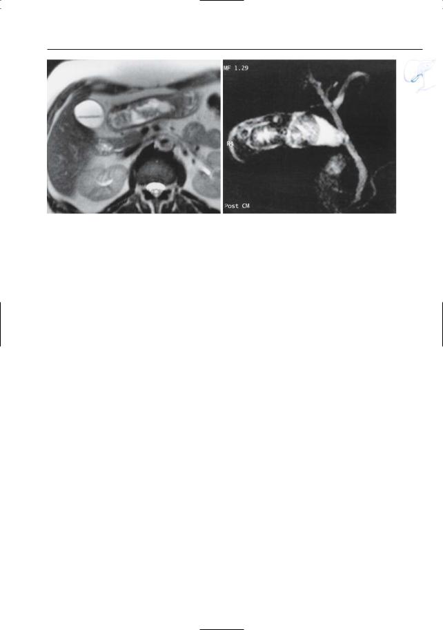
Fig. 94. a, b T2-weighted image (a) showing a gallstone as a rounded hypointense structure in the gallbladder lumen (arrow). Note the thick, hyperintense gallbladder wall. b T1-weighted image showing a gallstone containing two layers: a hyperintense outer layer and a hypointense (calcified) core.
The outer hypointense layer corresponds to the thickened wall of the gallbladder. c, d Different patient. T2-weighted HASTE image (c) and projective image (d) showing multiple small gallstones (arrow). Note several common bile duct stones
(arrowheads)

200 4.2 Benign Nontraumatic Abnormalities
#95 Cholecystolithiasis,
Variant Appearance
KEY FACTS: MRI
●Floating stones (Fig. 95a): causes and mechanisms:
–Relatively pure cholesterol stones
–Gas-containing stones
–Rise in specific gravity of bile
●Gas-containing stones:
–Dehydration of older stones Æ internal shrinkage Æ nitrogen gas
filling (“crow-foot” or “MercedesBenz” sign) Æ hypointense central core
●The central core may also contain water and appear as a hyperintense “star” (Fig. 95b)
●Gallstones may be missed if they are not surrounded by fluid:
–Microgallbladder
–Chronic cholecystitis
–Small stones impacted in the infundibulum/cystic duct
References
Baron RL, Shuman WP, Lee SP et al. (1989) MRI appearance of gallstones in vitro at 1.5 T: correlation with chemical composition. AJR Am J Roentgenol 153 : 497–502
Bret P, Reinhold C (1997) MRI of the gallbladder. In: Rossi P (ed) Biliary tract radiology. Springer, Berlin Heidelberg New York, pp 59–69
Moeser PM, Julian S, Karstaedt N, Sterchi M (1988) Unusual presentation of cholelithiasis on T1weighted MR imaging. J Comput Assist Tomogr 12 : 150–152
4 Gallbladder and Cystic Duct 201
a |
b |
Fig. 95. a Axial T2-weighted image showing floating stones. b Projective MR image showing gallstones with hyperintense central core

202 4.2 Benign Nontraumatic Abnormalities
#96 Acute Cholecystitis
KEY FACTS: DISEASE
●Acute inflammation of the gallbladder
●Age peak: fifth to sixth decade
●Types/mechanism:
–Calculous cholecystitis (90%): related to cystic duct obstruction by impacted stone
–Acalculous cholecystitis (10%): gallbladder wall ischemia secondary to distention (e.g.,associated with severe trauma, prolonged fasting)
●Predisposing factor: cholecystolithiasis
●Diagnosis: usually clinical
●Complications:
–Intramural abscess (see #98)
–Gangrene (see #99)
–Perforation (see # 100)
–Empyema
KEY FACTS: MRI (FIG. 96)
●T2-weighted images:
–Wall thickening (intramural edema)
–Increased signal intensity of wall (intramural edema)
–Hydrops
●T1-weighted images:
–Usually (not always) low signal intensity of bile, related to high fluid content (sign with differentiating value only in patients fasting for 8–12 h; Pu et al. 1994)
●Contrast-enhanced images:
–Marked enhancement of the gallbladder wall (not specific)
–Transient enhancement of adjacent liver parenchyma
●Differential diagnosis with chronic cholecystitis: high signal intensity of the gallbladder wall on heavily T2-weighted images (Fig. 96)
References
Kim KW, Park M,Yu J, et al. (2003) Acute cholecystitis at T2-weighted and manganese-enhanced T1-weighted MR cholangiography: preliminary study. Radiology 227: 580–584
Loud PA, Semelka RC, Kettritz U, Brown JJ, Reinhold C (1996) MRI of acute cholecystitis: comparison with the normal gallbladder and other entities. Magn Reson Imaging 14 : 349–355
Pu Y, Yamamoto F, Igimi H et al. (1994) A comparative study of the usefulness of magnetic resonance imaging in the diagnosis of acute cholecystitis. J Gastroenterol 29 : 192–198
Weissleder R, Stark DD, Compton CC, Simeone JF, Ferruci JT (1988) Cholecystitis: diagnosis by MR imaging. Magn Reson Imaging 6 : 345–348
4 Gallbladder and Cystic Duct 203
a
c
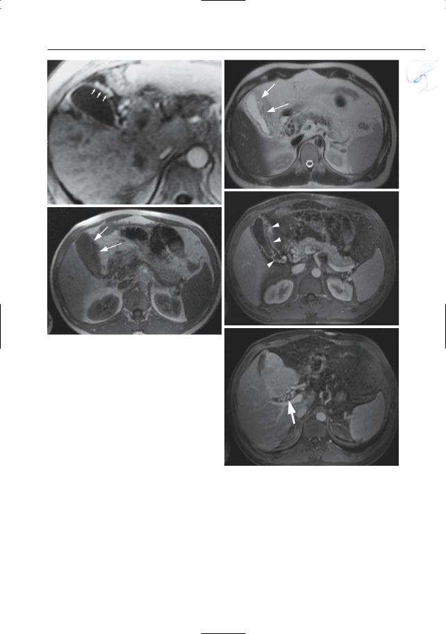
Fig. 96 a–c. Axial and coronal T2-weighted images showing thickening of the gallbladder wall with intramural edema (arrows in a) and a small amount of fluid in or adjacent to the wall (b). c Heavily T2weighted coronal image confirming the presence of fluid (arrows). d, e Different patient. T2-weighted HASTE image (d) and projective image (e) showing thickening of the gallbladder wall with intramural edema (arrows)
b
d
e

204 4.2 Benign Nontraumatic Abnormalities
#97 Acute Cholecystitis
with Pericholecystitis
KEY FACTS: DISEASE
●Acute cholecystitis with transmural extension of inflammation
KEY FACTS: MRI (FIG. 97)
●In patients with acute cholecystitis, the following features are commonly seen:
–Moderate hyperintensity of the adjacent fat (edema, inflammation)
–Fluid around the gallbladder
●The inflammation may extend to the common bile duct, duodenum, and/or hepatic flexure of colon
●Note: Detection of small amounts of fluid requires the use of heavily T2-weighted images (see double echo technique, #2)
References
Weissleder R, Stark DD, Compton CC, Simeone JF, Ferruci JT (1988) Cholecystitis: diagnosis by MR imaging. Magn Reson Imaging 6 : 345–348

4 Gallbladder and Cystic Duct 205
a
c
Fig. 97 a–c. a Moderately and b heavily T2weighted images showing a large exudate around the acutely inflamed gallbladder (arrows). c Projective image showing spread of exudate around the extrahepatic bile duct (arrows). d, e Different patient. Coronal T2-weighted HASTE image (d) and projective image (e) showing a large exudate around this inflamed gallbladder
b
d
e
206 4.2 Benign Nontraumatic Abnormalities
#98 Complications of Acute
Cholecystitis (1):
Intramural Abscess
KEY FACTS: DISEASE
●Acute cholecystitis with the presence of pus in the gallbladder wall
●Incidence: 2%–20%
●Mechanism: focal necrosis
●Other common locations of abscesses include:
–Gallbladder bed
–Peritoneal cavity
a
c
KEY FACTS: MRI
●Signs of acute cholecystitis plus a welldelineated fluid-containing area in the gallbladder wall (Fig. 98)
●Sometimes, circumferential intramural collections are observed with associated mucosal desquamation (Fig. 98 e–h)
References
Weissleder R, Stark DD, Compton CC, Simeone JF, Ferruci JT (1988) Cholecystitis: diagnosis by MR imaging. Magn Reson Imaging 6 : 345–348
b
d
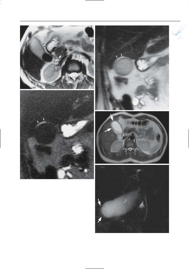
Fig. 98 a , b. Moderately (a) and heavily (b) T2weighted HASTE images showing multiple intramural fluid collections: small intramural abscesses (arrows). Note that, in this case, the imaging features could be confused with those of focal adenomyomatosis (see Fig. 109). c–e Different patient. Pro-
jective image (c), coronal (d) and axial (e) T2weighted HASTE images showing a large intramural fluid collection (large arrow) and an extensive exudate around the gallbladder (small arrows) due to a severe acute cholecystitis with intramural abscess.

4 Gallbladder and Cystic Duct 207
e
f
g
h
Fig. 98 f– i. Different patient. Axial (f) and coronal (g) T2-weighted HASTE images (TE 60) showing marked thickening of the gallbladder wall with intramural fluid and mucosal dissection (arrows). h T1-weighted FLASH image showing slightly hyperintense areas (arrows) along the internal contour, probably representing hemorrhage. i Projective image showing marked thickening of the gallbladder wall with mucosal dissection (arrows). Note that the external contours remain sharply defined, which is i an argument against diffuse mural necrosis

4 Gallbladder and Cystic Duct 209
#100 Complications of Acute
Cholecystitis (3): Perforation
KEY FACTS: DISEASE
●Focal rupture of the gallbladder wall
●Incidence: 3%–15% of patients with acute cholecystitis
●Types:
–Contained (localized) perforation (contained by omentum or adhesions produced by recurrent inflammation)
–Free perforation (less common)
●Location of wall defect: usually in the fundus
●Relative surgical emergency; percutaneous drainage can be useful
●Free perforation is associated with a high mortality rate (up to 30%)
KEY FACTS: MRI (FIG. 100)
●Signs of acute cholecystitis plus focal defect of wall
●Appearance of abscess:
–Well-demarcated fluid-containing lesion
–Usually (not always) relative thick wall
–Communication with the gallbladder lumen may be visible
References
Siskind BN, Hawkins HB, Cinti DC, Zeman RK, Burrell MI (1987) Gallbladder perforation. An imaging analysis. J Clin Gastroenterol 9 : 670–678
Sood B, Jain M, Khandelwal N, Singh P, Suri S (2002) MRI of perforated gall bladder. Australas Radiol 46 : 438–440
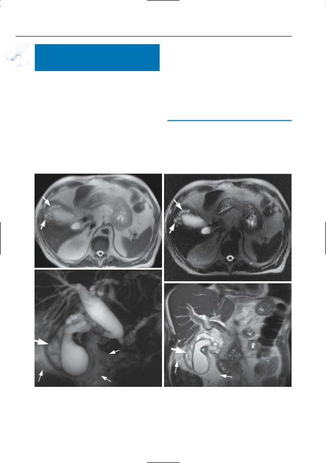
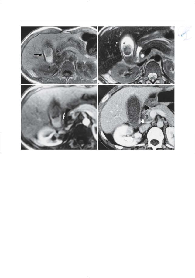
Fig. 100. Sagittal T2-weighted MR image showing fluid collection located between the gallbladder and liver (arrowheads) and communicating with the gallbladder lumen: perforated acute cholecystitis with abscess
210 4.2 Benign Nontraumatic Abnormalities
#101 Acute Emphysematous
Cholecystitis
KEY FACTS: DISEASE
●Acute inflammation with ischemia of the gallbladder wall and complicated by infection with gas-producing organisms
●Organism: Clostridium perfringens
●Mechanism: occlusion of the cystic artery secondary to cystic duct obstruction with edema
●Predisposing factors: diabetes (20%– 30% of cases of acute emphysematous cholecystitis)
●Complications:
–Gangrene (75%)
–Perforation (20%)
–Mortality (15%)
a
KEY FACTS: MRI (FIG. 101)
●Presence of gas:
–In the gallbladder wall
–In the gallbladder lumen
–In the pericholecystic tissue (if perforation has occurred)
–In bile ducts (not obligate)
●Differential diagnosis of intraluminal gas:
–Cholecystoenteric fistula (see # 103)
–Incompetent sphincter of Oddi
–After biliary surgery/ERCP
●Differential diagnosis of intramural gas: air-containing abscess with or without fistula
References
Koenig T, Tamm EP, Kawashima A (2004) Magnetic resonance imaging findings in emphysematous cholecystitis. Clin Radiol 59(5): 455–458
McMillin K (1985) Computed tomography of emphysematous cholecystitis. J Comp Assist Tomogr 9 : 330–332
b
Fig. 101 a, b. a Axial T2-weighted MR image showing a gallbladder with a large stone and an air– fluid level. Also note pericholecystic fluid (arrowheads) and low signal intensity of gallbladder wall
(arrows). b CT image showing the intramural air more clearly (arrows). In this patient, acute emphysematous cholecystitis was complicated by perforation
4 Gallbladder and Cystic Duct 211
#102 Peridiverticulitis
of the Gallbladder
KEY FACTS: DISEASE
●Definition: inflammation of true gallbladder diverticula (i.e., diverticula containing all mural layers, including muscle)
●Incidence: very rare
KEY FACTS: MRI
●Typical features (Fig. 102):
–Signs of acute inflammation (fluid, edema, wall thickening)
–Visualization of one or more diverticula
●Differential diagnosis: adenomyomatosis associated with acute cholecystitis
a
References
Chan-Wilde C, Chew R, Foong WC, Wee A (1990) Adenomyomatosis of the gallbladder: the NUH experience. Ann Acad Med Singapore 19 : 389– 392
Gore RM, Ghahremani GG, Fernbach SK (1994) Gallbladder: anomalies and anatomic variants. In: Gore RM, Levine MS, Laufer I (eds) Textbook of gastrointestinal radiology. Saunders, Philadelphia, pp 1621–1635
b
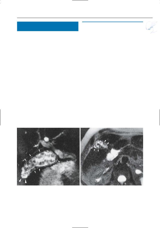
Fig. 102 a, b. a Projective image showing a gallbladder filled with stones. Note the presence of multiple small fluid “collections” within the wall (arrows) and large diverticula in the fundus (arrowheads). b Same patient. Heavily T2-weighted axial image showing diverticular outpouchings in the fundus (arrows) and pericholecystic fluid (arrowheads).
Pathology revealed gallbladder diverticulosis with diverticulitis and mural necrosis. The largest outpouchings located in the fundus probably represent diverticula. The smaller hyperintense areas seen elsewhere in the gallbladder wall may represent either diverticula or areas of necrosis

212 4.2 Benign Nontraumatic Abnormalities
#103 Cholecystoenteric Fistula
KEY FACTS: DISEASE
●Abnormal communication between the gallbladder and digestive tract
●Usually older patients (> 65 years)
●Most common causes:
–Cholecystolithiasis with or without acute cholecystitis (90%)
–Peptic ulcer disease (6%)
–Carcinoma
–Colonic diverticulitis
●Communication with duodenum in 70%, with colon in 26%
●Gallstone ileus is a possible complication:
–Erosion of stone in the gastrointestinal tract
–Stones may be “hung up” at narrow portions of the gastrointestinal tract, e.g.,ligament of Treitz,ileocecal valve, sigmoid colon
KEY FACTS: MRI (FIG. 103)
●Typical features:
–Air in the gallbladder lumen
–Air in the biliary tract if the cystic duct is patent
–Focal or diffuse thickening of the gallbladder wall (depending on underlying disease)
–Inflammatory changes around the gallbladder
–Direct visualization of the fistula
References
Beltran MA, Csendes A (2005) Mirizzi syndrome and gallstone ileus: an unusual presentation of gallstone disease. J Gastrointest Surg 9 : 686–689 Haff RC, Wise L, Ballinger WF (1971) Biliary-enteric
fistulas. Surg Gynecol Obstet 133 : 84–88
4 Gallbladder and Cystic Duct 213
a
b
c |
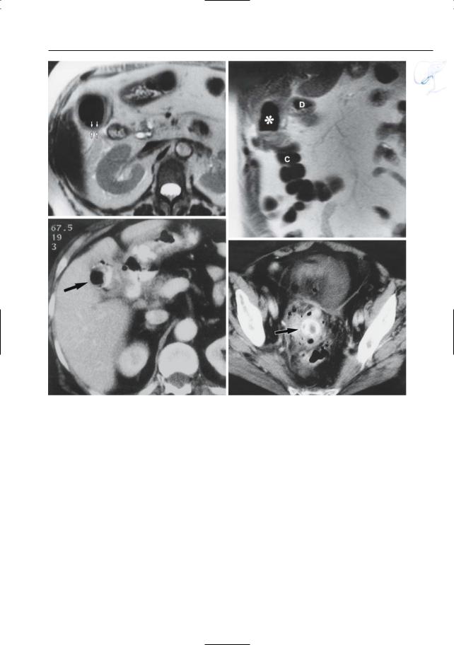
d |
Fig. 103 a–d. Patient with antecedent sigmoid diverticulitis now presenting with signs of obstruction. a Axial and b coronal T2-weighted images showing the gallbladder with a thickened wall (arrows in a) and a markedly hypointense lumen. Note that neither the colon (C) nor the duodenum (D) can be separated from the gallbladder wall in b. Asterisk, air in the gallbladder lumen. c Same patient. CT image showing the gallbladder (arrow) containing
air and orally administered contrast medium, thus suggesting the presence of a cholecystoduodenal fistula. d Same patient. CT image of pelvis showing gallstone impacted in the sigmoid colon (arrow). Note the presence of diverticula and inflammatory changes. The final diagnosis was cholecystitis with cholecystoduodenal fistula and spontaneous migration of a gallstone into the colon

214 4.2 Benign Nontraumatic Abnormalities
#104 Chronic Cholecystitis
KEY FACTS: DISEASE
●Chronic inflammation of the gallbladder wall
●Histology: chronic inflammatory cell infiltrate throughout all the layers of the wall which becomes thickened with fibrosis
●Most common form of gallbladder inflammation
●Causes/associated disease:
–Cholecystolithiasis
–(Intermittent) cystic duct obstruction
●Symptoms:
–Patients may be asymptomatic
–Recurrent right upper quadrant pain
KEY FACTS: MRI (FIG. 104)
●Morphologic features:
–Smooth or irregular thickening of the gallbladder wall
–Gallbladder shrinkage may be a prominent feature (Fig. 104c, d)
●Signal intensity: hypointense aspect of the gallbladder wall and adjacent fat on heavily T2-weighted images (no edema/ fluid; Fig. 104a, b)
●Dynamic contrast-enhanced MRI: smoothly delineated early enhancement of mucosa and muscle and more delayed enhancement of subserosa (fibrosis) (Demachi et al. 1997)
●Decreased contraction after cholecystokinin administration (see # 19)
●Differential diagnosis:
–Acute cholecystitis: presence of fluid/ edema (best seen on heavily T2weighted images)
–Carcinoma: more heterogeneous ! aspect; irregularly delineated enhancement; secondary signs of malignancy
References
Brugge WR, Brand DL,Atkins HL, Lane BP,Abel WG (1986) Gallbladder dyskinesia in chronic acalculous cholecystitis. Dig Dis Sci 31 : 461–467
Demachi H, Matsui O, Hoshiba K et al. (1997) Dynamic MRI using a surface coil in chronic cholecystitis and gallbladder carcinoma: radiologic and histopathologic correlation. J Comp Assist Tomogr 21 : 643–51

4 Gallbladder and Cystic Duct 215
a |
b |
c |
d |
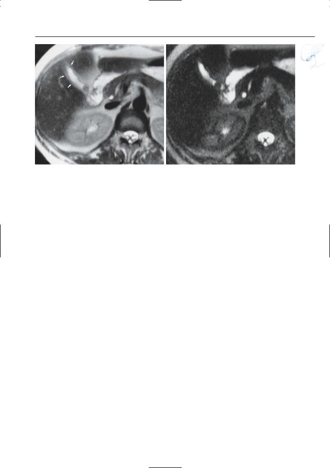
Fig. 104. a, b Moderately T2-weighted MR image showing a thickened gallbladder wall. Note the presence of a thin, hyperintense halo around the gallbladder (arrowheads), possibly fluid or fat. b Heavily T2-weighted image showing that this“halo”and the surrounding fat are isointense, which excludes fluid and suggests chronic (rather than acute)
inflammation. c, d Other patient. c Projective MR image showing a normal cystic duct. The gallbladder is invisible, however (arrow). d Cross-sectional T2-weighted image obtained in the coronal plane showing a shrunken gallbladder with a thickened wall and nearly no intraluminal fluid (arrow), thus explaining the findings in Fig. 104c

216 4.2 Benign Nontraumatic Abnormalities
#105 Xanthogranulomatous
Cholecystitis
KEY FACTS: DISEASE
●Chronic inflammatory disorder of the gallbladder
●Histology:
–Multiple yellow–brown intramural formations, severe proliferative fibrosis, foamy histiocytes
–Newer lesions often contain necrotic areas
●Incidence: 0.7%–10.6%
●Etiology (hypotheses):
–Intramural extravasation of bile with xanthogranulomatous reaction
–Chronic infection
●Clinical presentation: similar to chronic cholecystitis
!● Significance: may simulate gallbladder carcinoma, both clinically and radiologically (Cossi et al. 1987)
KEY FACTS: MRI (FIG. 105)
●Marked wall thickening, usually diffuse
●Signal intensity:
–Dependent on histologic composition
–T1: relatively high signal intensity (fat) (Joerg et al. 1989)
–Areas of isoto slightly high signal on T2-weighted images correspond with areas of abundant xanthogranulomas at pathologic examination
–Areas with very high signal itensity on T2-weighted images correspond with necrosis and/or abscesses
●Contrast-enhanced images: heterogeneous enhancement of the wall
● Differential diagnosis: |
! |
|||
|
|
|||
– |
Abscess (more focal) |
|
|
|
– Tumor (features |
pointing to |
xan- |
|
|
|
thogranulomatous |
cholecystitis |
are |
|
|
the presence of necrotic areas, diffuse |
|||
|
involvement of the gallbladder wall, |
|||
|
and preservation of the mucosal line) |
|||
– |
Adenomymatosis (presence of pseu- |
|||
|
dodiverticula) |
|
|
|
– |
Cholesterolosis |
|
|
|
References
Chun KA, Ha HK,Yu ES et al. (1997) Xanthogranulomatous cholecystitis: CT features with emphasis on differentiation from gallbladder carcinoma. Radiology 203 : 93–97
Cossi AF, Scholz FJ, Aretz HT, Larsen CR (1987) Computed tomography of xanthogranulomatous cholecystitis. Gastrointest Radiol 12 : 154–155
Joerg A, Cussenot O, Houlle D, Despoisse JM, Le Duc A (1989) La pyelonephrite xanthogranulomateuse. Interêt de l’imagerie par resonance magnetique. Ann Urol Paris 23 : 232–235
Shuto R, Kiyosue H, Komatsu E, Matsumoto S, Kawano K, Kondo Y, Yokoyama S, Mori H (2004) CT and MR imaging findings of xanthogranulomatous cholecystitis: correlation with pathologic findings. Eur Radiol 14 : 440–446, Epub 2003 Aug 6

4 Gallbladder and Cystic Duct 217
a |
b |
c |
d |
e |
f |
Fig. 105 a–f. a T1-weighted image showing a thickened gallbladder wall with relatively high signal intensity (arrows). b Heavily T2-weighted image showing several hyperintense foci in the wall (arrows). Also note fluid in the gallbladder fossa, caused by superimposed acute inflammation. c, d Different patient. Axial T2-weighted HASTE
images (TE 60) (c, d) showing marked focal wall thickening of the fundus of the gallbladder (arrows), with intramural fluid collections (arrowheads). e, f Projective images showing focal narrowing of the gallbladder lumen (arrows) near the fundus with intramural fluid collections (arrow-
heads)

218 4.2 Benign Nontraumatic Abnormalities
#106 Cholesterolosis
KEY FACTS: DISEASE
●Benign non-neoplastic, noninflammatory gallbladder abnormality
●Histology:
–Accumulation of foamy, cholesterolladen histiocytes in the subepithelial lamina propria, producing villus-like mucosal protrusions
–The mucosal protrusions may become sufficiently distended to produce tiny macroscopic excrescences or small polyps
●Incidence: common (found in up to 30% of patients undergoing routine cholecystectomy)
●Age/sex distribution unknown
●Types:
–Strawberry gallbladder = diffuse cholesterolosis = lipid cholecystitis = planar form (no luminal protrusions)
–Cholesterol polyp = polypoid form (see # 107)
●No relationship with blood cholesterol or atherosclerosis
●May be a cause of chronic biliary pain (Kmiot et al. 1994)
KEY FACTS: MRI
●No specific MRI features have been described
References
Berk RN,Van der Vegt JH, Lichtenstein JE (1983) The hyperplastic cholecystoses: cholesterolosis and adenomyomatosis. Radiology 146 : 593–601
Kmiot WA, Perry EP, Donovan IA et al. (1994) Cholesterolosis in patients with chronic alcalculous biliary pain. Br J Surg 81 : 112–115
Lubera RJ, Climie ARW, Kling GE (1967) Cholecystitis and the hyperplastic cholesteroloses: a clinical, radiological and pathology study. Am J Dig Dis 12 : 696–704
Salmenkivi K (1964) Cholesterolosis of the gallbladder: a clinical study based on 269 cholecystectomies. Acta Chir Scand 324 [Suppl]: 1–93
4 Gallbladder and Cystic Duct 219
a |
b |
Fig. 106. a Moderately T2-weighted image showing a small gallbladder with a thickened wall with the same intensity as fat (arrows).b Same patient.Heavily T2-weighted image reveals that the wall has a low
signal intensity, thus ruling out acute inflammation. Histology revealed diffuse cholesterolosis. It is un– clear whether the high signal intensity of the gallbladder wall in a is related to the increased fat content
220 4.2 Benign Nontraumatic Abnormalities
#107 Cholesterol Polyp
KEY FACTS: DISEASE
●Polypoid form of cholesterolosis (see # 106)
●90% of all gallbladder polyps
●No malignant potential
KEY FACTS: MRI (FIG. 107)
●Polypoid-like intraluminal lesion, single or (more frequently) multiple
●The diagnosis is especially likely if the lesion is small (< 1 cm) and if multiple polyps are present
●Signal intensity: not known
●Differential diagnosis of solitary cholesterol polyp:
–Adenoma (most common benign neoplasm)
–Carcinoma (15%–25% patients present with a polypoid mass, usually greater than 2 cm)
–Adherent stone
–Metastasis
–Neurinoma, granuloma, hemangioma, etc.
References
Berk RN,Van der Vegt JH, Lichtenstein JE (1983) The hyperplastic cholecystoses: cholesterolosis and adenomyomatosis. Radiology 146 : 593–601
Price RP, Stewart ET, Foley WD, Dodds WJ (1982) Sonography of polypoid cholesterolosis.AJR Am J Roentgenol 139 : 1197–1198
4 Gallbladder and Cystic Duct 221
a
c
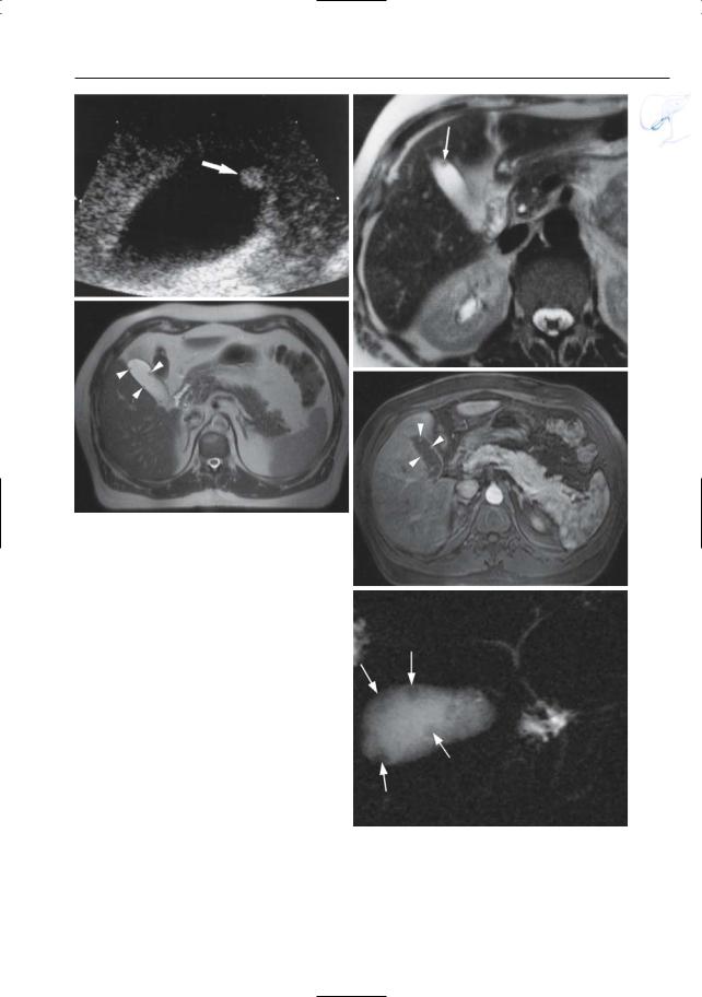
Fig. 107 a, b. Ultrasonography (a) showing a small, nonmobile polypoid structure in the gallbladder lumen (arrow). Note the absence of acoustic shadowing. b T2-weighted MR image showing the lesion as a hypointense nodule in a nondependent portion of the gallbladder lumen (arrow). On this image, it cannot be differentiated from an adherent stone. c–e Different patient. Axial T2-weighted HASTE image (c) showing multiple small polypoid-like lesions in the gallbladder (arrowheads). d On the post gadolinium T1-weighted VIBE image obtained in the arterial phase these lesions show contrastenhancement (arrowheads). e Projective image confirming the presence of multiple intraluminal polyps (arrows)
b
d
e

222 4.2 Benign Nontraumatic Abnormalities
|
|
|
|
|
|
|
KEY FACTS: MRI |
|
|||
|
|
#108 Diffuse Adenomyomatosis |
|
|
|||||||
|
|
|
|
|
|
|
|
||||
|
|
KEY FACTS: DISEASE |
|
|
|
● The diagnosis depends on the simulta- |
|
||||
|
|
|
|
|
|
neous demonstration of: |
|
||||
|
|
|
|
|
|
|
|
|
|||
|
|
|
|
|
|
|
|
|
|||
|
|
● Benign non-neoplastic, noninflamma- |
|
– |
Mural thickening |
|
|||||
|
|
|
– |
Pseudodiverticula |
|
||||||
|
|
tory gallbladder abnormality |
|
|
|
|
|||||
|
|
|
|
|
|
(“pearl necklace sign”) |
|
||||
|
|
● Histology: |
|
|
|
|
|
|
|||
|
|
|
|
|
● |
Visualization of small pseudodiverticula |
|
||||
|
|
– |
Exaggeration of |
the normal |
infold- |
! |
|||||
|
|
|
is only possible when heavily T2-weighted |
||||||||
|
|
|
ings of the luminal epithelium (Roki- |
|
|||||||
|
|
|
|
images are obtained (effective TE as large |
|||||||
|
|
|
tanski-Asshoff |
sinuses) associated |
|
|
|||||
|
|
|
|
as possible; Fig. 108) |
|
||||||
|
|
|
with proliferation of the smooth mus- |
|
|
||||||
|
|
|
● |
Note: If the intramural diverticula are |
|
||||||
|
|
|
cle |
|
|
|
|
||||
|
|
|
|
|
|
|
too small to be visualized by MRI, the |
|
|||
|
|
– |
These infoldings correspond to intra- |
|
|
||||||
|
|
|
appearance is nonspecific |
|
|||||||
|
|
|
mural ‘pseudo’-diverticula (i.e., out- |
|
|
||||||
|
|
|
|
|
|
|
|
||||
|
|
|
pouchings not containing all mural |
|
|
|
|
|
|||
|
|
|
layers) |
|
|
|
|
|
|
|
|
|
|
|
|
|
|
References |
|
||||
|
|
– Occasionally, the diverticula may pass |
|
||||||||
|
|
|
|
|
|
|
|||||
|
|
|
all the way through the wall |
|
|
Haradome H, Ichikawa T, Sou H et al. (2003) The |
|
||||
|
|
● Incidence: 5% of cholecystectomies |
|
pearl necklace sign: an imaging sign of adeno- |
|
||||||
|
|
● Develops with increasing age |
|
|
|
myomatosis of the gallbladder at MR cholan- |
|
||||
|
|
● Ratio of women to men: 3 : 1 |
|
|
|
giopancreatography. Radiology 227 : 80–88 |
|
||||
|
|
|
|
|
|
|
|
|
|||
●Associations:
–Gallstones
–Cholesterolosis in 33%

4 Gallbladder and Cystic Duct 223
a
Fig. 108 a–c. a Ultrasonography showing a thickened gallbladder wall with multiple hyperechoic intramural foci, probably representing diverticula filled with bile concretions (arrowheads). b Same patient. Heavily T2-weighted MR image showing a thickened and remarkably hyperintense gallbladder wall (arrowheads). c Projective image showing several tiny pseudodiverticula (arrow)
b
c

224 4.2 Benign Nontraumatic Abnormalities
#109 Focal Adenomyomatosis
KEY FACTS: DISEASE
●More common than diffuse adenomyomatosis
●Usually located in the fundus
●Cystic spaces are arranged in a circle and may communicate with the lumen through a central umbilicated depression
●Associated disease: gallbladder cancer (found in 6.4% of patients in one series; Ohtani et al. 1992)
KEY FACTS: MRI
●Characteristic signs:
–Multiple diverticular outpouchings at the fundus (Fig. 109)
–Small central sessile “mass” may be seen
–Focal wall thickening
References
Boukadoum M, Siddiky MA, Zerhouni EA, Stitik FP (1984) CT demonstration of adenomyomatosis of the gallbladder. J Comput Assist Tomogr 8 : 177 Gerard PS, Berman D, Zafaranloo S (1990) CT and ultrasound of gallbladder adenomyomatosis mimicking carcinoma. J Comput Assist Tomogr
14 : 490–491
Ohtani T, Shirai Y, Tsukada K, Muto T (1992) Relationship between gallbladder carcinoma and the segmental type of adenomyomatosis of the gallbladder. Cancer 69 : 2647–2652
4 Gallbladder and Cystic Duct 225
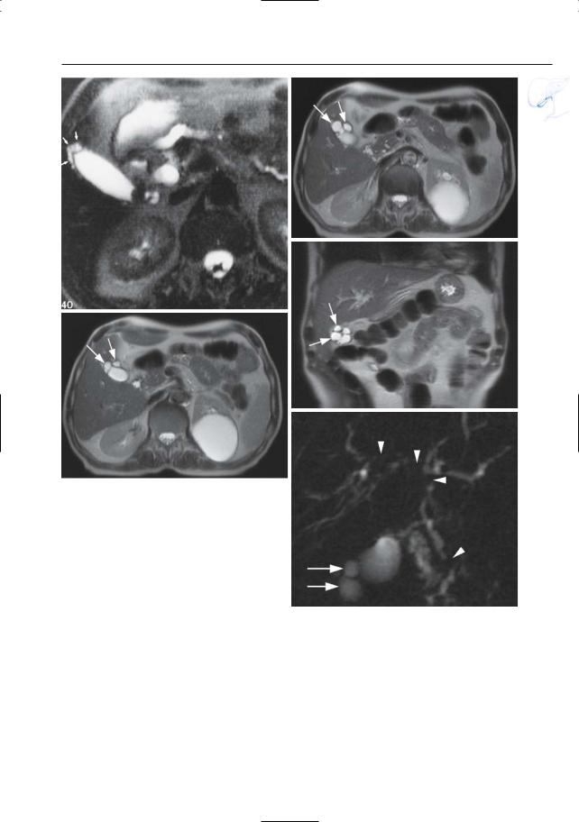
a
c
Fig. 109. a Heavily T2-weighted image showing multiple diverticular outpouchings at the fundus (arrows). b–e Different patient. Axial (b, c) and coronal (d) T2-weighted HASTE images (TE 60) showing multiple cystic spaces located at the fundus of the gallbladder (arrows). e Projective image also showing these cystic spaces (arrows). Note the multiple intraand extrahepatic bile duct stenoses (arrowheads) with segmental irregularity and narrowing (patient with type II PSC)
b
d
e

226 4.2 Benign Nontraumatic Abnormalities
|
|
|
|
|
|
KEY FACTS: MRI |
|
||
|
|
|
#110 Porcelain Gallbladder |
|
|
|
|||
|
|
|
|
|
|
|
|
|
|
|
|
KEY FACTS: DISEASE |
|
|
● If there is marked mural calcification, |
|
|||
|
|
|
|
|
the gallbladder wall appears hypo- |
|
|||
|
|
|
|
|
|
|
|
||
|
|
|
|
|
|
|
|
||
|
|
● |
Chronic calcifying cholecystitis |
|
|
|
intense on T1and T2-weighted images |
|
|
|
|
|
|
|
(Fig. 110) |
|
|||
|
|
● |
Histology: calcium incrustation of the |
|
|
||||
|
|
● |
Note: MRI is less sensitive than CT for |
|
|||||
|
|
|
gallbladder wall, either continuous or |
! |
|||||
|
|
|
|
detecting calcifications (Fig. 110). More- |
|||||
|
|
|
punctate |
|
|
|
|||
|
|
● |
Incidence: 0.6%–0.8% of cholecystec- |
|
over, MRI is less specific: a thickened |
|
|||
|
|
|
wall with low signal intensity may corre- |
|
|||||
|
|
|
tomy patients |
|
|
|
|
||
|
|
|
|
|
|
spond to fibrosis, intramural air (see |
|
||
|
|
● |
Ratio of women to men: 5 : 1 |
|
|
|
|
||
|
|
|
|
|
# 101), and calcifications |
|
|||
|
|
● Association: gallstones nearly |
always |
|
|
||||
|
|
|
|
|
|
||||
|
|
|
present |
|
|
|
|
|
|
|
|
● |
Symptoms: minimal |
|
|
|
|
|
|
|
|
|
|
References |
|
||||
|
|
● Complication: gallbladder carcinoma |
|
||||||
|
|
|
|
|
|
||||
|
|
|
(up to 22%; see # 115) |
|
|
Berk RN, Armbuster TG, Salzstein SL (193) Carci- |
|
||
|
|
|
|
|
|
|
noma in the porcelain gallbladder. Radiology |
|
|
|
|
|
|
|
|
|
106 : 29–31 |
|
|
|
|
|
|
|
|
Kane RA, Jacobs R, Katz J, Costello P (1984) Porce- |
|
||
lain gallbladder: ultrasound and CT appearance. Radiology 152 : 137–141
Rickes S, Ocran K (2002) Images in clinical medicine. Porcelain gallbladder. N Engl J Med 346 : e4

4 Gallbladder and Cystic Duct 227
a |
b |
Fig. 110. a T2-weighted MR image showing a thikkened gallbladder wall with very low signal intensity (arrows). b Corresponding CT image showing
extensive calcification of the gallbladder wall. Note ascites as an incidental finding

228 4.2 Benign Nontraumatic Abnormalities
#111 Reactive Thickening
of the Gallbladder Wall
Related topics: #34 (acute hepatitis), #36 (cirrhosis), # 151–157 (acute pancreatitis)
KEY FACTS: DISEASE
●Extrinsic causes of wall thickening include:
–Acute hepatitis (see #34)
–Hypoalbuminemia
–Ascites
–Cirrhosis (see #36)
–Veno-occlusive disease
–Heart failure
–Acute pancreatitis
●Differential diagnosis: contracted gallbladder after eating, cholecystitis, tumor,AIDS, primary sclerosing cholangitis, …
KEY FACTS: MRI (FIG. 111)
●Typical features (reflecting diffuse mural edema):
–Homogeneously thickened wall
–High signal intensity on T2-weighted images
–Preservation of mucosal line
●Differential diagnosis: distinguishing features:
–With tumor and xanthogranulomatous cholecystitis: homogeneous aspect of thickened wall
–With acute cholecystitis: concentric “symmetric” aspect of thickened wall; more homogeneous aspect; absence of pericholecystitic fluid; small size of the gallbladder lumen
●Note: diffuse mural edema may be caused by a shift of fluid from the intravascular to the extravascular space (venous congestion, hypoproteinemia) or to passive diffusion of extracellular fluid
References
Shlaer W, Leopold GR, Scheible FW (1981) Sonography of thickened gallbladder wall: a non-specific finding. AJR Am J Roentgenol 136 : 337–339

4 Gallbladder and Cystic Duct 229
a
Fig. 111 a–b. Patient with acute hepatitis. a T1and b T2-weighted images showing marked thickening and edema of the gallbladder wall (arrowheads). c Different patient.Axial T2-weighted HASTE image (TE 60) show diffuse ascites with secondary thickening of the gallbladder wall
b
c
230 4.2 Benign Nontraumatic Abnormalities
#112 Varices
in the Gallbladder Wall
Related topic: # 79 (extrahepatic bile duct, other benign causes of bile duct narrowing)
KEY FACTS: DISEASE
●Causes:
–Portal vein thrombosis
–Chronic portal hypertension
●Mechanisms:
–Increased “backward” pressure in the cystic vein
–Veins in the gallbladder wall may act as a bypass around a focally thrombosed extrahepatic segment of the portal vein (blood flows from the cystic vein to gallbladder varices to hepatic parenchyma)
KEY FACTS: MRI (FIG. 112)
●Features on contrast-enhanced images:
–Thickening of the gallbladder wall
–Presence of multiple serpiginous vessels
References
Marchal G, Van Holsbeeck M, Tshibwabwa-Ntumba E et al. (1985) Dilation of the cystic veins in portal hypertension: sonographic demonstration. Radiology 154 : 187–189
West MS (1991) Gallbladder varices: imaging findings in patients with portal hypertension. Radiology 179 : 179–182
4 Gallbladder and Cystic Duct 231
a
c
Fig. 112. a Patient with thrombosis of the portal vein. Contrast-enhanced T1-weighted image showing venous collaterals within the wall of the gallbladder (arrows). b–e Different patient. Axial T2weighted HASTE image (b) and T1-weighted FLASH image (c) showing diffuse thickening of the gallbladder wall (arrows). Axial post-gadolinium T1weighted VIBE image obtained in the venous phase (d) showing multiple small serpiginous vessels in the gallbladder wall (arrowheads). Axial post-gado- linium T1-weighted VIBE image obtained at the level of the liver hilum (e) showing collateral venous branches [arrow in (e)] together with an apparently normal portal venous segment
b
d
e

232 4.3 Traumatic, Postoperative, and Iatrogenic Abnormalities
4.3Traumatic, Postoperative, and Iatrogenic Abnormalities
#113 Blunt Gallbladder Trauma
Related topics: #48 (sequelae of direct liver trauma), # 181 (pancreatic duct injury)
KEY FACTS: DISEASE
●Gallbladder injury occurs in 2%–3% of patients with severe blunt abdominal trauma
●Types of injury:
–Contusion (i.e., intramural/intraluminal bleeding without rupture)
–Laceration (i.e., perforation of the wall)
–Avulsion (i.e., disconnection of the vascular pedicle)
●Associations:
–Liver laceration
–Duodenal rupture
KEY FACTS: MRI
●Common findings:
–Blood in the gallbladder lumen (signal intensity: see #51; Fig. 113)
–Ill-defined contour of the gallbladder wall with or without focal defect
–Fluid/blood around the gallbladder
●Other possible findings:
–Absence of enhancement of the gallbladder wall (avulsion)
–Pseudoaneurysm (rare) (Fig. 113)
–Collapsed gallbladder (rupture)
References
Jeffrey RB Jr, Federle MP, Laing FC,Wing VW (1986) Computed tomography of blunt trauma to the gallbladder. J Comput Assist Tomogr 10 : 756–758 Kim PN, Lee KS, Kim IV, Bae WK, Lee BH (1994) Gallbladder perforation: comparison of US find-
ings with CT. Abdom Imaging 19 : 239–242
4 Gallbladder and Cystic Duct 233
a |
b |
c |
d |
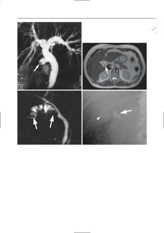
Fig. 113 a–d. Patient who was victim of a blunt abdominal trauma. a T1and b T2-weighted images showing mixed signal intensity of the gallbladder content: hematoma with clot formation (arrow). Note marked thickening of the gallbladder wall in b (arrowheads). c Contrast-enhanced MR image
obtained in the late arterial phase showing an enhancing structure in the gallbladder wall (arrow): small post-traumatic pseudoaneurysm. d CT confirming the presence of a small pseudoaneurysm (arrow). (Figure courtesy of D. Perdieus)

234 4.3 Traumatic, Postoperative, and Iatrogenic Abnormalities
#114 After Cholecystectomy: Complications
with Cystic Duct Remnant
Related topics: #52 (after cholecystectomy: stricture/transection of an aberrant bile duct), #82, 83 (extrahepatic duct: complications after cholecystectomy), #84 (postcholecystectomy syndrome)
KEY FACTS: DISEASE
●After cholecystectomy, almost 50% of patients have a cystic duct remnant measuring between 1 and 6 cm in length
●Complications with the cystic duct remnant account for 10% of reoperations after cholecystectomy
●Most common problems in the early postoperative phase:
–Retained lithiasis
–Fistula
–Bile leakage
●Most common problems in the late postoperative phase:
–Retained/recurrent lithiasis
–Neurinoma, suture granuloma
–“Recurrent gallbladder”: large cystic duct stumps can function like a residual gallbladder with inflammatory changes. This typically causes lateonset pain several months or years after cholecystectomy
KEY FACTS: MRI
●Most common imaging findings:
–Early phase: fluid collection around the cystic duct remnant (leakage)
–Early and late phase: stone in the cystic duct remnant (Fig. 114)
References
Bodvall B, Overgaard B (1966) Cystic duct remnant after cholecystectomy: incidence studied by cholangiography in 500 cases and significance in 103 reoperations. Ann Surg 163 : 382–390
Rubini G, Dimonte M (1999) Postcholecystectomy syndrome: evaluation by biliary cholescintigraphy and MR cholangiopancreatography. Clin Nucl Med 24 : 784–788
4 Gallbladder and Cystic Duct 235
a |
b |
c |
d |
Fig. 114. a Projective MR image showing a small stone in the cystic duct remnant (arrow). b–d Different patient. Axial T2-weighted HASTE image (b), projective image (c), and conventional X-ray image
(d), showing multiple stones in the cystic duct remnant after cholecystectomy (arrows). Note the operation clips in (d) (arrowhead)