

44 |
3 Imaging Differential Diagnosis of Pediatric CNS Tumors |
responsive tumors in a delicate localization. Secreting and mixed germ cell tumors usually contain teratoma parts, which are inhomogeneous tumors containing larger cysts (Fig. 3.22i, j). After treatment usually the teratoma part persists without further change and has to be resected to achieve complete response. However, surgery is usually easier after a significant reduction of size [92].
A frequent and characteristic way of dissemination is an extension along the ventricular borders subependymally or along the optic nerve sheaths [93] (Fig. 3.22k, l). Germ cell tumors with a similar extension are considered as disseminated in the ongoing SIOP CNS-GCT study even if there is a direct contact to the primary tumor.
As germ cell tumors affect structures that physiologically enhance, like the pituitary stalk and the pineal gland which do not have a clear cutoff to a normal size in an individual patient, it is sometimes hard or even impossible to separate a very good partial response with a nearly complete regression of the tumor from a true complete response. We solve this problem for the ongoing SIOP-CNS-GCT study with a follow-up examination. If there is a further reduction during treatment we then retrospectively can diagnose a very good partial response. But if the lesion remains in the same size we can conclude that this was a true complete response already at the time of the previous MRI (Fig. 3.22m–o). Contrast enhancement is reliable and if enhancement is completely gone we diagnose a complete remission even if T2-signal abnormalities persist and are then considered scar tissue.
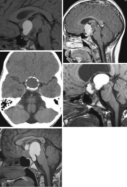
Germ cell tumors of the suprasellar region may be slowly growing and frequently cause a disturbance of the posterior pituitary lobe resulting in the clinical picture of a diabetes insipidus [94–96]. As the size of the pituitary stalk cannot be predicted in an individual patient, sometimes a slowly growing borderline or slightly thickened stalk is seen on MRI [97]. An indication of tumor growth in the pituitary stalk is the loss of the normal bright signal of the posterior pituitary lobe on unenhanced T1-weighted images [98] (Fig. 3.22p, q). We have the impression that small germ cell tumors arise from the posterior pituitary lobe and grow from there upward to the floor of the third ventricle. The displaced and compressed anterior pituitary lobe can be visualized in front of the tumor in early cases (Fig. 3.22r, s). In our database we did not find the posterior bright spot in nearly all germ cell tumors in suprasellar localization. Unfortunately, this diagnostic tool can only be used if imaging is performed according to our guideline of an ideal imaging in suprasellar lesions including sagittal thin slice T1-sequences before the injection of contrast medium. We prefer SE or TSE in 2–3 mm thickness to MPR sequences even if thinner slice lengths are possible. (See Sect. 3.2.)
3.6Craniopharyngiomas
In the suprasellar region, craniopharyngiomas are the most frequent nonglial tumors in children. Boys and girls are equally affected and the peak incidence is in the first decade. Craniopharyngiomas are rare tumors representing only 1.2–4.6 % [99] of intracranial tumors at all. They are divided into two histological subtypes with
3.6 Craniopharyngiomas |
45 |
different origin, development, and morphology: the adamantinous and the papillary subtype. The later is presumably induced by metaplastic changes of buccal mucosa premordia in the Rathke cleft and pouch and affects only adults with a peak incidence in the fifth and sixth decade. Adamantinous craniopharyngiomas are found in all ages but are the only craniopharyngiomas in children and adolescents. Their origin is cell remnants of teeth premordia in the Rathke pouch and duct, a tubular later in development involuted structure. Through Rathke’s duct, the anterior pituitary forms meeting the posterior pituitary an evagination from the floor of the third ventricle to form the united pituitary gland [100]. The typical localization of craniopharyngiomas is the intraand suprasellar region. Less frequently they arise only in the suprasellar position and even rarer exclusively intrasellarly (intrasellar 4 %, suprasellar 25 %, and intra-/suprasellar 71 % in our database). Exceptionally rare and unexplained are anecdotal reports of craniopharyngiomas arising in other localizations as, e.g., the fourth ventricle. Craniopharyngiomas are malformative tumors and do not disseminate. Rare reports on second tumors after resection mainly in the surgical access are thought to arise from implantation of tumor during surgery [101]. This can also explain drop “metastases” in the spinal canal. Adamantinous craniopharyngiomas are predominantly cystic tumors. Macroscopically the cysts frequently contain a fluid resembling machine-oil called colloid. On MRI colloid is characterized by a hyperintense signal on T1-weighted images before contrast enhancement and is quite characteristic (Fig. 3.23a). Cyst walls usually enhance and the histological hallmark of adamantinous craniopharyngiomas so-called wet keratin eventually calcifies resulting to a high proportion of calcifications (91 % in our database; Fig. 3.23b). A so-called 90 %-rule defines 90 % of cysts, calcifications, and cyst wall enhancement in adamantinous craniopharyngiomas [102] (Fig. 3.23c). On the contrary papillary tumors, which do not occur during childhood, do not calcify and are predominantly solid [59].
As in the other suprasellar tumors the bright pituitary spot is of differential diagnostic value. However, it depends on the individual localization and the size of the tumor if the bright posterior spot on unenhanced T1-weighted images can be seen (Fig. 3.23d, e). Usually the size of the pituitary is abnormally atrophic corresponding to a smaller size of children compared to normal controls already several years before the diagnosis of the tumor [103].
Rarely other lesions affect the pituitary and the stalk. Inflammations like hypophysitis are rare and cannot be diagnosed without histology. Langerhans cell histiocytosis (LCH) can affect the pituitary stalk and is an unclear disease (Fig. 3.24a). In case of a suspicion, a whole body MRI should be initiated to rule out the most frequently coexisting bone lesions. If they are not found, LCH is not a probable diagnosis. Hypothalamic hamartomas are rare malformations resembling abnormal brain with signal intensities comparable to gray matter (Fig. 3.24b).
The differential diagnoses to craniopharyngiomas besides true tumors like chiasmal LGGs and germ cell tumors are Rathke-cleft cysts and xanthogranulomas. Rathke cleft cysts can imitate small craniopharyngiomas perfectly and are cystic lesions in an intra-/suprasellar position (Fig. 3.24c). Xanthogranulomas are not considered tumors [104] and therefore not included in the WHO classi-
46 |
3 Imaging Differential Diagnosis of Pediatric CNS Tumors |
a |
b |
c
d
e
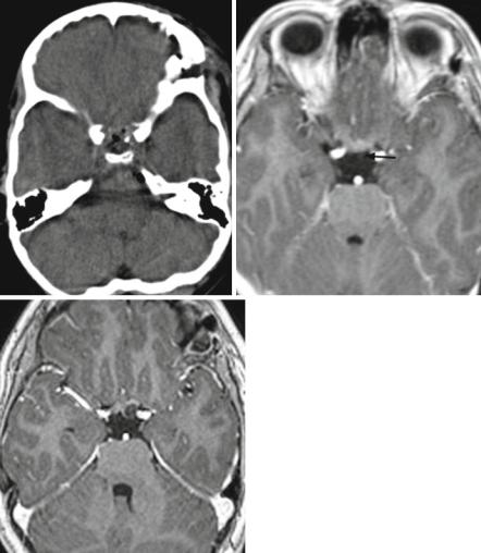
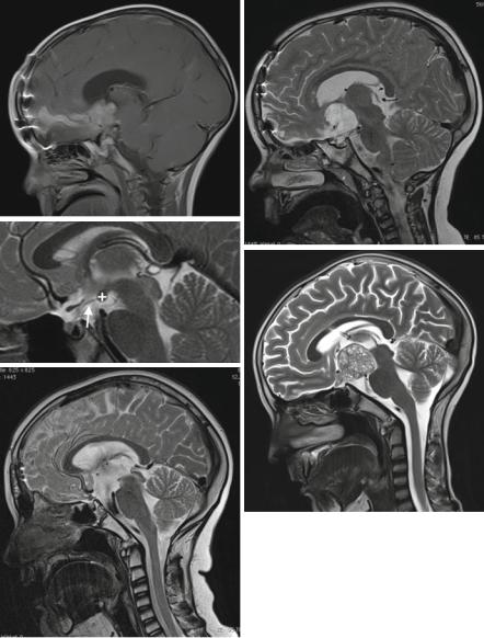
Fig. 3.23 Colloid is the content of one of the multiple cysts in a large craniopharyngioma (a). The walls of cysts show enhancement (b). Thick ring-like calcification on CT (c) in the cyst walls. Coarse calcifications are characteristic of adamantinous craniopharyngiomas. The loss of the bright posterior pituitary spot in a very atrophic pituitary gland is caused by a suprasellar craniopharyngioma (d). In another patient despite a large suprasellar craniopharyngioma the bright spot on unenhanced T1-weighted MRI (arrow) is preserved (e)

3.6 Craniopharyngiomas |
47 |
fication. They can be found in other parts of the brain, skull base, and body and are characterized by residues of bleeding leading to a primarily high signal on T1-weighted images in all our cases (Fig. 3.24d). However, a differentiation from colloid is not possible and small craniopharyngiomas cannot be distin-
a |
b |
c
d
Fig. 3.24 A Langerhans cell histiocytosis in the suprasellar area cannot be differentiated from a germ cell tumor (a). Note also the pituitary atrophy. Search for additional bone and organ lesions. In a hypothalamic hamartoma, a mass with irregular gyrus-like pattern mimics abnormal brain (b). Rathke cysts (c) are intraand suprasellar cystic lesions that cannot be differentiated from small purely cystic craniopharyngiomas. In xanthogranulomas (d) of the intraand suprasellar region the high signal on unenhanced T1-weighted MRI is characteristic. On pathology this is explained by blood degradation products (met-hemoglobin). Development of a recurrence around a tiny residual calcification, visible on a plain CT (e) but not seen on the postoperative MRI (f) after resection of an adamantinous craniopharyngioma. The cystic recurrence (g) evolved exactly in the position of the residual calcification several months after surgery
48 |
3 Imaging Differential Diagnosis of Pediatric CNS Tumors |
e |
f |
g
Fig. 3.24 (continued)
guished. In a report from Japan [105], a lack of calcification in five xanthogranulomas was thought to be a possibility for a differentiation from craniopharyngiomas. However, in our eight CTs of xanthogranulomas, we found five without and three containing calcifications. Thus the lack or existence of calcifications does not seem to help.
The regular early postoperative MRI (performed within 24–48 h after surgery, see also the chapter postoperative MRI) is frequently rather unclear, because craniopharyngiomas like pituitary adenomas or meningeomas are extraaxial
3.6 Craniopharyngiomas |
49 |
tumors and the dura in and surrounding the surgical region shows immediate enhancement and may lead to the false impression of a residual tumor. We postpone the decision on a residual tumor in unclear MRI images to 2–3 months after surgery. Even if the postoperative MRI does not show a suspicion of a residual tumor, a residual calcification may remain undetected by MRI. The guidelines for imaging in the ongoing craniopharyngioma study contain the advice to perform a postoperative, unenhanced CT only of the tumor region avoiding to touch the eye lenses. With the means of this CT in a tumor free postoperative MRI a persisting calcification should be picked up under the assumption that a residual calcification signifies a residual tumor. Ellioth and coworkers [106] discussed this problem and reported that small residual calcifications (<2 mm) do not lead to an increased rate of relapses compared to postoperative sites without residual calcifications. We cannot contradict because in our database postoperative CTs are only scarce despite the fact that the postoperative MRI did not show a residual tumor. Contrary to the study guidelines, CTs are not performed fearing the inherent radiation in pediatric patients. However, we saw one patient with a tiny calcification, which was not detectable on his tumor-free postoperative MRI. In this patient, after some months a relapse occurred exactly around the tiny calcification (Fig. 3.24e–g).
Craniopharnygiomas tend to compromise the hypothalamus. Damage to this brain area either by the tumor itself or the surgical resection frequently leads to severe impairment of the quality of life by development of massive obesity. Adipositas together with hormonal deficiencies leads to severe metabolic syndromes and early deaths mostly from cardiovascular complications. It was recognized that patients with a compromise of the posterior hypothalamic nuclei are in danger of more severe side effects than those with a compression or damage of the anterior hypothalamus [107]. The surgical attitude to achieve a complete resection (whenever possible) is now changing to a staged resection or cysts treatment without touching the posterior hypothalamus (whenever possible) to avoid these devastating consequences [108] (Fig. 3.25a, b). Several classifications exist to identify the high-risk group [109, 110]. In the German craniopharyngioma-studies we use the level of the mammillary bodies to separate the anterior from the posterior hypothalamus (Fig. 3.25c). The increase in the body mass index (BMI) correlates well to the thickness of the nuchal fat fold that can be measured on T1-weighted images on MRI (personal communication Prof. H. Müller Oldenburg, Germany, leader of the German craniopharyngioma study). Unfortunately, we also see increasingly frequently such a rapidly developing subcutaneous fat depositions in children after partial resections of chiasmatic gliomas or germ cell tumors (Fig. 3.25d, e). The rapid development of adipositas in operated chiasmal glioma or germ cell tumor patients is probably also due to a damage of the hypothalamic nuclei in analogy to craniopharyngiomas.
50 |
3 Imaging Differential Diagnosis of Pediatric CNS Tumors |
a |
b |
c
d
e