

C H A P T E R 1 1
Diencephalon
Objectives
1.List the thalamic nuclei and attribute at least one main functional association to each.
2.Describe the internal capsule’s parts and what fibers travel within each.
3.Identify the internal capsule and surrounding structures on an image or diagram.
4.List the hypothalamic nuclei and attribute at least one main functional association to each.
5.Describe the anatomy of the hypothalamus, its boundaries and various divisions.
6.Describe the “systems” associated with hypothalamic regions, for example, the heat regulation or satiety centers.
IIntroduction. The diencephalon is divided into four parts: the subthalamus,
epithalamus, dorsal thalamus (i.e., the thalamus), and the hypothalamus. The epithalamus includes the pineal gland, which in humans has a role in circadian rhythms and reproductive cycles and the habenula, which has connections between the basal nuclei, limbic system, and brainstem reticular formation. The subthalamus is region that is essentially a continuation of the midbrain tegmentum, the main component is the subthalamic nucleus, which functions as
part of the basal nuclei.
II |
The thalamus is the largest division of the diencephalon. It plays an important role in |
|
the integration of the sensory and motor systems. |
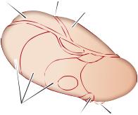
Major Thalamic Nuclei and Their Connections (Figure 11-1)
A.The anterior nucleus receives hypothalamic input from the mammillary nucleus through the mammillothalamic tract. It projects to the cingulate gyrus and is part of the Papez circuit of emotion of the limbic system.
B.The mediodorsal (dorsomedial) nucleus is reciprocally connected to the prefrontal
cortex. It has abundant connections with the intralaminar nuclei. It receives input from the amygdala, substantia nigra, and temporal neocortex. When it is destroyed, memory loss occurs (Wernicke– Korsakoff syndrome). The mediodorsal nucleus plays a role in the expression of affect, emotion, and behavior (limbic function).
C.The centromedian nucleus is the largest intralaminar nucleus. It is reciprocally connected to the motor cortex (Brodmann area 4). The centromedian nucleus receives input from the globus pallidus. It projects to the striatum (caudate nucleus and putamen) and projects diffusely to the entire neocortex.
92
Diencephalon 93
Ant. nuclear group |
Internal medullary lamina |
|
|
|
Mediodorsal nucleus |
VA |
MD |
LD |
|
|
VL |
LP |
|
|
||
VPL |
|
|
VPM |
|
|
Ventral tier nuclei |
Medial geniculate body |
|
Lat. geniculate body |
||
|
Figure 11-1 Major thalamic nuclei and their connections. A. Dorsolateral aspect and major nuclei. LD, lateral dorsal nucleus; LP, lateral posterior nucleus; MD, medial dorsal nucleus; VA, ventral anterior nucleus; VL, ventral lateral nucleus; VPL, ventral posterior lateral nucleus; VPM, ventral posterior medial nucleus.
D.The pulvinar is the largest thalamic nucleus. It has reciprocal connections with the association cortex
of the occipital, parietal, and posterior temporal lobes. It receives input from the lateral and medial geniculate bodies and the superior colliculus. It plays a role in the integration of visual, auditory, and somesthetic input. Destruction of the pulvinar may result in sensory dysphasia.
E.Ventral Tier Nuclei
1.The ventral anterior nucleus receives input from the globus pallidus and substantia nigra. It projects diffusely to the prefrontal cortex, orbital cortex, and premotor cortex (Brodmann area 6).
2.The ventral lateral nucleus receives input from the cerebellum (dentate nucleus), globus pallidus, and substantia nigra. It projects to the motor cortex (Brodmann area 4) and the supplementary motor cortex (Brodmann area 6).
3.The ventral posterior nucleus is the nucleus of termination of general somatic afferent (touch, pain, and temperature) and special visceral afferent (taste) fibers. It has two subnuclei:
a.Ventral posterolateral nucleus receives the spinothalamic tracts and the medial lemniscus. It projects to the somesthetic (sensory) cortex (Brodmann areas 3, 1, and 2);
b.Ventral posteromedial (VPM) nucleus receives the trigeminothalamic tracts and projects to the somesthetic (sensory) cortex (Brodmann areas 3, 1, and 2). The gustatory (taste) pathway originates in the solitary nucleus and projects via the central tegmental tract to the VPM and thence to the gustatory cortex of the postcentral gyrus, of the frontal operculum, and of the insular cortex. The taste pathway is ipsilateral.
4.The lateral geniculate body is a visual relay nucleus. It receives retinal input through the optic tract and projects to the primary visual cortex (Brodmann area 17).
5.The medial geniculate body is an auditory relay nucleus. It receives auditory input through the brachium of the inferior colliculus and projects to the primary auditory cortex (Brodmann areas 41 and 42).
F.The reticular nucleus of the thalamus surrounds the thalamus as a thin layer of γ-amino- butyric acid (GABA)-ergic neurons. It lies between the external medullary lamina and the internal capsule. It receives excitatory collateral input from corticothalamic and thalamocortical fibers. It projects inhibitory fibers to thalamic nuclei, from which it receives input.
94 Chapter 11
IIIBlood Supply. The thalamus is irrigated by the following three arteries (see Figure 4-1):
A.Posterior Communicating Artery
B.Posterior Cerebral Artery
C.Anterior Choroidal Artery (Lateral Geniculate Body)
IV The Internal Capsule (Figure 11-2) is a layer of white matter (myelinated axons) that separates the caudate nucleus and the thalamus medially from the lentiform nucleus laterally. It can be divided into five parts, the:
1.Anterior limb is located between the caudate nucleus and the lentiform nucleus (globus pallidus and putamen). It contains fibers that interconnect the anterior nucleus and the cingulate gyrus, as well as fibers connecting the dorsomedial nucleus with the prefrontal cortex. Finally, it contains frontopontine fibers.
2.Genu is located near the interventricular foramen and contains corticonuclear fibers.
3.Posterior limb is located between the thalamus and the lentiform nucleus. It contains fibers
that connect the VA and VL nuclei with motor cortex, as well as fibers connecting the VP nuclei to somatosensory cortex. Descending fibers include corticospinal (pyramid) and corticonuclear fibers.
4. Retrolenticular part is composed of fibers passing posteriorly to the lentiform nucleus. This includes the optic radiations and fibers that interconnect the pulvinar nucleus with
parietal and occipital association cortices.
Caudate nucleus |
Anterior limb |
|
|
Genu |
Globus pallidus |
|
|
Corticonuclear fibers |
|
|
Putamen |
Posterior limb |
|
|
Corticospinal fibers |
Thalamus |
Auditory radiation (sublenticular |
|
|
Sensory radiations from |
part of internal capsule) to |
VP nucleus to areas 3, 1, 2 |
superior temporal gyrus |
|
(areas 41 and 42) |
Medial geniculate body |
|
(audition) |
|
Lateral geniculate body |
Visual radiation (retrolenticular portion |
(vision) |
of internal capsule) to striate cortex of |
|
occiptal lobe (area 17) |
Figure 11-2 Horizontal section of the right internal capsule showing the major fiber projections. Clinically important tracts lie in the genu and posterior limb. Lesions of the internal capsule cause contralateral hemiparesis and contralateral hemianopia. VP, ventral posterior.

Diencephalon 95
5.Sublenticular part is located inferior to the lentiform nucleus. Sublenticular fibers are composed of the remaining optic radiations, auditory radiations, and interconnections between the temporal association cortices and the pulvinar.
D.Blood Supply to the Internal Capsule
1.The anterior limb is irrigated by the medial striate branches of the anterior cerebral artery and the lateral striate (lenticulostriate) branches of the middle cerebral artery.
2.The genu is perfused either by direct branches from the internal carotid artery or by pallidal branches of the anterior choroidal artery.
3.The posterior limb is supplied by branches of the anterior choroidal artery and lenticulostriate branches of the middle cerebral arteries.
4.The anterior choroidal supplies most of the blood to the retroand sublenticular parts of the internal capsule.
VThe hypothalamus, the inferiormost division of the diencephalon, subserves
three systems: the autonomic nervous system, the endocrine system, and the limbic system. The hypothalamus helps to maintain homeostasis. It is bilateral structure, with the inferior recess of the third ventricle intervening between its left and right sides.
A.Major Hypothalamic Nuclei and Their Functions
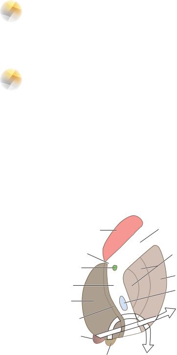
1.The medial preoptic nucleus (Figure 11-3) regulates the release of gonadotropic hormones from the adenohypophysis. It contains the sexually dimorphic nucleus, the development of which depends on testosterone levels.
2.The suprachiasmatic nucleus receives direct input from the retina. It plays a role in the regulation of circadian rhythms.
3.The anterior nucleus plays a role in temperature regulation. It stimulates the parasympathetic nervous system. Destruction results in hyperthermia.
Paraventricular and supraoptic nuclei ssREGULATE waTEREBALANCe ssPRODUCED!$($AND oXYTOCIN ssdestrUCTION CAUSES DIABETES INSIPIDUS ssparavENTRICULAR NUCLEUS PROJECTSETO AUTONOMICNNUCLEI OF BRAINSTEMEAND SPINAL CORD
Anterior nucleus ssTHERMALAREGULATIONDISSIPATION OFAHEAT) ssSTIMULATES PARASYMPATHETIC .3
ssdestrUCTION RESULTS INEHYpertHERMIA
Preoptic area 
ssCONTAINS SEXUALLY DIMORPHIC NUCLEUS ssREGULATESLRELEASESOF GONADOTROPIC HORMONEs
Suprachiasmatic nucleus ssRECEIVES INPUT FROMURETINa ssCONTROLS CIRCADIAN RHYTHMS
Dorsomedial nucleus
ssSTIMULATIONLRESULTSOIN OBESITY AND SAvAGEGBEHAVIOr
Posterior nucleus
ssTHERMALAREGULATION CONSERvaTION OFNHEAT) ssdestrUCTION RESULTS INEINABILITYSTO THERMOREGULATe ssSTIMULATES THE SYMPATHETIC .3
Lateral nucleus ssSTIMULATIONLINDUCESOEATINg ssdestrUCTION RESULTS INESTArvATION
Mammillary body ssRECEIVES INPUT FROM HIPPOCAMPALCFOrMATION VIAIFORNIX
-IDBRAIN ssPROJECTS TOCANTERIORONUCLEUS OF THALAMUS
ssCONTAINS HEMORRHAGIC LESIONs
IN WerNICKE’s ENCEPHALOPATHY
#. )))
Ventromedial nucleus
PoNS ssSATIETYICENTER
 ssdestrUCTION RESULTS INEOBESITy
ANDNSAvAGEGBEHAVIOr
Arcuate nucleus
ssPRODUCES HYPOTHALAMICARELEASINGCFACTORs
ssCONTAINS $/PA-ergIC NEURONS THATOINHIBITTPROLACTINIRELEASe
Figure 11-3 Major hypothalamic nuclei and their functions. ADH, antidiuretic hormone; CN, cranial nerve; DOPA, dopamine; NS, nervous system.

96 Chapter 11
 Paraventricular nucleus
Supraoptic nucleus 

Optic chiasm
Superior hypophyseal artery
Hypophyseal portal veins
Anterior lobe (adenohypophysis) 
Third
ventricle
Arcuate (tuberal) nucleus
 Tuberohypophseal tract
 Supraopticohypophseal tract
 Infundibulum
 Sinusoids of infundibular stem
Oxytocin ADH
 Posterior lobe (neurohypohysis)
 Hypophyseal vein
Inferior hypophyseal artery
Figure 11-4 The hypophyseal portal system. The paraventricular and supraoptic nuclei produce antidiuretic hormone (ADH) and oxytocin and transport them through the supraopticohypophysial tract to the capillary bed of the neurohypophysis. The arcuate nucleus of the infundibulum transports hypothalamic-stimulating hormones through the tuberohypophysial tract to the sinusoids of the infundibular stem. These sinusoids then drain into the secondary capillary plexus in the adenohypophysis.
4.The paraventricular nucleus (Figure 11-4) synthesizes antidiuretic hormone (ADH), oxytocin, and corticotropin-releasing hormone. It gives rise to the supraopticohypophyseal tract, which projects to the neurohypophysis. It regulates water balance (conservation) and projects directly to the autonomic nuclei of the brain stem and all levels of the spinal cord. Destruction results in diabetes insipidus.
5.The supraoptic nucleus synthesizes ADH and oxytocin (similar to the paraventricular nucleus).
6.The dorsomedial nucleus, when stimulated in animals, results in savage behavior.
7.The ventromedial nucleus is considered a satiety center. When stimulated, it inhibits the urge to eat. Bilateral destruction results in hyperphagia, obesity, and savage behavior.
8.The arcuate (infundibular) nucleus contains neurons that produce factors that stimulate or inhibit the action of the hypothalamus. This nucleus gives rise to the tuberohypophysial tract, which terminates in the hypophyseal portal system (see Figure 11-4) of the infundibulum (medium eminence). It contains neurons that produce dopamine.
9.The mammillary nucleus receives input from the hippocampal formation through the postcommissural fornix. It projects to the anterior nucleus of the thalamus through the mammillothalamic tract (part of the Papez circuit). Patients with Wernicke encephalopathy, which is a thiamine
(vitamin B1) deficiency, have lesions in the mammillary nucleus. Lesions are also associated with alcoholism.
10.The posterior hypothalamic nucleus plays a role in thermal regulation (i.e., conservation and increased production of heat). Lesions result in poikilothermia (i.e., inability to thermoregulate).
11.The lateral hypothalamic nucleus induces eating when stimulated. Lesions cause anorexia and starvation.
Diencephalon 97
B.Major Fiber Systems of the Hypothalamus
1.The fornix is the largest projection to the hypothalamus. It projects from the hippocampal formation to the mammillary nucleus, anterior nucleus of the thalamus, and septal area. The fornix then projects from the septal area to the hippocampal formation.
2.The medial forebrain bundle traverses the entire lateral hypothalamic area. It interconnects the orbitofrontal cortex, septal area, hypothalamus, and midbrain.
3.The mammillothalamic tract projects from the mammillary nuclei to the anterior nucleus of the thalamus (part of the Papez circuit).
4.The stria terminalis is the major pathway from the amygdala. It interconnects the septal area, hypothalamus, and amygdala.
5.The supraopticohypophysial tract conducts fibers from the supraoptic and paraventricular nuclei to the neurohypophysis, which is the release site for ADH and oxytocin.
6.The tuberohypophysial (tuberoinfundibular) tract conducts fibers from the arcuate nucleus to the hypophyseal portal system (see Figure 11-4).
7.The hypothalamospinal tract contains direct descending autonomic fibers. These fibers influence the preganglionic sympathetic neurons of the intermediolateral cell column and preganglionic neurons of the sacral parasympathetic nucleus. Interruption above the first thoracic segment (T-1) causes Horner syndrome.
C.Hypothalamic Functional Regions
1.Autonomic function
a.The anterior hypothalamus has an excitatory effect on the parasympathetic nervous system. Lesion results in unopposed sympathetic activation.
b.The posterior hypothalamus has an excitatory effect on the sympathetic nervous system. Lesion results in unopposed parasympathetic activation.
2.Temperature regulation
a.The anterior hypothalamus regulates and maintains body temperature. Destruction causes hyperthermia.
b.The posterior hypothalamus helps to produce and conserve heat. Destruction causes hypothermia.
3.Water balance regulation. The paraventricular nucleus synthesizes ADH, which controls water excretion by the kidneys.
4.Food intake regulation. Two hypothalamic nuclei play a role in the control of appetite.
a.When stimulated, the ventromedial nucleus inhibits the urge to eat. Bilateral destruction results in hyperphagia, obesity, and savage behavior.
b.When stimulated, the lateral hypothalamic nucleus induces the urge to eat. Destruction causes starvation and emaciation.
D.Hypothalamic Clinical Correlations
1.Diabetes insipidus, characterized by polyuria and polydipsia, results from lesions of the ADH pathways to the posterior lobe of the pituitary gland.
2.The syndrome of inappropriate ADH secretion may be caused by lung tumors or drug therapy (e.g., carbamazepine, chlorpromazine) and results in hyponatremia.
3.Craniopharyngioma is a congenital tumor that originates from remnants of Rathke pouch (see Chapter 2). The tumor is usually calcified. It is the most common supratentorial tumor in children and the most common cause of hypopituitarism in children.
a.Pressure on the optic chiasm results in bitemporal hemianopia.
b.Pressure on the hypothalamus causes hypothalamic syndrome (i.e., adiposity, diabetes insipidus, disturbance of temperature regulation, and somnolence).
E.Pituitary Adenomas account for 15% of clinical symptomatic intracranial tumors. They are rarely seen in children. When pituitary adenomas are endocrine-active, they cause endocrine abnormalities (e.g., amenorrhea and galactorrhea from a prolactin-secreting adenoma, the most common type).
1.Pressure on the optic chiasm results in bitemporal hemianopia.
2.Pressure on the hypothalamus may cause hypothalamic syndrome (Figure 11-5).
98 |
|
Chapter 11 |
|
|
|
|
VL |
F X |
Thalamus |
F X |
VL |
|
Mammillothalamic |
||||
|
|
|
|||
|
|
|
|
|
|
|
|
MD |
tract |
MD |
|
|
|
Dorsal hypothalamic |
|
||
|
VP |
|
|
||
|
area |
|
|
||
|
|
|
|
|
|
|
|
|
Dorsomedial nucleus |
|
|
|
|
|
Lateral hypothalamic |
|
|
|
|
|
area |
|
OT |
|
|
|
|
|
|
|
|
|
Supraoptic nucleus |
|
|
|
|
|
Ventromedial nucleus |
|
|
|
Lesions (black) in |
|
Lesions (black) in extreme |
|
|
|
ventromedial nuclei |
Voracious appetite |
lateral part of hypothalamus |
Loss of |
|
|
|
|
|
||
|
|
|
(and rage) |
|
appetite |
Fornix (body) |
|
|
|
Third ventricle |
F X |
VL |
|
Medial nuclei |
|||
|
|||
|
|
||
Of thalamus Intralaminar nuclei |
MD |
Reticular nucleus of thalamus |
|
Lateral nuclei |
|
VP |
|
Mammillothalamic tract |
|
|
|
Internal capsule |
|
Stimulation of this region |
|
Dorsal hypothalamic area |
|
(dorsomedial nucleus) |
|
|
or |
||
Fornix (column) |
|
||
|
Destruction of this region |
||
Third ventricle |
|
||
|
(ventromedial nucleus) |
||
Lateral nucleus |
|
||
|
|
||
Arcuate nucleus |
|
Produces |
|
|
|
||
Median eminence |
|
|
|
|
|
Rage |
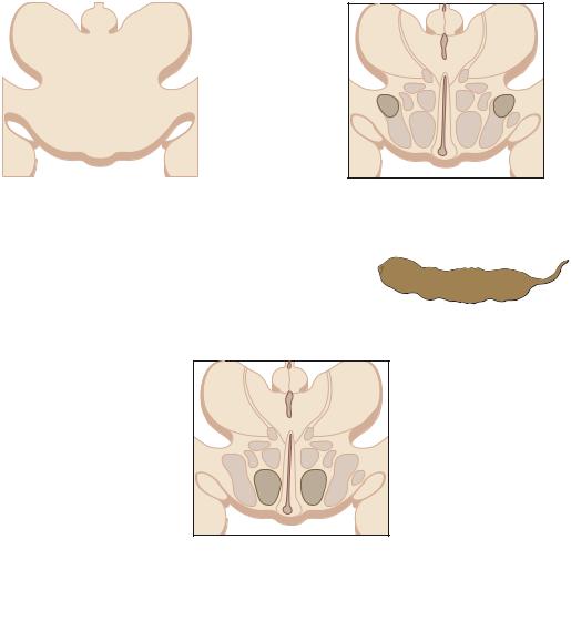
Figure 11-5 Coronal section through the hypothalamus at the level of the dorsomedial, ventromedial, and lateral hypothalamic nuclei. Lesions or stimulation of these nuclei result in obesity, cachexia, and rage. The column of the fornix separates the medial from the lateral hypothalamic zones. A lesion of the optic tract results in a contralateral hemianopia. FX, fornix; DM, medial dorsal nucleus of thalamus; OT, optic tract; VL, ventral lateral nucleus of thalamus; VP, ventral posterior nucleus of thalamus. (Reprinted from Fix JD. BRS Neuroanatomy. 3rd ed. Baltimore, MD: Williams & Wilkins; 1996:313, with permission.)

Diencephalon 99
CASE 11-1
A 90-year-old woman complains of an intense burning sensation on the left side of her neck and upper limb. The patient has a history of high blood pressure and diabetes. What is the most likely diagnosis?
Differentials
● Hypoglycemia; middle cerebral artery stroke; migraine
Relevant Physical Exam Findings
●Unilateral sensory loss is observed. Though the patient may complain of weakness on the affected side, no weakness is found on examination.
Relevant Lab Findings
●Normal serum glucose levels
●Thrombocytopenia
●Ischemic infarction of posterior cerebral artery seen on computed tomography scan
Diagnosis
●Infarction of the ventral posterolateral nucleus of the thalamus results in pure hemisensory loss contralateral to the lesion.
CASE 11-2
A 65-year-old diabetic man was hospitalized after an auto accident with lethargy and progressive confusion. Laboratory tests results revealed low sodium levels. The patient was discharged after serum sodium levels were elevated to 130 mmol/L.
Differentials
● Diabetes
Relevant Physical Exam Findings
●Fatigue and depression
●Slowed mental processing time
●Slowed pulse and hypothermia
●Hyporeflexia and hypotonia
Relevant Lab Findings
●Normal hepatic, renal, and cardiac function
●Hyponatremia that worsens with fluid load
●Serum hypo-osmolality
●Urine hyperosmolarity
Diagnosis
●Inappropriate secretion of antidiuretic hormone by the hypothalamus. As many as 50% of traumatic brain injury patients experience endocrine complications that may result in intracerebral osmotic fluid shifts and brain edema, affecting hypothalamic function.