

Anaphylactic_Shock
.pdfWe are IntechOpen,
the world’s leading publisher of Open Access books
Built by scientists, for scientists
4,300 116,000 130M
Open access books available |
International authors and editors |
Downloads |
Our authors are among the
154 |
TOP 1% |
12.2% |
||
Countries delivered to |
most cited scientists |
Contributors from top 500 universities |
||
|
||||
|
|
|
|
|
|
|
|
|
|
|
|
|
|
|
Selection of our books indexed in the Book Citation Index in Web of Science™ Core Collection (BKCI)
Interested in publishing with us? Contact book.department@intechopen.com
Numbers displayed above are based on latest data collected.
For more information visit www.intechopen.com

Chapter
Anaphylactic Shock
Stephen DeTurk, Shravan Reddy, Anna Ng Pellegrino
and John Wilson
Abstract
Anaphylaxis is a life threatening hypersensitivity reaction that can cause shock. Epidemiology studies show anaphylaxis and anaphylactic shock is relatively rare, but its incidence is increasing. A review of the pathophysiology of anaphylaxis can provide insight into clinical decisions. Diagnosing anaphylaxis can be difficult as symptoms and history are not always obvious. Diagnostic guidelines provide an objective tool to assess for anaphylaxis. Early intervention during anaphylaxis may prevent development of shock. Management is focused on circulation support with epinephrine and IV fluids, and airway maintenance. Following an acute anaphylactic reaction, patients should be provided with a referral for follow up and educated on avoidance of triggers and use of epinephrine autoinjectors.
Keywords: anaphylaxis, anaphylactic shock, distributive shock, management
1. Introduction
In this chapter, we will learn the differences between anaphylaxis and anaphylactic shock. Epidemiology of anaphylactic shock will be reviewed. We will also discuss the biochemical markers and mediators most noted to triggering an anaphylactic reaction. Lastly, we will provide a review on clinical manifestations and management of an anaphylactic reaction in a nonclinical setting and in a clinical setting.
Throughout this chapter anaphylaxis will be defined according to the World Allergy Organization (WAO) definition ‘a severe, life-threatening generalized or systemic hypersensitivity reaction’ [1]. This includes Gell-Combs Type I hypersensitivity, as well anaphylactoid reactions that are immunologically and nonimmunological mediated.
There are few studies on anaphylactic shock, and most recommendations for anaphylactic shock management come from major allergy organizations: WAO, AAAA/ACAAI, and EAACI. The recommendations of these groups for management of anaphylactic shock are presented in this chapter. Recent changes in anaphylaxis definitions require a review of the immunologic and nonimmunologic biochemical pathways of anaphylaxis.
1.1 Definitions
1.1.1 Anaphylactoid reactions
Reactions that occur via mechanisms other than IgE mediated mast cell degranulation have been referred to as Anaphylactoid reactions. However, the WAO, AAAA/ ACAAI, and EAACI have recommended stoppage of the term [2]. Throughout this chapter reactions that would be included in this term will be referred to as anaphylaxis.
1

Clinical Management of Shock - The Science and Art of Physiological Restoration
1.1.2 Anaphylaxis
Anaphylaxis is typically taught as Gell-Combs classification type 1 hypersensitivity, that is IgE mediated. However, the World Allergy Organization (WAO) proposed a new expanded definition of anaphylaxis ‘a severe, life-threatening generalized or systemic hypersensitivity reaction’ [3]. This new definition of anaphylaxis now includes reactions previously categorized as ‘anaphylactoid reactions’.
1.1.3 Anaphylactic shock
End manifestation of anaphylaxis, occurs when there is inadequate tissue perfusion causing end organ damage.
2. Epidemiology
Studies have estimated that the incidence of anaphylaxis is between 0.05 and 2.0% of the population [4], although the actual incidence is not clear. Issues previously identified with epidemiologic studies include variations in definitions, under reporting of anaphylaxis, and unclear use of incidence and prevalence of disease [5]. Although the actual incidence is not clear, there have been multiple studies showing
that the incidence of anaphylaxis in the United States has increased in recent years [6–9], although the case fatality rate has decreased [8]. Similar findings are reported in other countries, with UK reporting increasing rates of anaphylaxis [10–12], but no increase in the incidence of fatal anaphylaxis [10]. In Australia the incidence of anaphylaxis [13–15] and fatal anaphylaxis has increased as well [16]. One study on the incidence of anaphylaxis with circulatory symptoms found a rate of approximately 8–9 cases per 100,000 persons per year [17]. Severe anaphylaxis, including respiratory or circulatory symptoms, occurs more frequently at a rate of about 1–3 per 10,000 people [18].
Factors that may affect the incidence of anaphylaxis include geography, seasonal variations, age, and gender [19]. Demographic factors associated with higher incidence include living in northern areas of US [7, 20].
3. Pathophysiology
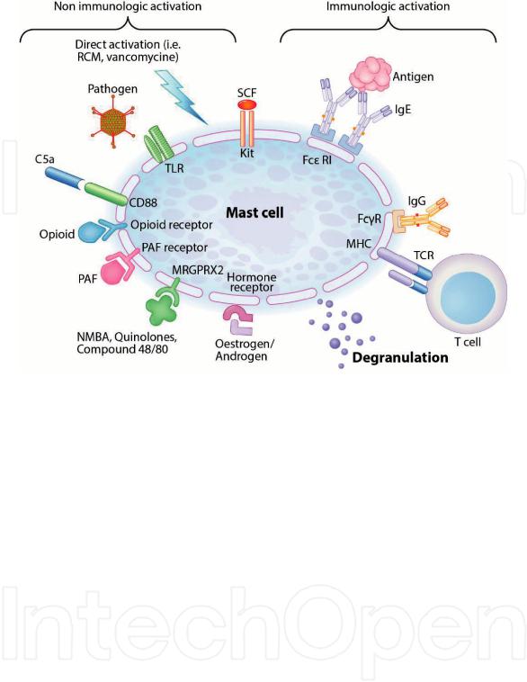
Anaphylaxis is caused by massive release of biochemical mediators from mast cell and basophils. Mast cells activation occurs mainly via antigen crosslinking of IgE bound to FcεRI receptors on cell membranes. However, other membrane receptors can activate mast cells as well or potentiate IgE activation [21]. The multiple activation pathways allow for immunologic (e.g. IgE mediated) and/or nonimmunologic activation (e.g. drug directly interacting with receptors) (Figure 1: mechanisms of mast cell degranulation). Some antigens may mediate effects via several mechanisms simultaneously (e.g. vespid venom, NSAIDs, opiates). In non-IgE mediated anaphylaxis, symptoms can occur on first exposure to an antigen as prior exposure and sensitization is not required.
Reproduced from Spoerl et al. [22] in agreement with publishing under terms of the Creative Commons Attribution (CC BY) license.
3.1 Triggers and cell activation
In IgE mediated anaphylaxis, an immunogen cross links membrane bound IgE in previously sensitized mast cell. Immunogens are typically large foreign proteins
2
Anaphylactic Shock
DOI: http://dx.doi.org/10.5772/intechopen.88284
Figure 1.
Mechanisms of mast cell degranulation. Abbreviations: RCM, radiocontrast media; TLR. Toll-like receptor; SCF, stem cell factor; FcεRI, high affinity IgE receptor; FcγR, IgG receptor; TCR, T-cell receptor; NMBA, neuromuscular blocking agent; PAF, platelet activating factor; MHC, major histocompatibility complex.
with multiple epitope binding sites (e.g. proteins in insect venom and certain foods) [23]. Antigens that are too small to cross link IgE (e.g. penicillin) must first bind to larger carrier molecules in order to elicit an immune response. Common triggers of IgE-mediated anaphylaxis include various food, venom and medications are summarized in Table 1: IgE-dependent triggers and discussed in more detail below.
3.1.1 Food
The most common foods causing anaphylaxis varies by region, in North America the most common food allergies includes milk, egg, peanuts, tree nuts, fish, shellfish, wheat, soy, and sesame [24]. Allergenic proteins have been identified for these common causes as well as in rice, barley, buckwheat, mustard, celery, potato, carrots, and apples [25].
Most cases of anaphylaxis due to food occurs in children or those with known food allergies, and fatal cases are rare [7, 8, 10]. When fatal, the cause of death is more often due to respiratory arrest, although shock can occur as well. Arrest
occurs later compared to medication or venom, typically occurring 25–35 min after exposure [26].
3.1.2 Venom
Stinging insects belonging to the order Hymenoptera produce venoms that can cause anaphylaxis. The important insects from this order include bees, vespids (wasps, yellow jackets, hornets) and stinging ants [27]. Vespid venom additionally activates complement in an IgE independent mechanism, which may potentiate anaphylaxis [28]. In addition to hymenoptera venom, bites from rattlesnakes [29],
3

Clinical Management of Shock - The Science and Art of Physiological Restoration
Foods |
Milk |
|
Eggs |
|
Peanuts |
|
Tree nuts |
|
Fish |
|
Shellfish |
|
Soy |
|
Wheat |
|
Sesame |
Bites/venoms |
Hymenoptera venom (bee, vespids*, ants) |
|
Tick |
|
Rattlesnake |
|
Hamster |
Medications |
Antibiotics |
|
NSAIDs* |
|
Chemotherapy |
|
Neuromuscular blocker* |
|
Radiocontrast* |
|
Opioids* |
*Non-IgE mediated mechanisms identified as alternative or additional mechanism.
Table 1.
IgE-dependent triggers [9, 24, 27, 29–32].
hamsters [30], and ticks [31] have been implicated as causes of anaphylaxis. Fatal cases due stings are more often due to shock than respiratory distress, arrest typically occurs 10–15 min after exposure [26].
3.1.3 Medications
The most common medications causing anaphylaxis are beta-lactam antibiotics, NSAIDs, neuromuscular blocking agents, and chemotherapy [9, 32] . However, nearly any medication has the potential to cause anaphylaxis, some drugs that have been implicated includes intravenous iron [33], gelatin found in a vaccines [34], dextran [35], and human serum albumin [36]. In addition to IgE mediated mechanisms, other mechanisms of inducing anaphylaxis have been identified for multiple drugs [37]. This includes complement or contact activation (e.g. radiocontrast media, pegylated compounds, liposomal drugs [38], and heparin contaminated with oversulfated chondroitin sulfate (OSCS) [39]) and direct mast cell activation (e.g. opiates and neuromuscular blockers [37]). There have been multiple reports of anaphylaxis occurring to biologic agents, where the patient had IgG but no detectable IgE antibody to the therapy [40]. In medication induced anaphylaxis, shock is more common in severe cases than respiratory distress [26, 41]. Arrest occurs can occur rapidly after exposure, most cases in less than 5 min [26].
3.2 Biochemical mediators and clinical pharmacology
Once activated, mast cells and basophils release a cascade of mediators which cause physiologic changes, activate other immunology pathways, and attract other immune cells. Preformed mediators are released immediately upon activation including histamine, tryptase, heparin, and chymase. Over several minutes additional mediators are generated including platelet activating factor, leukotrienes, and prostaglandins (Table 2). Various cytokines and chemokines are generated over several hours further propagate the inflammatory response [37, 42].
4

Anaphylactic Shock
DOI: http://dx.doi.org/10.5772/intechopen.88284
Immediate |
Histamine |
Vasodilation, edema, bronchoconstriction, mucus secretion, |
release |
|
change myocardial contractility |
|
Tryptase |
Vasodilation, edema, bronchoconstriction |
|
Chymase |
Vasodilation, edema, mucus secretion |
|
Heparin |
Initiates formation of bradykinin causing edema |
|
TNFa |
Bronchoconstriction |
Rapid generation |
PAF |
Vasodilation, edema, bronchoconstriction, platelet activation, |
(min) |
|
decrease myocardial contractility |
|
LTs C4, D4 |
Potent vasoactive and spasmogenic agents |
|
Prostaglandin D2 |
Bronchospasm and increase mucus secretion |
Table 2.
Physiologic effects of mediators [42, 49].
Histamine has long been considered the principal mediator of anaphylaxis, and concentrations of histamine correlate with symptom severity [42]. Histamine acts on receptors to cause vasodilation and increased permeability [43, 44], bronchoconstriction, and increase mucus secretion. In the heart H2 receptors have positive chronotropic and ionotropic effects and causes coronary vasodilation, while the H1 receptor causes coronary vasoconstriction [45].
Platelet activating factor (PAF) has been increasingly recognized as important in the pathophysiology of anaphylaxis. In PAF receptor knockout mice, symptoms of anaphylaxis are reduced [46]. In humans PAF levels in the serum directly correlates with the severity of anaphylaxis symptoms [47]. In addition to activating platelets, PAF causes vasodilation, increased vascular permeability, decreased myocardial contractility, bronchoconstriction, and initiates allergic response through stimulation of other mediators [48, 49].
4. Signs and Symptoms
Anaphylaxis causes a generalized systemic reaction affecting multiple organ systems, symptoms involving the skin occur in 80–90% of cases, respiratory tract in 70%, GI in 45%, CV in 45%, and CNS involvement in 15% [50, 51]. The cardiovascular and respiratory systems are the principal shock organs in fatal anaphylaxis. Death occurs in most often due to shock or acute respiratory distress, but DIC and epinephrine overdose have also been cited as cause of death [26, 52–55]. Most fatal cases of anaphylaxis due to medication or venoms are a result of shock, in food related anaphylaxis respiratory involvement is the main cause of death although shock is still possible [26, 56].
Anaphylaxis develops rapidly with symptoms developing in minutes. Biphasic reactions, where symptoms resolve and then reappear later occurs around 20% of the time [57]. A systematic review of biphasic reactions found the medium time between resolution of initial symptoms and onset of delayed symptoms to be 11 h, with a range of 0.2–72 h [58].
4.1 Cardiovascular changes
Cardiovascular manifestations of anaphylaxis develop due to direct and indirect effects of mediators on the vasculature and heart. Increased vascular permeability causes rapid fluid extravasation, with up to 35% of plasma volume shift occurring
5

Clinical Management of Shock - The Science and Art of Physiological Restoration
in a matter of minutes [59, 60]. Vascular smooth muscle relaxation causes vasodilation and a rapid decrease in SVR [61, 62]. Rapid drop in measured CVP suggests that venodilation plays a major as well. The combined effects as extravasation and venodilation cause significant reduction in preload. This can be severe enough to cause Empty ventricle syndrome, has been documented in autopsies of patients who died from anaphylaxis [63].
Arrhythmias and myocardial infarction can also be seen in anaphylaxis. Arrhythmias may develop due to the combined direct effect of mediators, and hemodynamic changes previously described. Myocardial infarctions seen in anaphylaxis may be due to decreased venous return and direct effects of mediators on coronary arteries causing vasospasm or disruption of atherosclerotic plaques [64, 65].
Compensatory response to these changes includes rise in heart rate, ejection fraction, and cardiac index [61, 62]. AlthoughTachycardia is typical in anaphylaxis, although bradycardia may be seen as well. Bradycardia occurs due to a compensatory Bezold-Jarisch reflex, and has been found to have lower mortality rates when compared to cases of anaphylactic shock with tachycardia [66, 67]. Atropine therefore should not be administered, as it would counteract this protective effect and may increase mortality [68].
4.2 Respiratory changes
The entire respiratory tract can be affected in anaphylaxis, involving the upper airway and/or lower airway [69]. Upper airway symptoms develop due to fluid extravasation causing mucosal edema [70]. Some symptoms of upper airway involvement include angioedema, stridor, dysphagia, and rhinorrhea [50, 71]. Lower airway obstruction occurs due to mucosal edema, bronchospasm, and mucous plugging [70]. Oxygen saturation may decrease secondary to respiratory effects of anaphylaxis limiting airflow. When there is diffuse lower respiratory tract involvement, decreased oxygen saturation can persist despite endotracheal intubation [72]. When PaO2 is adequate, tissue oxygenation can still be compromised causing anaerobic metabolism. In one study on rats, tissue oxygenation of muscle decreased faster in anaphylaxis compared to nicardipine induced hypotension. The anaphylactic group also showed a greater increase in lactate and lactate-pyruvate ratio indicating depletion of intracellular energy storage [73].
4.3 Mucocutaneous, gastrointestinal, and neurologic changes
Mucocutaneous symptoms (e.g. flushing, pruritus, angioedema, and urticaria) are common in anaphylaxis, however in cases of shock cutaneous symptoms are often absent. Vasodilation and increased vasculature permeability leads to flushing and angioedema [74]. In addition to vascular changes, urticaria develops due inflammatory cell infiltration and neuropeptide release from sensory nerves [75]. Gastrointestinal symptoms include nausea, vomiting, diarrhea, and abdominal pain [76]. These symptoms are likely due to intestinal smooth muscle contraction and alterations in water and electrolyte absorption [77, 78]. Neurologic changes are mostly secondary to hypotension and decreased perfusion and may manifest as dizziness, confusion, syncope/presyncope, or headache [50]. More serious effects including seizure and stroke may also be seen but are rare [74, 79].
5. Diagnosing
It is often difficult to recognize anaphylaxis, and many cases go undiagnosed [80, 81]. Early intervention in acute anaphylaxis reduces risk of severe reaction and
6

Anaphylactic Shock
DOI: http://dx.doi.org/10.5772/intechopen.88284
need for hospitalization [82]. In order to aid in diagnosis of anaphylaxis, diagnostic criteria have been developed (Table 3). In the clinic or emergency department, it may be unclear if the patient had been exposed to an allergen. In the ICU or operating room, rapid development of symptoms after administering medication makes recognizing anaphylaxis easier. Following the acute phase of anaphylaxis, serum tryptase and histamine should be measured to aid immunologist in confirming the diagnosis of anaphylaxis during follow-up care [83, 84]. Plasma tryptase remains elevated for 6 h following the onset of symptoms, but histamine levels remain elevated for just 1 h [85, 86]. Urinary histamine metabolites remain elevated for a longer period and may therefore be more useful than plasma histamine for confirming anaphylaxis [86].
5.1 Diagnostic criteria
Anaphylaxis is likely when one of the following criteria is fulfilled:
1.Acute onset of an illness (minutes to several hours) with involvement of the skin and/or mucosal tissue and at least one:
a.Respiratory compromise
b.Reduced blood pressure or symptoms of end-organ damage
2.Two or more of the following that occur rapidly after exposure to a likely allergen for that patient
a.Involvement of the skin-mucosal tissue
b.Respiratory compromise
c.Reduced blood pressure or associated symptoms
d.Persistent gastrointestinal symptoms
3.Reduced blood pressure after exposure to known allergen for that patient (minutes to several hours)
a.Adults: systolic blood pressure of less than 90 mm Hg or greater than 30% decrease from that person’s baseline
b.Infants and children: low systolic blood pressure (age-specific) or greater than 30% decrease in systolic blood pressure
Table 3.
Clinical criteria for diagnosing anaphylaxis, modified from [3, 87].
5.2 Differential diagnosis
There are several conditions which may mimic certain characteristics of anaphylaxis. Acute anxiety can present as dyspnea and near syncope with hyperventilation. Hypoglycemia can precipitate an altered sensorium and syncope. Vasovagal episodes can involve nausea with vomiting, hypotension, pallor, bradycardia, diaphoresis and syncope. Additional considerations include severe reactive airway disease, vocal cord dysfunction and non-allergy mediated angioedema. Vasovagal episodes can involve nausea with vomiting, hypotension, pallor, bradycardia, diaphoresis and syncope [50, 88].
6. Management
There is a general lack of evidence basis for the treatment of anaphylaxis [89], but multiple expert guidelines highlight the chief treatment as epinephrine, oxygen, and fluids [86, 90, 91].
6.1 Principal treatment
Epinephrine is the first line treatment of anaphylaxis, and delayed administration increases the likelihood of poor outcomes [54, 55, 92]. Despite this, use of
7

Clinical Management of Shock - The Science and Art of Physiological Restoration
epinephrine in anaphylaxis is infrequent and often delayed [80]. There are no contraindications for use of epinephrine in anaphylaxis, and it should still be administered in patients with history of heart disease [93].
Epinephrine exerts its effects via alpha and beta adrenergic receptors in a dosedependent response where beta receptors effects are dominant at low doses, but alpha receptors effects are seen at higher doses. The α1 receptors cause vasoconstriction increasing peripheral vascular resistance and blood pressure and improving coronary and cerebral perfusion. The β1 receptors exert positive chronotropic and inotropic effects which improves cardiac output and increases blood pressure. In the respiratory system, β2 receptors stimulation results in bronchodilation, and relief and respiratory symptoms. β receptors also inhibit release of mediators from mast cells and basophils, via increased cAMP production [94].
In most situations intramuscular administration is preferred, but IV epinephrine can be used in the ICU. IM epinephrine should be given in 0.2–0.5 mg doses (1:1000 dilution), and repeated every 5 min depending on the resolution of symptoms [86]. Studies showed that peak epinephrine concentrations were higher when epinephrine was given IM into the thigh, compared to IM administration in the arm or subcutaneous administration [95]. Multiple doses of epinephrine may be required to reverse symptoms [96]. Care should be taken to closely monitor pulse and blood pressure when epinephrine is administered intravenously as there is greater risk of severe adverse effects compared to intramuscular administration including arrhythmias and myocardial infarctions [97].
Following epinephrine administration, patients should continue to be assessed for worsening signs of anaphylaxis. Patients should be placed on pulse oximeter and given high flow oxygen as needed. Patients in anaphylactic shock should be placed in supine or Trendelenburg position to increase blood return to the heart. Patients who are sitting upright can have a significant decrease in preload leading to empty ventricle syndrome and PEA [98].
Due to the intravascular depletion, fluid is often necessary to maintain pressure. Crystalloids or colloids may be used, although physicians should be aware of the anaphylactic potential of some colloid solutions. Normal saline (0.9% saline) should be chosen over other crystalloids as it remains in the intravascular space longer than dextrose solutions [86, 99]. Caution should taken while giving fluids to patients with heart failure to prevent fluid overload.
6.2 Adjunct treatments
Antihistamines are often given as adjunct therapy in anaphylaxis, however there is no evidence to support or advise against their use in anaphylaxis [100]. Steroids may be used to prevent biphasic anaphylaxis [86], although there is no evidence to support the use of steroids in acute treatment of anaphylaxis [101, 102].
Nebulized albuterol or other beta-2 agonists may be useful to treat respiratory distress due to bronchoconstriction. While there is no clinical trial on use of these medications in anaphylaxis, their effectiveness in treating other allergic diseases offers some basis for their use [90].
6.3 Additional consideration
Beta blockers may complicate the treatment of anaphylaxis, as some of the beneficial effects of epinephrine will be diminished [103]. In patients on betablockers who do not respond to epinephrine and fluids, other vasopressors should be considered. Glucagon has been reported to be a successful treatment in several case reports of patients on beta blockers who experienced anaphylactic shock [104].
8

Anaphylactic Shock
DOI: http://dx.doi.org/10.5772/intechopen.88284
Glucagon mechanism of action is via direct activation of adenylate cyclase, bypassing the blocked adrenergic receptors.
Vasopressin or phenylephrine can be used to increase systemic vascular resistance without further increasing heart rate. Dopamine or norepinephrine can be added in cases of relative bradycardia [105]. Mechanical support with ECMO has been reported to be successful at treating refractory anaphylaxis with profound myocardial dysfunction [106].
Methylene blue has been reported to be an effective treatment in cases of severe anaphylaxis not responding to epinephrine [88], as well as cases of anaphylaxis without hypotension. Methylene blue inhibits NO mediated vasodilation via competitive inhibition of guanylate cyclase decreasing cyclic GMP production and subsequent vasodilation [107]. This mechanism acts independent of adrenergic receptors, and may be effective in patients with refractory anaphylactic shock [108].
6.4 Follow up
Before discharging a patient that experienced anaphylaxis, they should be referred to an immunologist for a thorough evaluation. Labs to assess for anaphylaxis (i.e. tryptase, histamine) should be drawn to assist allergist in making a diagnosis. Patients should also receive a prescription for an epinephrine autoinjector, along with education on how to use it [87].
7. Conclusions
Anaphylaxis is a rapidly acting life-threatening hypersensitivity reaction. Diagnosis of anaphylaxis can be difficult, and early recognition and treatment is essential to prevent development of shock. Shock is more common in cases due to medication compared to food, although shock can occur. The primary treatment in anaphylactic shock is epinephrine, fluids, and oxygen. Additional medications including antihistamines, steroids, and inhaled beta-agonist should be used as needed. In patients who do not respond to epinephrine, other vasopressors or mechanical support can be used.
Abbreviations
AAAAI |
American Academy of Allergy, Asthma and Immunology |
ACAAI |
American College of Allergy, Asthma and Immunology |
EAACI |
European Academy of Allergy and Clinical Immunology |
WAO |
World Allergy Organization |
Ig |
immunoglobulin |
Antigen |
molecule capable of interacting with Ig, includes immunogens and |
|
haptens |
Immunogen |
molecule that can interact with Ig and cause an immune response |
Hapten |
molecule that can interact with Ig, but cannot cause immune |
|
response on its own |
9