

Книги по МРТ КТ на английском языке / Thomas R., Connelly J., Burke C. - 100 cases in radiology - 2012
.pdfANSWER 64
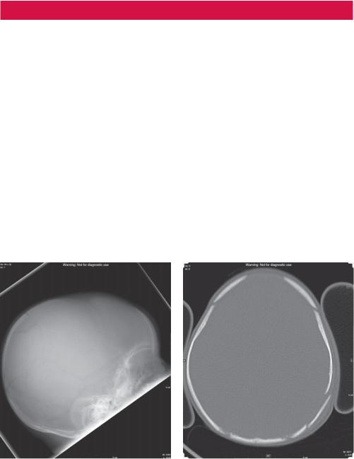
Figure 64.1 shows a single slice of a cranial CT scan taken at the level of the caudate heads. There is asymmetry demonstrated with an area of increased density on the left side lying underneath the left temporal bone. This abnormality is more dense than adjacent brain parenchyma, but less dense than the skull bones in keeping with acute blood. It has a biconvex elliptical shape and is homogeneous in appearance. This collection of blood exerts mass effect on adjacent brain parenchyma with total effacement of the adjacent lateral ventricle and loss of normal left cerebral sulci. There is midline shift to the right of approximately 3 mm. The quadrageminal cistern is effaced but the right lateral ventricle remains of expected size.
The same image rewindowed to emphasize bony pathology (Figure 64.2) demonstrates a vertical fracture of the left temporal bone (arrow). There is some subcutaneous soft tissue thickening at this site, which corresponds to the site of impact of the cricket ball. This represents an acute extradural haemorrhage with mass effect compressing the brain within the skull.
Extradural haemorrhage is defined as a collection of blood within the space between the inner table of the skull and the dura mater of the meninges. Blood collects in the extradural space running along the inside of the skull. It is strongly associated with direct head trauma and skull fracture, with bony fragments lacerating the meningeal vessels (commonly middle meningeal). Depending on whether the lacerated vessels are arteries or veins, the rate of haematoma expansion dictates whether the patient presents hours or days after the incident. Common symptoms are of gradually increasing drowsiness, which can progress to coma as the haematoma has focal mass effect compressing the brain within the tight skull. Some patients can demonstrate a third nerve palsy or hemiparesis as a sign of cerebral herniation.
Figure 64.2 CT scan rewindowed to emphasize bony pathology.
An unenhanced cranial CT usually demonstrates an acute hyperdense collection of blood, but if a few days old, can have varied attenuation appearances. It is differentiated from other extra-axial collections by its shape. The expanding haematoma will have a biconvex elliptical shape as tracking blood is limited by the tethered cranial sutures. There may be a degree of mass effect, and image review with bone windows has a high sensitivity for resolving the underlying skull fracture. All patients will require urgent neurosurgical discussion and neurologically compromised patients will require urgent decompression and evacuation.
KEY POINTS
•In an extradural haemorrhage, blood collects between the inner table of the skull and the dura mater.
•It classically presents with reduced conscious level several hours after a traumatic incident.
•A characteristic concave elliptical haematoma is seen on CT with tracking blood limited by cranial sutures.
172

CASE 65: A CHESTY INFANT
History
A 3-month-old boy was referred to the paediatric department by his general practitioner (GP) with respiratory problems. He has had a cough and runny nose for 2 days and has become progressively more chesty. He now has problems feeding, at least one episode of vomiting that may be cough related and dry nappies. Older siblings in the family also have coryzal symptoms. The patient was born 2 weeks prematurely with no significant neonatal problems. The mother is on treatment for asthma and there is no other significant medical or family history.
Examination
The initial examination showed a febrile infant with copious nasal secretions, conjunctivitis and noisy breathing (grunting and wheezing). There is tachypnoea (58 breaths per minute) with intercostal and subcostal retraction. On listening to the chest there are fine crackles and wheeze throughout. The rest of the examination is normal.
Over the next 24 hours, the patient did not improve on standard treatment and continuous positive airways pressure (CPAP) and eventually required intubation. At this point a chest radiograph (Figure 65.1) was done to check the lungs and position of the tube (an earlier radiograph had been unremarkable).
Six hours later, problems with ventilation developed and a further radiograph was done (Figure 65.2).
Figure 65.1 Anterior–posterior (AP) |
Figure 65.2 Later radiograph. |
radiograph. |
|
Questions
•What differentials are you considering?
•What does the first radiograph show?
•What complication has developed in the second radiograph and why?
173

ANSWER 65
Given the age and presentation, the most likely diagnosis is bronchiolitis although pneumonia and aspiration should also be considered. Undiagnosed congenital cardiac or lung disease and foreign body may need to be ruled out.
The first radiograph (Figure 65.1) shows hyperinflation with flattened diaphragm. The lung volumes are large due to air trapping rather than fortuitous timing of the radiograph. Coarsened lung markings that reflect patchy airspace and interstitial infiltrates are seen. There is also some thickening of the perihilar bronchial walls (cuffing). No focal consolidation, cardiac abnormality or foreign body is seen. The appearance is consistent with severe bronchiolitis, although similar appearances are also seen in atypical pneumonia and aspiration. The tip of the endotracheal (ET) tube is well above the carina and the tip of the nasogastric (NG) tube is below the diaphragm.
The second radiograph (Figure 65.2) shows a low ET tube in the right main bronchus resulting in left lung occlusion and collapse. Some compensatory hyperexpansion of the right lung across the midline is seen. The problem is remedied by withdrawing the tube tip (ideally to the level of the clavicles). Remember to comment on tubes and complications on every image.
The most common complication is bacterial coinfection. This was suspected in this patient’s case and shown on a subsequent radiograph (Figure 65.3) that also showed re-expansion of the left lung.
Bronchiolitis is usually diagnosed clinically and a chest radiograph is only needed if the course of the illness is atypical or to rule out other causes or complications. The small airways become infected, resulting in narrowing and obstruction. The most common cause is the respiratory syncytial virus (RSV) and occasionally adenovirus and parainfluenza viruses. This may be confirmed by a nasopharyngeal aspirate. The illness can usually be managed at home, but 2–3 per cent of cases require admission to hospital of which 3–7 per cent require ventilation. Children
with underlying cardiopulmonary disease, infants with a history of gestational age less than 34 weeks, infants younger than 6 weeks, and infants with congenital or acquired immunodeficiency are at high risk for severe RSV infection.
KEY POINTS
•Bronchiolitis is largely diagnosed and managed without imaging.
•A chest radiograph is helpful to check for other causes and complications.
•The radiograph appearance of bronchiolitis is non-specific and may consist of hyperexpanded lungs only, although patchy infiltrates may also be seen.
174
CASE 66: DIFFICULTY IN SWALLOWING
History
A 47-year-old mother has been referred for a barium swallow study by her general practitioner (GP). She complains of increasing discomfort when eating and finds that food sometimes gets stuck in her throat. This is more often the case when eating meat or bread, and she finds that if she does not chew her food carefully, then each mouthful needs to be washed down with a glass of water. She has recently taken to eating soup for most meals, and does not find this too much of a problem. These symptoms have been getting very gradually worse over the past 5 months, and despite her change in diet she denies any significant weight loss.
There is no relevant past medical history, but her GP has recently prescribed iron tablets following a blood test for feelings of increasing lethargy. She attributes this to her heavy periods. She is a non-smoker and only drinks socially.
Examination
Her barium swallow study results are shown in Figure 66.1.
Figure 66.1 Barium swallow image.
Questions
•What does this view from the barium swallow show?
•What is the likely diagnosis?
175

ANSWER 66
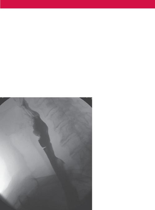
For a barium swallow study, the patient is positioned between a fluoroscopy machine containing an X-ray tube and an image intensifier. This provides a real-time image that can be collimated to emphasize organs of the patient’s neck and chest. The patient is asked to drink a contrast agent (e.g. barium) and hold it in the mouth. In coordination with the radiologist, asking the patient to swallow on command allows for good opacification of the oesophagus and direct real-time observation of the passage the contrast takes. Although many fluoroscopy studies are rarely used these days (e.g. barium enemas), swallow studies using barium or iodinated contrast medium are the ‘workhorse’ of foregut imaging. As a relatively non-invasive technique, it allows for accurate visualization of the oesophageal diameter and mucosa, and is often used to confirm the presence of persistent oesophageal leaks post-operatively.
This single lateral image is centred on the hypopharynx and upper oesophagus. Contrast passes freely from the mouth to the mid-oesophagus with no evidence of hold-up or obstruction. There is no evidence of aspiration. Within the proximal oesophagus at the level of C5, there is an anterior ‘shelf-like’ filling defect seen. This arises at right angles to the anterior oesophageal wall and appears to encroach into the oesophageal canal by approximately one-third. There is no transition delay of contrast, and no evidence of prestenotic dilatation or oesophageal diverticulosis. These features are in keeping with an oesophageal web.
Oesophageal webs are thin membranes of normal oesophageal squamous tissue that grow out from the anterior mucosal wall. Measuring approximately 1–2 mm in thickness, they can cause complete or incomplete circumferential narrowing of the oesophageal lumen and are most commonly seen in middle-aged white women. Symptoms are dependent on the degree of obstruction, with patients complaining of dysphagia for solids rather than liquids. Often found incidentally when patients present with feelings of globus or suffering food bolus impaction, symptoms can also include pain (odynophagia).
The aetiology of oesophageal webs is uncertain but can be both congenital, or more commonly, acquired. They are associated with chronic inflammatory conditions of the oesophagus including epidermolysis bullosa, pemphigus and bullous pemphigoid, and are also seen in coeliac and graft-versus-host disease. There is a strong association with Plummer–Vinson syndrome (PVS), in which patients experience concomitant glossitis, angular stomatitis and iron-deficiency anaemia. Image findings should always be correlated with a full blood count to exclude this.
Treatment of oesophageal webs is routinely performed with balloon dilatation, and is often a common byproduct of upper gastrointestinal endoscopy by bougienage. There is an increased association with oesophageal cancer, and recurrence of symptoms should be investigated early.
KEY POINTS
•A barium swallow investigation is recommended for assessment of dysphagia.
•Oesophageal webs are thin membranes of normal oesophageal squamous tissue that grow out from the anterior mucosal wall.
•There is a strong correlation between oesophageal webs and other chronic inflammatory conditions.
176

CASE 67: PREGNANT WOMAN WITH VOMITING
History
A 24-year-old pregnant woman with hyperemesis presents to the accident and emergency department with chest pain and swelling around the neck after 3 days of worsening vomiting. She is dehydrated and breathless. She is in the 10th week of a confirmed intrauterine twin pregnancy and has already attended the early pregnancy unit for treatment for vomiting. There has been no per vaginal (PV) bleeding or abnormal discharge. None of her close contacts have similar symptoms. Her medical history is otherwise unremarkable.
Examination
She appears dehydrated and mildly agitated but otherwise fit. Swelling over the lower neck and upper chest is noted that on palpation causes crepitus. She has mild tachycardia but no pyrexia. More crepitus is heard over the lungs. The heart sounds are normal. The abdomen is soft and mildly diffusely tender.
You arrange a chest radiograph (Figure 67.1) and blood tests.
Figure 67.1 Posterior–anterior (PA) chest radiograph.
Questions
•What does the X-ray show?
•What imaging would you do next?
•What about the pregnancy?
177
ANSWER 67
The chest radiograph shows free gas within the upper mediastinum (i.e. a pneumomediastinum outlining the aorta and the trachea). There is also subcutaneous free gas (i.e. surgical emphysema), extending from the neck down over the upper thorax. No pneumothorax is seen. The lungs and heart appear normal.
Pneumomediastinum can be difficult to see on chest X-rays and is often missed if small. The characteristic appearances are streaky lucencies in the mediastinum, gas outlining structures such as vessels, pulmonary ligament and producing lucency between the hemidiaphragms. There may be associated pneumothoraces, pleural effusions, pneumopericardium or subcutaneous emphysema.
In terms of causes, it is useful to consider where the gas has come from. If it is intrathoracic, it could be from:
•oesophagus perforation secondary to foreign body, tumour or persistent vomiting (Boerhaave’s syndrome);
•trachea or major bronchi – usually the result of blunt chest trauma, pneumothorax or tumours;
•lungs – due to alveolar rupture secondary to asthma in children, lung disease, ventilation or inhaled recreational drugs;
•pleural space – pneumothorax;
•iatrogenic causes – surgery, endoscopy or positive pressure ventilation;
•spontaneous – a cause is not found, typically in older patients.
If it is extrathoracic, it could be:
•head and neck – trauma, tumour, surgery;
•subdiaphragmatic – due to perforation of a viscus with gas tracking superiorly and through diaphragmatic openings.
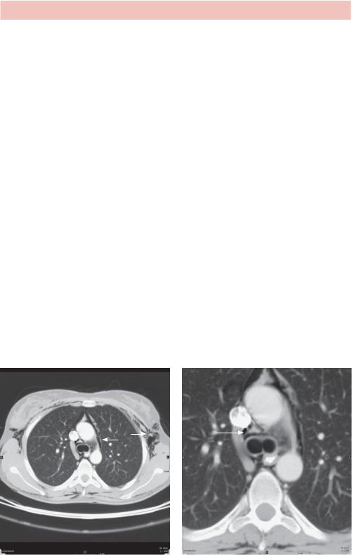
Computed tomography (CT) is usually done if there is uncertainty about the radiograph and to see the extent of free gas (Figure 67.2).
(a) |
(b) |
Figure 67.2 (a) Axial CT of the mediastinum showing mediastinal gas outlining the vessels and trachea and in the subcutaneous tissues (arrows); (b) enlarged view of the mediastinum.
178
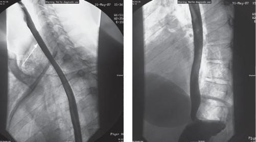
If there is suspicion of oesophageal rupture, a water-soluble contrast swallow test should be done as there is a high risk for mediastinitis which has a poor prognosis (Figure 67.3).
(a) |
(b) |
Figure 67.3 (a) Lateral and (b) water-soluble contrast swallow. Adjacent outline of the tracheal wall is seen due to interposed gas (arrow).
In Boerhaave’s syndrome, where there is oesophageal rupture secondary to vomiting, the point of injury is typically in the left posterolateral wall a few centimetres above the gastro-oesophageal junction, and the largest amount of gas may be in the left side of the mediastinum. Neither of these signs is seen in Figure 67.3.
Rules governing licensing of radiology departments, which in the United Kingdom go under the name IRMER (Ionising Radiation (Medical Exposure) Regulations 2000), require a physician to justify the investigation, a technical member of the department to perform the investigation and a qualified person to review and report the outcome. In addition is the principle of the ALARP (‘as low as reasonably possible’) dose. In routine practice, people tend to think most about this when performing X-ray investigations on children or pregnant women. The reason is that certain types of tissue, typically rapidly growing or metabolically active tissue, is the most radiosensitive. It is worth remembering that some adult tissue remains more radiosensitive, in particular breast, gonads and thyroid.
The process of calculating radiation doses and including tissue sensitivities to produce an exposure that can be used in calculating risks is complicated and is often not required in such detail because the clinical risk associated with an uncertain diagnosis may be much higher (e.g. in pulmonary embolism). A few numbers are helpful to get a feel for comparative doses. Annual background is about 3 mSv, chest X-ray 0.02 mSv, abdomen 0.7 mSv, head CT 2 mSv, chest CT 8 mSv, abdominal/pelvis CT 10 mSv.
Justification for an investigation usually rests on the clinical risk of complications of a missed diagnosis, which are typically much larger than the long-term risk of X-ray exposure. In this case the surgical risk of mediastinitis and miscarriage justifies the exposure. The ALARP principle may have been used in deciding to do the contrast swallow first as a CT may not have been necessary if a leak had been demonstrated.
179
KEY POINTS
•Pneumomediastinum is easy to miss on a chest radiograph. Look for gas outlining the upper mediastinal structures.
•If oesophageal rupture is suspected, a contrast swallow should be performed as there is a high rate of mediastinitis and mortality.
•You need to be able to justify your investigations from a clinical and X-ray exposure point of view.
180
CASE 68: INFANT WITH A HEAD INJURY
History
A 4-month-old boy is brought into the paediatric accident and emergency department by his parents with a head injury. The mother gives a history of the child falling from the changing table earlier in the day and explains the delay in coming to hospital on difficulties getting transport. The child otherwise has an unremarkable medical and neonatal history.
Examination
The child is listless but with normal observations. He responds to voice by eye opening but appears to move very little. He is probably a little under weight. There is a hard irregularity in the left parietal scalp but no laceration. The fontanelles are not bulging. There are no focal neurological signs. There appears to be some bruising over the left side of the chest and a swollen left finger.
You are suspicious about the extent of the injuries and about the behaviour of the father who appears to be keen to downplay the incident and leave. You discuss your concerns with the registrar who admits the patient, arranges skull and chest radiographs (Figure 68.1 and 68.2) and contacts the on-call social services to see if the child is on the at-risk register.
(a) |
(b) |
Figure 68.1 Skull X-ray and subsequent computed tomography (CT) scan (bone window) of the head.
181