

новая папка 1 / 220883
.pdfCopyright ОАО «ЦКБ «БИБКОМ» & ООО «Aгентство Kнига-Cервис»
2013. 54, 2 – . 336 – 338
UDC 541.6:546.47:546.21
CHARACTERIZATION OF PURE PHASE Zn(II) OXIDE NANOPARTICLES
VIA THERMAL DECOMPOSITION OF TWO ZINC(II) COMPLEXES OF
THE 6,6 -DIMETHYL-2,2 -BIPYRIDINE LIGAND
M. Hosseinifard1, L. Hashemi2, V. Amani1, A. Morsali2
1Department of Chemistry, Faculty of Sciences, Islamic Azad University, Shahr-e-Rey Branch, Tehran, Iran 2Department of Chemistry, Faculty of Sciences, Tarbiat Modares University, Tehran, Iran
E-mail: morsali_a@modares.ac.ir
Received October, 7, 2011 |
Revised — July, 1, 2012 |
Two zinc(II) complexes [Zn(6,6 -dimethyl-2,2 -bipy)Cl2]n (1) and [Zn(6,6 -dimethyl-2,2 - bipy)I2]n (2) are synthesized from the reaction of the 6,6 -dimethyl-2,2 -bipy ligand with ZnCl2 and ZnI2. Zinc(II) oxide nanoparticles are synthesized by the thermolysis of [Zn(6,6 -dimethyl- 2,2 -bipy)Cl2]n (1) and [Zn(6,6 -dimethyl-2,2 -bipy)I2]n (2) at two different temperatures. The ZnO nanoparticles are characterized by X-ray diffraction and scanning electron microscopy (SEM). SEM images show the average size of the ZnO nanoparticles produced of 50 nm and 60 nm in compounds 1 and 2 respectively.
K e y w o r d s: nanoparticle, 6,6 -dimethyl-2,2 -bipy, Zn(II), surfactant.
INTRODUCTION
ZnO is polar inorganic crystalline material with many applications due to its interesting properties such as nontoxicity, good electrical, optical and piezoelectric behavior, stability in a hydrogen plasma atmosphere and low price [ 1—10 ]. ZnO is a well-known semiconductor with a wide direct band gap (3.37 eV), a large excitation binding energy of 60 meV at room temperature [ 4, 5 ] and a wide range of applications such as solar cells, luminescent, electrical and acoustic devices, gas and chemical sensors, coatings, catalysts, micro lasers, memory arrays and biomedical applications [ 6 ]. Many methods have already been developed to synthesize zinc(II) oxide and sulfide nanocrystals such as vapor phase growth [ 7 ], vapor liquid-solid process [ 2 ], soft chemical method [ 9 ], electrophoretic deposition [ 10 ], sol-gel process [ 11, 12 ], homogeneous precipitation [ 13 ], etc. The use of supramolecular compounds as precursors for the preparation of inorganic nanomaterials such as zinc(II) oxide has not yet been investigated thoroughly. In this paper we describe a simple method for preparing nanoparticles of ZnO with the use of [Zn(6,6 -dimethyl-2,2 -bipy)Cl2]n (1) and [Zn(6,6 -dimethyl-2,2 -bipy)I2]n (2) as precursors.
EXPERIMENTAL
Materials and physical techniques. All reagents and solvents for the synthesis and analysis were commercially available and used as received. X-ray powder diffraction (XRD) measurements were performed using a Philips X pert diffractometer with monochromated CuK radiation ( = 1.54056 Å), a step size of 0.05 degrees and a counting time of 5 s per step. The samples were characterized with a scanning electron microscope (SEM) (Philips XL 30) with gold coating.
Preparation of [Zn(6,6 -dimethyl-2,2 -bipy)Cl2]n (1). According to the literature [ 14 ], for the preparation of compound [Zn(6,6 -dimethyl-2,2 -bipy)Cl2]n (1), a solution of 6,6 -dimethyl-2,2 -bi-
© Hosseinifard M., Hashemi L., Amani V., Morsali A., 2013

. 2013. . 54, 2 |
337 |
pyridine (0.20 g, 1.10 mmol) in methanol (10 ml) was added to a |
solution of ZnCl2 (0.15 g, |
0.88 mmol) in acetonitrile (10 ml) and the resulting colorless solution was stirred for 20 min at 313 K, and then left to evaporate slowly at room temperature. After one week, colorless prisms were isolated.
Preparation of [Zn(6,6 -dimethyl-2,2 -bipy)I2]n (2). According to the reported method [ 15 ], for the preparation of compound [Zn(6,6 -dimethyl-2,2 -bipy)I2]n (2), a solution of 6,6 -dimethyl-2,2 - bipyridine (0.20 g, 1.10 mmol) in methanol (10 ml) was added to a solution of ZnI2 (0.15 g, 0.88 mmol) in acetonitrile (10 ml) and the resulting colorless solution was stirred for 20 min at 313 , and then left to evaporate slowly at room temperature. After one week, colorless needles were isolated.
Preparation of ZnO nanoparticles. Two precursors [Zn(6,6 -dimethyl-2,2 -bipy)Cl2]n (1) and [Zn(6,6 -dimethyl-2,2 -bipy)I2]n (2) (0.1 mmol) were heated to 600 C for 4 h. Also precursor (1) (0.1 mmol) was dissolved immediately in 1.58 ml of oleic acid as a surfactant and formed a light yellow solution. This solution was degassed for 20 min and then heated to 180 C for 2 h. At the end of the reaction, a black precipitate was formed. A small amount of toluene and a large excess of EtOH were added to the reaction solution and ZnO nanoparticles were separated by centrifugation. The solids were washed with EtOH and dried in the air.
RESULTS AND DISCUSSION
The reaction of the 6,6 -dimethyl-2,2 -bipy ligand with ZnCl2 and ZnI2 leads to the formation of two zinc(II) complexes: [Zn(6,6 -dimethyl-2,2 -bipy)Cl2]n (1) and [Zn(6,6 -dimethyl-2,2 -bipy)I2]n (2) respectively. The crystal data and refinement of compounds 1—2 were reported ago [ 14, 15 ].
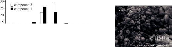
ZnO nanoparticles were synthesized from the decomposition of precursors 1—2 in 600 C in the air for 4 h (Fig. 1). The morphology and size of the as-prepared ZnO samples were further investigated using scanning electron microscopy (SEM). The histographs showed that the average size of ZnO nanoparticles in two compounds is 50—60 nm and 60—70 nm respectively (Fig. 2). Surfactants are often used to synthesize nanostructures in order to change the morphology and particle size. To investigate the role of surfactant on the particle size of zinc(II) oxide nanostructures produced by compound 1, the calcination of compound 1 was performed in the presence of oleic acid as a surfactant at 180 C and in the absence of surfactant at 600 C (Fig. 3). Comparison of the SEM images of ZnO nanoparticles at two different methods and temparatures (180 C and 600 C) shows that the higher temperature (600 C) results in an increasing of agglomeration and thus small and spherical ZnO particles with good separation were produced by the thermolysis of compound 1 at 180 C and by the use of surfactant. The histograph of the ZnO nanostructure shows that the maximum dispersion range size is about 40—50 nm in this method. The final product obtained by the decomposition of compounds 1 and 2 is hexagonal ZnO, according to their XRD patterns. The phase purity of the as-prepared hexagonal ZnO nano-particles is completely obvious and all diffraction peaks are perfectly indexed to the hexagonal ZnO structure with the lattice parameters a = 3.24982 Å, c = 5.20661 Å, Z = 2 and S.G = P63mc, which are in JCPDS card file No. 36-1451. No characteristic peaks of impurities are detected in the XRD pattern.
CONCLUSIONS
Small and spherical ZnO particles with good separation were produced by the thermolysis of compounds 1—2 with and without the use of surfactant. This study demonstrates that zinc(II) com-
Fig. 1. SEM photograph of ZnO nanoparticles produced by calcination of a) precursor 1 and b) precursor 2 at 600 C
338 |
M. HOSSEINIFARD, L. HASHEMI, V. AMANI, A. MORSALI |
Fig. 2. Histograph of ZnO nanoparticles produced by |
Fig. 3. SEM image of ZnO nanostructure pre- |
calcination of precursors 1—2 at 600 C |
pared by the thermal decomposition of compound |
|
1 with oleic acid as a surfactant at 180 C |
plexes may be suitable precursors for the preparation of nanoscale materials and do not need special conditions such as high temperature, long times and pressure controlling [ 16—18 ]. This method of preparation may have some advantages: it proceeds within shorter reaction times, produces better yields and is also likely to produce nano-sized particles of the complex.
Acknowledgments. Support of this investigation by Islamic Azad University (Shahr-e-Rey branch) and Tarbiat Modares University are gratefully acknowledged.
REFERENCES
1.Ennaoui A., Weber M., Scheer R., Lewerenz H.J. // Sol. Energy Mater. Sol. Cells. – 1998. – 54. – P. 277 – 302.
2.Liqiang J., Baiq W., Baifu X., Shudan L., Keying S., Weimin C., Honggang F. // J. Solid State Chem. – 2004.
–177. – P. 4221 – 4227.
3.Shinde V.R., Gujar T.P., Lokhande C.D. // Sens. Actuators B. – 2007. – 120. – P. 551 – 559.
4.Ayudhya S.K.N., Tonto P., Mekasuwandumrong O., Pavarajarn V., Praserthdam P. // Cryst Growth Des.
–2006. – 6. – P. 2446 – 2450.
5.Vafaee M., Ghamsari M.S. // Mater. Lett. – 2007. – 61. – P. 3265 – 3268.
6.Kim Y.S., Tai W.P., Shu S.J. // Thin Solid Films. – 2005. – 491. – P. 153 – 160.
7.Liu J.Z., Yan P.X., Yue G.H., Kong L.B., Zhuo R.F. // Mater. Lett. – 2006. – 60. – P. 3471 – 3476.
8.He Y., Wang J. // Mater. Lett. – 2008. – 62. – P. 1379 – 1383.
9.Gao P.X., Wang Z.L. // Appl. Phys. Lett. – 2004. – 84. – P. 2883 – 2885.
10.Vayssieres L. // Adv. Mater. – 2003. – 15. – P. 464 – 466.
11.Liu C.H., Zapien J.A., Yao Y., Meng X.M., Lee C.S., Fan S.S., Lifshitz Y., Lee S.T. // Adv. Mater. – 2003.
–15. – P. 838 – 841.
12.Zhang H., Ma X.Y., Xu J., Niu J., Yang D. // Nanotechnology. – 2003. – 14, N 4. – P. 423 – 426.
13.Bigdeli F., Morsali A. // Mater. Lett. – 2010. – 64. – P. 4 – 5.
14.Alizadeh R., Kalateh Kh., Ebadi A., Ahmadi R., Amani V. // Acta Crystallogr. – 2009. – E65. – P. m1250.
15. Alizadeh R., Kalateh Kh., Khoshtarkib Z., Ahmadi R., Amani V. // Acta Crystallogr. – 2009. – E65.
– P. m1439 – m1440.
16.Rashidi Z., Morsali A. // J. Mol. Struct. – 2009. – 936, N 1-3. – P. 206 – 212.
17.Hashemi L., Morsali A. // J. Inorg. Organomet. Polym. – 2010. – 20, N 4. – P. 856 – 861.
18.Hashemi L., Morsali A., Retailleau P. // J. Inorg. Chim. Acta. – 2011. – 367. – P. 207 – 211.