

Budras_Анатомия лошади
.pdfInguinal Rings and Vicinity in the Stallion ( )
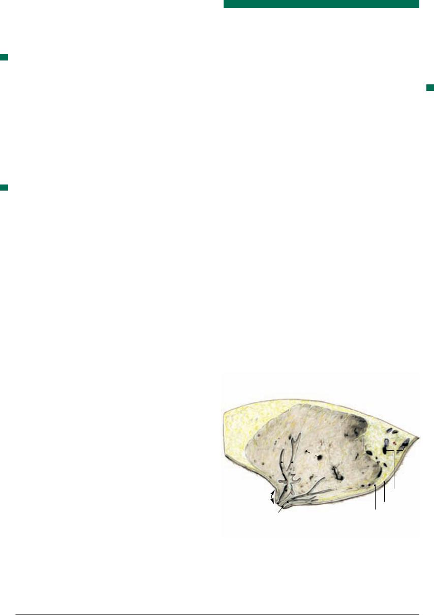
)
|
i |
|
|
|
|
|
|
|
|
|
|
|
|
|
|
|
|
|
|
||||||||
|
1 |
|
|
|
|
|
|
|
|
|
|
|
|
|
|
|
|
|
|
|
|
|
|
|
|
|
|
|
4 |
|
|
|
|
|
|
|
|
||||
|
|
|
|
|
|
|
|
|
|||||
|
5 |
|
|
|
|
|
|||||||
|
|
|
|
|
|
|
|||||||
|
|
|
|
|
|
|
|
||||||
|
6' |
|
6'' |
||||||||||
|
6 |
||||||||||||
|
8 |
|
|
|
|
|
|||||||
|
|
|
|
|
|
||||||||
a |
Iliocostalis |
8'' |
|
|
|||||||||
b |
Longissimus dorsi |
|
|
||||||||||
|
|
|
|||||||||||
c |
Multifidus |
8' |
|
|
|||||||||
|
|
|
|
|
|
|
|
|
|
|
|
|
|
dPsoas major
ePsoas minor
fCaudal vena cava
gAbdominal aorta
hUreter
iCoxal tuber
jInternal iliac fascia
kDescending mesocolon
l Descending colon |
9 |
mLeft colic vessels
nBladder
oLat. lig. of bladder
pPenis
qEpididymis
rTestis
sScrotum
b
|
|
a |
|
|
|
|
|
|
|
d |
|
1 |
Inguinal ligament |
j |
h |
|
|
|
|||||
i |
|
|
|
||
2 |
Lumbar nerve L2 |
|
|
|
|
3 |
Genitofemoral nerve |
|
|
|
|
4 |
Cremaster |
|
|
|
l |
|
|
|
|
|
|
5Internal abdominal oblique
6Deep inguinal ring
6' Lateral angle
6'' Medial angle
6'
7 Deferent duct
14
8 Abdominal tunic
8' External spermatic fascia
8'' Femoral lamina 8' 7
8''' Suspensory lig. of penis
8''
8'''
9 Skin
13
18
 15
15
8'''
12'
p
20
c
vL3
f g
3
k
o
n
6''
13'
13''
16
s
13'
5
10'
10''
12'
4
p
13''
q
r
s
12
10
10 External abdominal oblique 10' Pelvic tendon
10'' Abdominal tendon
11 Transversus abdominis
12 Transverse fascia
12' Internal spermatic fascia
13Peritoneum 13' Vaginal ring
13'' Vaginal process
14External pudendal vessels
15Rectus abdominis
16Supf. inguinal ring
17Testicular vessels
18Linea alba
19 Fascia lata
20 Scrotal septum
(vide p. 73, 77)
75
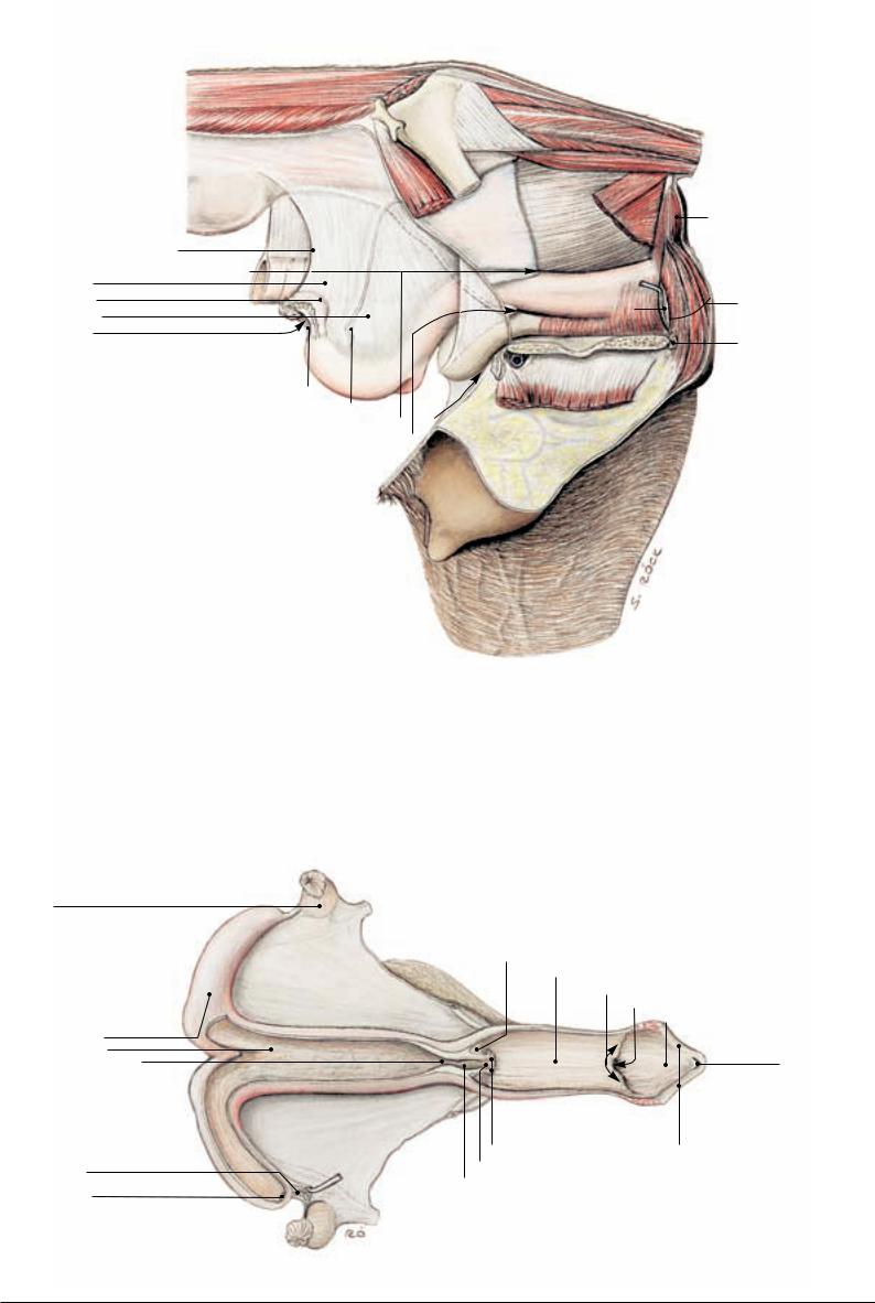
3.Prepubic Tendon, Inguinal Canal of the Mare, Nerves of the Lumbar Plexus, Hypaxial Lumbar Muscles, and Udder
a)PREPUBIC TENDON
1The prepubic tendon (2) is a strong, transversely oriented, fibrous mass set against the pecten of the pubis and extending from one iliopubic eminence to the other. The prepubic tendon is formed principally by the right and left pectineus (and adductor longus) muscles (5) whose tendons of origin, after arising from the pecten on both sides of the median as far laterally as the iliopubic eminences, decussate ventromedial to the pelvic symphysis. (The tendinous cranial parts of the gracilis and external obturator muscles also take part in its formation.)
The inguinal ligament (1) together with the pelvic tendon of the external abdominal oblique join the lateral parts of the prepubic tendon. The accessory ligament (4), the insertion tendon of the rectus abdominis, perforates, but also attaches itself to, the prepubic tendon and eventually passes via the acetabular notch to the fovea on the femoral head, accompanied the last few cm by the ligament of the femoral head (3).
2The linea alba (14) is formed by the convergence of the aponeuroses of the flat abdominal muscles in the ventral midline. It is several cm wide near the umbilicus, but narrows to a few mm as it joins the prepubic tendon between the bellies of the rectus muscles.
b)STRUCTURES ASSOCIATED WITH THE INGUINAL CANAL OF THE MARE
The evaginations of the peritoneum (19, not shown on this side but at 12), the transverse fascia (12), and the abdominal tunic (13) are much less prominent than in the male, not always present, and difficult to demonstrate. The round ligament of the uterus (11) contains small amounts of smooth and striated muscle and presents a small, pendulous appendage near its origin from the uterine horn.
It and a rudimentary cremaster muscle can be traced through the inguinal canal to where they end at the base of the udder. (In the specimen from which the lower Fig. on the facing page was drawn, the cremaster [see p. 75.4] was relatively well developed.)
c) THE NERVES OF THE LUMBAR PLEXUS (VENTRAL BRANCHES OF L2
TO L6)
Nerve L1 continues solely the ventral branch of the first lumbar nerve and thus cannot be regarded as belonging to the lumbar plexus, though it is often described under that heading. It takes a subperitoneal course to the vicinity of the deep inguinal ring and takes part in the innervation of the caudal parts of the internal abdominal oblique and the transversus abdominis muscles. Its sometimes double lateral cutaneous branch perforates (and also supplies) the abdominal muscles to innervate the skin and the fold of the flank.
Nerve L2 (18; with contribution from L3) also supplies the skin and the fold of the flank with its lateral cutaneous branch. It crosses the ventral surface of the deep circumflex iliac vessels (9), shortly before the latter penetrate the flank, to pass with one or two branches through the lateral part of the supf. inguinal ring.
The lateral cutaneous femoral nerve (10; from L3 and 4) obliquely crosses the dorsal surface of the deep circumflex iliac vessels and then accompanies their caudal branch through the body wall to the flank fold.
The genitofemoral nerve (20; from L2 to 4) crosses the deep circumflex iliac vessels more medially with one or two branches and passes through the medial part of the supf. inguinal ring.
The femoral and, its branch, the saphenous nerves (see p. 19.12; 19.25; from L4 to 6) were described on p 18.
Also the obturator nerve (see p. 19.5; from L4 to 6) was already described on p. 18.
Clinical and Functional Anatomy p. 173
d) HYPAXIAL LUMBAR MUSCLES
For psoas major (7), psoas minor (8), and quadratus lumborum
(not shown) consult the Tables on the musculature on p. 99.
e) UDDER
The udder of the mare lies ventral to the area where the ventral 3 abdominal wall joins the bony pelvis. Its shape and size are related
to the functional state of the ovaries. It is a relatively small gland, hardly noticeable in the virgin mare but rounded and semispherical during the latter part of pregnancy and subsequent lactation. One hair (or more) protruding from the teat orifice, as well as other less obvious features remind us that the mammary gland is an enormously enlarged apocrine sweat gland. The hair disappears at the beginning of lactation or is worn off by the foal during suckling. Secretions of both sebaceous and sweat glands in the tip of the teat (or perhaps precocious colostrum) fill the two teat orifices and cover the tip of the teat with a waxy material whose presence indicates that foaling is imminent.
The udder (see Fig. on this page) comprises right and left mammary glands (15) each surmounted by a teat (17). Each gland accommodates two (sometimes three) duct systems which channel the milk through increasingly larger lactiferous ducts (B) into a lactiferous sinus (A) in the base of the teat and in the teat itself (teat sinus). Two papillary ducts (C) ending at the teat orifices (D) convey the milk to the outside. Though the two duct systems in each half of the udder are separate, the glandular tissue belonging to each can only be demonstrated by injections of different color suspensions into the two orifices. (For blood and nerve supply of the udder see p. 80.)
The udder is supported by a suspensory apparatus (21) that comprises medial and lateral laminae. The paired medial lamina (21') is elastic and arises from the abdominal tunic near the linea alba and separates the two halves of the udder along the prominent intermammary groove (16). The lateral laminae (21'') are less elastic though they arise also from abdominal tunic but in the vicinity of the supf. inguinal rings from where they pass lateral to the glandular tissue and separate it from a layer of adjacent fat.
Udder, sagittal section through a teat
(Cranial end) |
(Caudal end) |
15
|
17 |
|
|
B |
|
|
Mammary blood |
||
|
|
|
|
||||||
|
|
|
|
|
|
|
A |
||
|
|
|
|
|
|
|
|||
|
|
|
|
|
|
|
|
||
|
|
|
|
|
|
|
|
vessels |
|
|
|
|
|
|
|
|
|
|
|
|
|
|
|
|
|
|
|
|
Skin |
|
|
|
|
|
|
|
|
|
|
|
|
|
|
|
|
|
|
|
Subcutis |
|
D |
|
C |
||||||
|
|
|
|||||||
A |
Lactiferous sinus |
|
|
|
|
|
C |
Papillary duct |
|
B |
Lactiferous duct |
|
|
|
|
|
D |
Teat orifice |
|
76
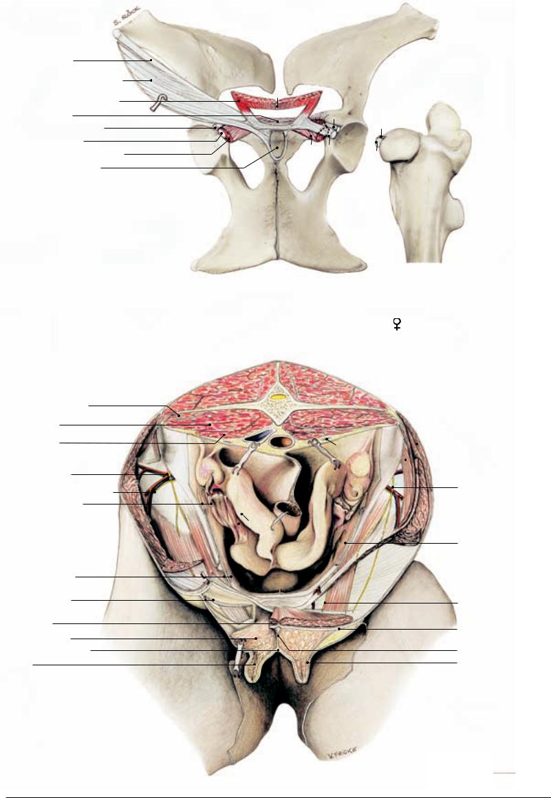
Prepubic Tendon, Inguinal Ligament, and Accessory Ligament
1 Inguinal ligament
Pelvic tendon of ext. abdominal oblique
1' Rectus abdominis, fenestrated
2Prepubic tendon
3Ligament of head of femur
4Accessory ligament
5Pectineus (and adductor longus) Symphysial tendon fossa
(ventral view)
14
3
3
5 4
4
|
Inguinal Rings and Vicinity of the Mare ( |
|
|
) |
|
|
a Suspensory lig. of ovary |
|||||||||||||
|
|
|
|
|
||||||||||||||||
|
|
|
|
|
b |
Ovary |
||||||||||||||
|
|
|
|
(cranioventral view) |
|
|
|
|
c Proper lig. of ovary |
|||||||||||
|
|
|
|
|
|
|
|
d Uterine horn and mesometrium |
||||||||||||
|
|
|
|
|
|
|
|
|
|
|
|
|
|
|
|
|
|
|
||
|
|
|
|
|
|
|
|
|
|
|
|
|
|
|
|
|
|
|
e |
Caudal epigastric vessels |
|
|
|
|
|
|
|
|
|
|
|
|
|
|
|
|
|
|
|
f |
External pudendal vessels |
|
|
|
|
|
|
|
|
|
|
|
|
|
|
|
|
|
|
|
g |
Ureter |
|
|
|
|
|
|
|
|
|
|
|
|
|
|
|
|
|
|
|
h |
Descending colon |
6 Quadratus lumborum |
|
|
|
|
|
|
|
|
|
|
|
|
|
|
|
|
|
|
i |
Bladder |
|
|
|
|
|
vL3 |
|
|
|
|
j |
Cremaster |
|||||||||
|
|
|
|
|
|
|
|
|
|
|||||||||||
|
|
|
|
|
|
|
|
|
|
k |
Internal abdominal oblique |
|||||||||
7 Psoas major |
|
|
|
|
|
|
|
|
|
|
|
|
|
|
|
|
|
|
||
|
|
|
|
|
|
|
|
|
|
|
|
|
|
|
|
|
|
l |
External abdominal oblique |
|
|
|
|
|
|
|
|
|
|
|
|
|
|
|
|
|
|
|
|
||
8 Psoas minor |
|
|
|
|
|
|
|
|
|
|
|
|
|
|
|
|
|
l |
m Transversus abdominis |
|
|
a |
|
|
|
|
|
|
|
|
|
g |
|
|
|
n |
Rectus abdominis |
||||
|
|
|
|
|
|
|
|
|
|
|
|
|
||||||||
|
|
|
|
|
|
|
|
|
|
|
|
|
|
|
||||||
|
|
|
|
|
|
|
|
|
|
|
|
|
|
|
|
|
|
|
|
|
9 Deep circumflex |
|
|
b |
|
|
|
|
|
|
m |
|
|
|
|
|
|
|
|||
|
|
|
|
|
|
|
|
|
|
|
||||||||||
|
|
|
|
|
|
|
|
|
|
|
|
|
|
|
|
|
||||
|
|
|
|
|
|
|
|
|
|
|
|
|
|
|
|
|
|
|
|
|
iliac vessels |
|
|
c |
|
|
|
|
|
|
|
|
|
|
|
|
|
|
18 Lumbar nerve L2 |
||
10 Lat. cutaneous femoral nerve |
|
|
|
|
|
|
|
|
|
|
|
|
|
|
|
|
||||
|
|
|
|
|
|
|
|
|
|
|
|
|
|
|
|
|
|
|
||
|
|
|
|
|
|
|
|
|
|
|
|
|
|
|
|
|
|
|
|
|
11 Round lig. of uterus |
|
|
|
|
|
|
|
|
|
|
|
|
|
|
|
|
|
|
|
|
|
j |
|
|
|
d |
|
|
h |
|
|
|
|
|
|
||||||
|
|
|
|
|
|
|
|
|
|
|
|
|
|
|
|
|
|
|
||
|
|
|
|
|
|
|
|
|
|
|
|
|
|
|
|
|
|
|
|
|
|
k |
|
|
|
|
|
|
|
|
|
|
|
|
|
|
|
|
|
|
19 Peritoneum |
|
|
|
|
|
|
|
|
|
|
|
|
|
|
|
|
|
|
|
|
|
12 Transverse fascia |
|
|
|
|
|
|
i |
|
|
|
|
|
|
|||||||
|
|
|
|
|
|
|
|
|
|
|
|
|
||||||||
13 Abdominal tunic |
|
|
|
|
|
|
|
f |
|
|
|
e |
|
|
|
|
|
20 Genitofemoral nerve |
||
|
|
|
|
|
|
|
|
|
|
|
|
|
|
|
||||||
|
|
|
|
|
|
|
|
|
|
|
|
|
|
|
|
|
|
|||
14 Linea alba |
|
|
|
|
|
|
|
|
|
|
|
|
|
|
||||||
|
|
|
|
|
|
|
n |
|
|
|
|
|
21 Suspensory apparatus |
|||||||
|
|
|
|
|
|
|
|
|
|
|
|
|||||||||
|
|
|
|
|
|
|
|
|
|
|
|
|
|
|
|
|
|
|
|
|
15 Mammary gland |
|
|
|
|
|
|
|
|
|
|
|
|
|
|
|
|
|
|
|
of udder |
16 Intermammary groove |
|
|
|
|
|
|
|
|
|
|
|
|
|
|
|
|
|
|
|
21' Medial lamina |
17 Teat |
|
|
|
|
|
|
|
|
|
|
|
|
|
|
|
|
|
|
|
21'' Lateral lamina |
(See p. 73 and 75)
77
1
2
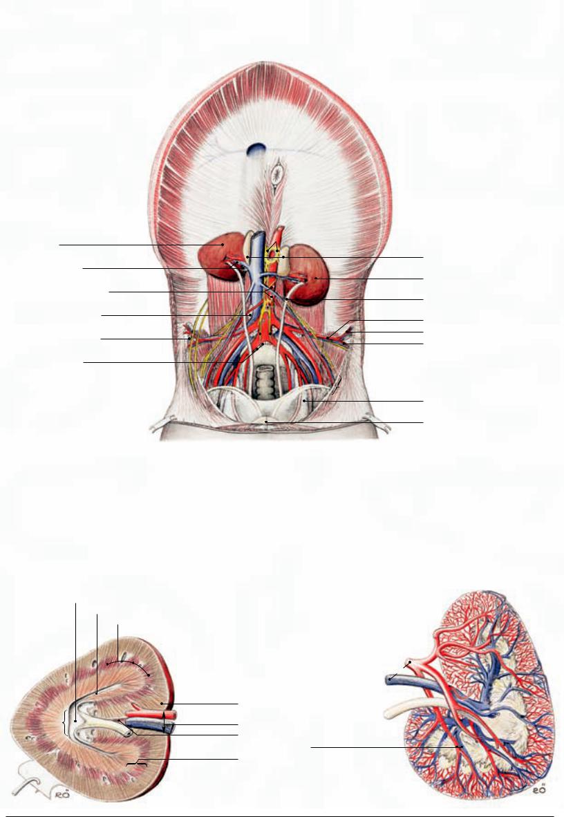
4. Lymphatics, Adrenal Glands, and Urinary Organs Clinical and Functional Anatomy p. 173–174
a) THE LYMPHATICS OF THE PELVIC AND LUMBAR AREAS
I. Lymph nodes outside the abdominal and pelvic cavities.
The supf. inguinal lymph nodes (see p. 85.s) in the male lie on each side of the penis and extend to the scrotum (scrotal lymph nodes); in the female they extend to the base of the udder (mammary lymph nodes).
The subiliac lymph nodes, which lie in the flank fold halfway between the coxal tuber and the patella, were described with the hindlimb (see p. 19.D) and with the abdominal wall (see p. 65.10).
The deep inguinal lymph nodes were mentioned in connection with the femoral triangle (see p. 19.B).
II. The retroperitoneal lymph nodes at the junction of abdomen and pelvis.
The sacral lymph nodes (6) lie caudal to the aortic bifurcation between the origins of right and left internal iliac arteries.
The medial iliac lymph nodes (4) surround the break-up of the abdominal aorta a little more cranial than the preceding nodes; some lie at the origin of the deep circumflex iliac vessels (10).
The lateral iliac lymph nodes (5) are located at the break-up of the deep circumflex iliac vessels into cranial (10') and caudal (10'') branches.
The lumbar aortic lymph nodes (3) continue the medial iliac nodes cranially along the abdominal aorta.
The renal lymph nodes (2) lie near the renal hilus between the branches of the renal vessels; they are not easily distinguished from neighboring lumbar aortic nodes.
Lymph passing through the afore-mentioned nodes enters the cisterna chyli which at the level of the kidneys accompanies the aorta on its right dorsal aspect. The cisterna is continued cranially by the thoracic duct which conveys the lymph to one of the veins at the thoracic inlet.
b)The paired ADRENAL GLANDS (7) are about 8 cm long, have an irregular surface, and are bilaterally compressed. Their yellowishbrown color distinguishes them from the more grayish-brown lymph nodes. The left gland lies against the left kidney cranial to its hilus. The right one lies between the hilus of the right kidney and the caudal vena cava.
c)THE URINARY ORGANS
The right kidney (1), shaped like the heart on a playing card, lies with its cranial pole in the renal impression of the liver, and makes contact dorsally the diaphragm. The base of the cecum is attached to its ventral surface. The bean-shaped left kidney (8) lies medial to the spleen to which it is bound by the renosplenic ligament (see p.
69.12). It is ventral to the last rib and the first two lumbar transverse processes and thus half a kidney's length caudal to the right kidney which lies ventral to the last two ribs and the first lumbar transverse process.
The kidneys are smooth on the surface. The renal lobes (15) have completely fused so that their original limits are revealed only by the course of the interlobar arteries and veins (19). When sectioned, the kidney discloses a granular reddish-brown cortex (16) which is easily distinguished from the dark-red external (20') and the paler internal (20”) part of the smooth medulla (20). The papillary ducts of the central parenchyma open on the small renal crest (20''') which juts into the equally small renal pelvis (13) located in the center of the organ. The papillary ducts near the poles of the kidney open into the two so-called terminal recesses (14). These are about 10 cm long, have a diameter of about 0.5 cm, and are cranial and caudal extensions of the renal pelvis, though they can also be regarded as overgrown collecting ducts without walls.
The renal sinus (18), the indentation on the medial border of the organ that gives passage to the ureter and the lymphand blood vessels, contains the renal pelvis at its depth and forms the hilus (17) where ureter and blood vessels emerge. Some branches of the renal arteries do not utilize the hilus but penetrate the renal parenchyma from the ventral surface.
I. The wall of the renal pelvis—and that of the initial segment of the ureter—contain mucous glands that can be detected with the naked eye; they give the horse's urine its cloudy and slimy quality.
II. The ureters (9), after emerging from the hilus, lie retroperitoneally on each side of the two great abdominal blood vessels (aorta and caudal vena cava). Their pelvic part descends towards the dorsal surface of the bladder whose wall it penetrates obliquely; the
pelvic part in the male briefly lies in the mesoductus deferens and 2 crosses the dorsal surface of the deferent duct.
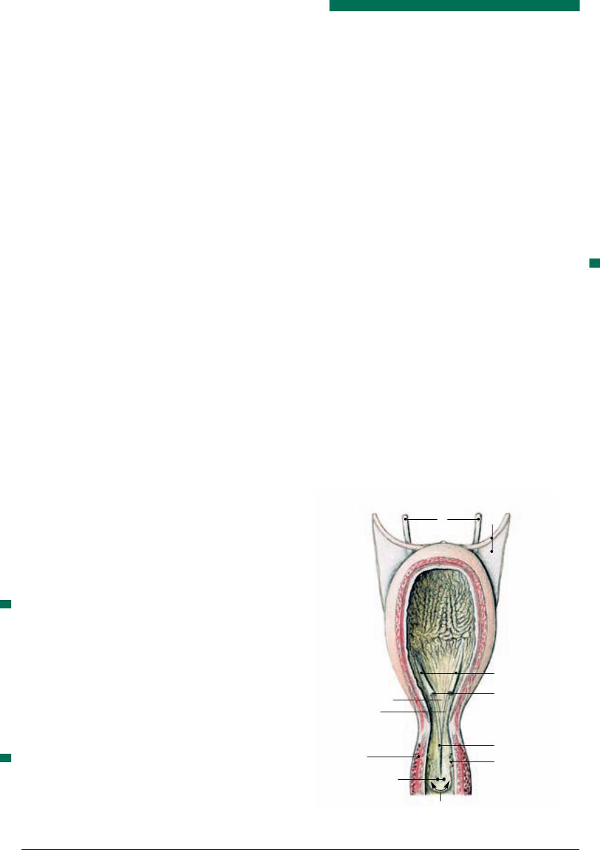
III. The bladder (see text Fig. below), when empty, has the size of a small fist and resides entirely within the pelvic cavity in a largely retroperitoneal position. When moderately filled, its apex (21) and body (22) hardly protrude into the abdominal cavity; but when filled to capacity, the apex may advance to the level of the umbilicus. The neck of the bladder (23), however, always remains within the pelvic cavity. The lateral ligaments of the bladder (11) carry the nearly obliterated umbilical artery (round ligament) in their free border (the artery arises from the cranial or caudal gluteal arteries). The median ligament of the bladder (12) connects the bladder to the pelvic floor and to the linea alba, and contains smooth muscle tissue.
IV. The male urethra (see p. 85.p) within the pelvic cavity (pelvic 3 part) is 12 cm long; its lumen is narrow at the level of the prostate and at the ischial arch where it is continuous with the spongy penile part.
V. The female urethra (see p. 83), only 6 cm long, is short but compensates with a wide lumen throughout. This, plus its ability to dilate, occasionally permits a prolapse of the bladder through the urethra when bladder mucosa becomes visible in the vestibule of the vagina.
3
Urinary Bladder, with Pelvic Part of Male Urethra
9 11
21
(ventral view) |
22 |
Ureteric columns

 Ureteric orifices Trigonum vesicae
Ureteric folds 
 23
23
Urethral crest
Urethralis
Prostatic ductules
Ejaculatory orifices
Colliculus seminalis
78
Abdominal Cavity and Urinary Organs
(ventral view)
a
1 |
Right kidney |
|
|
|
|
|
c |
|
|
|
|
|
|
||
2 |
Renal lymph nodes |
|
|
|
|
|
b |
|
|
|
|
|
|
|
|
3 |
Lumbar aortic lymph nodes |
|
|
|
|
|
|
4 |
d |
|
|
h |
|
||
Medial iliac lymph nodes |
|
|
|
|
|
|
|
|
e |
f |
|
|
|
|
|
|
|
|
|
i |
|
|
|
5 |
Lateral iliac lymph nodes |
|
|
|
|
|
|
|
|
|
|
|
|||
k |
l |
|
|||||
|
|
j |
|||||
|
|
g |
|
|
|
|
|
6 |
Sacral lymph nodes |
|
|
|
|
|
|
|
|
|
|
m |
p |
||
|
|
|
|
n o |
|||
q
7Adrenal glands
8Left kidney
9Ureter
10Deep circumflex iliac vessels 10' Cranial branch
10'' Caudal branch
11Lateral and round ligaments of bladder
12Median ligament of bladder
(See p. 63 and 69)
a |
Caval foramen |
f |
Lat. cutaneous femoral nerve |
k |
Ext. iliac artery |
p |
Int. pudendal vessels |
b |
Renal vessels |
g |
Testicular vessels |
l |
Int. iliac artery |
q |
Deferent duct |
c |
Celiac and cran. mesenteric ganglia |
h |
Caud. mesenteric artery and ganglion |
m |
Rectum |
r |
Deep inguinal ring |
d |
Iliohypogastric nerve (L1) |
i |
Hypogastric nerves |
n |
Obturator vessels |
s |
Interlobular vessels |
e |
Ilioinguinal nerve (L2) |
j |
Genitofemoral nerve |
o |
Umbilical artery |
|
|
Right Kidney
sectioned through hilus and poles
13 Renal pelvis
14 Terminal recesses
15 Renal lobes
20''' |
9 |
|
20'' 14
20'
16 Renal cortex
17 Renal hilus
18 Renal sinus
19 Interlobar vessels
20 Medulla
20' External part 20'' Internal part 20''' Renal crest
Left Kidney
cast of renal vessels and renal pelvis
b
9
13
s
79

5. Arteries, Veins, and Nerves of the Pelvic Cavity Clinical and Functional Anatomy p. 174
1a) The ABDOMINAL AORTA (2) begins its break-up already at the level of the 5th lumbar vertebra. It gives rise to the paired external and internal iliac arteries, and to the unpaired median sacral artery which is occasiomnally absent.
The external iliac artery (10), seldom the internal iliac, in the mare releases the uterine artery (11), the principal artery of the uterus, and in the stallion it releases the cremasteric artery (11) which accompanies the testicular artery (from the aorta) through the vaginal ring. Before entering the vascular lacuna to gain the thigh, the external iliac gives rise to the deep femoral artery (12) which in turn gives off the pudendoepigastric trunk (13). This immediately gives off the external pudendal artery (15) which passes through the inguinal canal. The external pudendal, among other branches, supplies vessels for the udder and the cranial artery of the penis (19) which anastomoses on the dorsal surface of the penis with the middle artery of the penis (46; from the obturator) and the dorsal artery of the penis (38; from the internal pudendal).
The internal iliac artery (22), as does the abdominal aorta and sometimes also the caudal gluteal, sends lumbar arteries (3) to the vertebral column and associated structures, and immediately (opposite the 6th lumbar vertebra) divides into the caudal gluteal (47) and internal pudendal arteries (23). Of these the former is destined predominantly for the pelvic wall, while the latter goes to the pelvic viscera.
The internal pudendal artery (23), close to its origin, gives off the umbilical artery (24) which, in the stallion, releases the small artery of the deferent duct (25), and in both sexes the cranial vesical artery (26) and ends as the round ligament of the bladder (27) in the free border of the lateral ligament of the bladder. The internal pudendal then courses along the medial surface of the sacrosciatic ligament in the vicinity of the pudendal nerve and with its next branch, the vaginal artery (prostatic in the male) (28), supplies most of the pelvic viscera: branch to deferent duct or uterine branch (29), caudal vesical artery (30), ureteric branch (31), urethral branch (32), middle rectal artery (33), ventral perineal artery (40), and the caudal rectal artery (41). The internal pudendal itself is continued by the artery of the penis (36); the clitoris receives its supply from the middle clitoral artery, a branch of the obturator artery.
The caudal gluteal artery (47) and its branches, especially the cranial gluteal artery (49) that exits by the greater sciatic foramen, supply the dorsolateral wall of the pelvis and the croup. The obturator artery (44), arising either from the caudal or cranial gluteal, is the exception: it passes caudoventrally on the medial aspect of the shaft of the ilium and leaves the pelvic cavity together with its satellite vein and nerve by the obturator foramen. Below the floor of the pelvis it detaches the middle artery of the penis (clitoris) (46).
b) The VEINS of the pelvic cavity by and large are satellite to the arteries; deviations are shown in the Figures on the opposite page.
The external pudendal vein (15) that accompanies its satellite artery through the inguinal canal is very thin. Its blood-return function is assumed by the accessory external pudendal vein (16) which collects most of the veins from penis or udder and delivers the blood to the ipsilateral deep femoral vein (branch of external iliac) at the cranial end of the pelvic symphysis. Right and left accessory external pudendal veins anastomose here (in the symphysial fossa; see p.
73.3) across the median plane; it is the transected anastomosis that attracts attention after the hind quarters of the cadaver are split (see p. 83). The large obturator vein (44) assists the accessory external pudendal vein in draining penis and udder.
The blood supply of penis and udder follows in greater detail.
c) The BLOOD SUPPLY OF THE PENIS derives from three sets of vessels: internal pudendal artery and vein, obturator artery and vein, and external pudendal artery and the accessory external pudendal vein. The internal pudendal artery (23) becomes the artery of the
penis (36) which gives rise to the artery of the bulb (37) for the bulb and corpus spongiosum, and to the dorsal artery of the penis (38) which anastomoses with the middle (46) and cranial (19) arteries of the penis on the dorsal surface of the organ. The deep artery of the penis (39) for the corpus cavernosum carries blood from the obturator artery. The veins form an extensive plexus dorsal and lateral to the penis whose blood enters the accessory external pudendal vein (16) but also the obturator (44) and internal (23) pudendal veins.
d) The BLOOD SUPPLY OF THE UDDER comes fom the external pudendal artery via the caudal supf. epigastric (cranial mammary artery; 20) and from the internal pudendal artery via the dorsal labial branch that anastomoses with the ventral labial branch (caudal mammary artery; 18) of the external pudendal.
The venous drainage is mainly by the accessory external pudendal vein (middle mammary vein; 16) into the deep femoral, but also by the caudal supf. epigastric vein (cranial mammary vein; 20) and by the ventral labial vein of the external pudendal (caudal mammary vein; 18) which anastomoses with the dorsal labial vein of the internal pudendal.
e)The BLOOD SUPPLY OF THE UTERUS comes principally from the uterine artery (11), a branch of the external (rarely internal) iliac.
The uterine artery anastomoses cranially with the uterine branch of the ovarian artery (5) and caudally with the uterine branch of the vaginal artery (29).
f)The SACRAL PLEXUS OF NERVES is the continuation of the lumbar plexus; together they form the lumbosacral plexus (see p. 76).
The cranial gluteal nerve (L6–S2; f) accompanies the like-named artery through the greater sciatic foramen to supply the gluteus medius, accessorius, and profundus, and also the tensor fasciae latae (see p. 19.8).
The sciatic nerve (L5–S2; g) also emerges from the greater sciatic foramen. Lying on the sacrosciatic ligament, it passes the hip joint dorsally and caudally, and as the largest nerve of the plexus enters the pelvic limb.
The caudal gluteal nerve (L6–S2; h) passes also through the greater sciatic foramen and accompanies like-named blood vessels into the gluteus supf. and into the vertebral heads of biceps and semitendinosus.
The caudal cutaneous femoral nerve (S1–S2; i) at first follows the dorsal border of the sciatic nerve but as the latter turns ventrally into the limb, it passes over the ischial tuber to end subcutaneously on the caudal surface of the thigh.
The pudendal nerve (S2–S4; j) at first lies on the medial surface of the sacrosciatic ligament, and then within the ligament, in which position it reaches the lesser sciatic foramen. Here it communicates with the caudal cutaneous femoral nerve. Before accompanying the internal pudendal vessels to the penis (clitoris), it gives rise to the deep perineal nerve (l) for the striated muscles of the perineum and the supf. perineal nerve (m) which is sensory to the vulva and perineal body.
The caudal rectal nerve (S4, S5; k) passes caudoventrally and supplies sensation to rectum, anal canal, and perineum; it also contributes to the motor innervation of the perineal musculature.
g) For the AUTONOMIC NERVOUS SYSTEM at the entrance to the pelvic cavity we need merely mention that the caudal mesenteric ganglion
(o) lies cranioproximal to the origin of the caudal mesenteric artery
(7), and that the hypogastric nerve (p), which begins here, at first follows the roof of the cavity and then descends to the pelvic plexus
(q) on the viscera. The parasympathetic pelvic nerves (r) arise from the roots of the pudendal and caudal rectal nerves, pass ventrally, and join the pelvic plexus.
80

Arteries, Veins, and Nerves of the Pelvic Cavity
1Caudal vena cava
2Abdominal aorta
3 Lumbar vessels |
L2 |
4Ovarian or testicular vessels
5Uterine branch
6Pampiniform plexus
7Caudal mesenteric artery
8Common iliac vein
9Deep circumflex iliac vessels
10External iliac vessels
11Uterine or cremasteric vessels
12Deep femoral vessels
13Pudendoepigastric arterial trunk
|
and pudendoepigastric vein |
Lvm 1 |
|
|
||
14 |
Caudal epigastric vessels |
|
|
|||
|
|
|||||
|
|
|
|
|
||
15 |
External pudendal vessels |
|
|
|
|
|
16 |
Accessory ext. pudendal vein |
|
|
|
|
|
|
[middle mammary vein] |
|
|
|
|
|
17 |
Venous plexus of penis |
|
|
|
|
|
18 |
Ventral labial (scrotal) arterial branch and |
|
|
|
|
|
|
v. [caudal mammary vessels] |
|
|
|
|
|
19 |
Cranial vessels of penis |
|
|
|
|
|
20 |
Caudal supf. epigastric vessels [cranial |
|
|
|
|
|
|
mammary vessels] |
|
|
|
|
|
21 |
Medial circumflex femoral vessels |
|
|
|
|
|
22 |
Internal iliac vessels |
|
|
|
|
|
23 |
Internal pudendal vessels |
|
|
|
|
|
24 |
Umbilical artery |
|
|
|
|
|
25 |
Vessels of deferent duct |
|
|
|
|
|
26 |
Cranial vesical artery |
|
|
|
|
|
27 |
Round ligament of bladder |
|
|
|
|
|
28 |
Vaginal or prostatic artery |
|
|
|
|
|
29 |
Uterine branch or branch to deferent duct |
|||||
30 |
Caudal vesical vessels |
|
|
|
|
|
31 |
Ureteric branches |
|
|
|
|
|
32 |
Urethral branches |
|
|
|
|
|
33 |
Middle rectal vessels |
|
|
|
|
|
34 |
Vestibular branch |
|
|
|
|
|
35 |
Vessels of the vestibular bulb |
|
|
|
|
|
36 |
Vessels of the penis |
|
|
|
|
|
37 |
Vessels of the bulb of the penis |
|
|
|
|
|
38 |
Dorsal vessels of the penis (clitoris) |
|
|
|
|
|
39 |
Deep vessels of the penis (clitoris) |
|
|
|
|
|
40 |
Ventral perineal vessels |
|
|
|
|
|
41 |
Caudal rectal vessels |
|
L2 |
|||
42 |
Dorsal labial branches |
|
||||
43 |
Iliolumbar vessels |
|
|
|
|
|
44 |
Obturator vessels |
|
|
|
|
|
45 |
Iliacofemoral vessels |
|
|
|
|
|
46 |
Middle vessels of the penis (clitoris) |
|
|
|
|
|
47 |
Caudal gluteal vessels |
|
|
|
|
|
48 |
Sacral branches |
|
|
|
|
|
49 |
Cranial gluteal vessels |
|
|
|
|
|
50 |
Median caudal vessels |
|
|
|
|
|
51 |
Ventrolateral caudal vessels |
|
|
|
|
|
52 |
Dorsolateral caudal vessels |
|
|
|
|
|
|
|
Lvm 1 |
|
|
||
|
|
|
||||
aIlioinguinal nerve (L2)
bGenitofemoral nerve
cLateral cutaneous femoral nerve
dFemoral nerve
eObturator nerve
fCranial gluteal nerve
gSciatic nerve
hCaudal gluteal nerve
iCaudal cutaneous femoral nerve
jPudendal nerve
kCaudal rectal nerves
lDeep perineal nerve
mSupf. perineal nerve
nDorsal nerve of penis (clitoris)
oCaudal mesenteric ganglion
pHypogastric nerve
qPelvic plexus
rPelvic nerves
L3 L4
3
2
1
a
4
L3 L4
3
2
1
a
L5
c
b
7 o
5 9
L5
c
b
o
7
9
4
L6 |
|
S1 |
S2 |
S |
S4 |
|
|
|
|
|
3 |
S5 |
|
|
|
|
|
|
|
|
|
|
|
|
48 |
|
|
|
43 |
|
|
|
|
k 47 |
d 8 |
e |
|
f |
r |
|
h |
49 |
|
|
||||
|
|
j |
||||
|
22 |
|
q |
g |
|
|
|
p |
|
23 |
|
i |
|
|
24 |
28 |
|
|
33 |
|
|
10 |
|
|
|||
|
|
|
|
|
|
|
|
|
45 |
|
|
|
|
|
|
29 |
|
|
|
|
|
|
|
30 |
|
|
|
|
11 |
31 |
|
32 |
|
|
|
27 |
|
|
44 |
||
|
|
|
26 |
|
|
|
|
|
|
|
|
|
|
|
|
|
12 |
|
|
|
13 |
|
21 |
|
|
|
|
15 |
16 |
|
|
14
20
L6 |
|
|
S1 |
S2 |
|
S3 |
S4 |
S5 |
|
|
|
|
|
|
|
|
|
|
|
|
|
|
|
48 |
|
|
|
43 |
|
|
|
|
|
|
47 |
d |
8 |
e |
49 |
f |
g |
r |
|
h |
|
|
|
j |
|||||
|
|
q |
|
|
i |
|||
p |
|
22 |
|
23 |
|
|||
|
10 |
24 |
|
|
|
|||
|
|
|
28 |
|
33 |
|||
|
|
|
45 |
|
|
|
29 |
|
|
|
|
|
|
|
|
|
|
|
|
11 |
|
|
31 |
|
32 |
|
|
|
|
27 |
|
|
|
|
|
|
|
|
|
|
30 |
|
44 |
|
|
|
|
|
26 |
|
|
||
|
|
|
|
|
|
|
||
|
|
|
|
12 |
|
|
|
|
|
|
|
|
|
13 15 |
|
21 |
|
|
|
|
|
|
16 |
|||
|
|
|
|
|
|
|
||
|
|
|
14 |
|
|
|
17 |
|
|
|
|
|
|
|
|
|
|
|
|
|
20 |
|
|
|
18 |
|
|
|
|
|
|
|
|
|
|
|
|
|
|
|
|
11 |
25 |
|
|
|
|
19 |
6 |
|
|
|
|
|
|
|
|
|
|
|
||
52 |
||
50 |
51 |
|
l |
41 |
|
m |
||
|
||
n |
40 |
|
34 |
|
35 |
42 |
|
|
||
|
|
|
|
|
|
|
38 |
|
46 |
|
39 |
|
|
|
|
|
|
18 |
|
52
50
51
k
l |
m 41 |
|
n40 |
36
37
46 39
38
81

6. Female Reproductive Organs
1a) The OVARY (10) is relatively very large (about 8 by 5 cm) lies about 10 cm caudal to the kidneys and, when projected onto the skin, the same distance cranioventral to the coxal tuber, i.e., at the level of the 5th lumbar vertebra. The ovary is suspended by the 15 cm long mesovarium (2) whose cranial border (suspensory ligament of the ovary; 1) extends on the sublumbar area towards the diaphagm. The mesovarium splits off the mesosalpinx (3) from its lateral surface and is continued caudally by the mesometrium (4). The three sheets taken together constitute the broad ligament.
The oval ovary of the filly gradually takes on the shape of a bean. This is brought about by regression of the cell masses in the stroma
(cessation of the hormonal influence), more rapid growth at the poles, and progressive indentation at the free border to form the ovulation fossa. The germinal epithelium is concentrated in the area of the fossa which thus is the only region on the surface of the organ where ovulation can occur. The remainder of the ovary is covered by peritoneum (flat mesothelial cells) which has a slightly different color than the germinal epithelium. The parenchymatous zone containing the follicles lies deep to the germinal epithelium and thus surrounds the depth of the ovulation fossa. (In the filly, before the fossa developed, the parenchymatous zone covered (cap-like) the free border of the ovary.) The vascular zone of the adult ovary is relegated to the poles and attached border. The concept of the parenchymatous zone forming a cortex and the vascular zone forming a medulla, as is the case in other domestic mammals, does not apply in the mare.
The surface of the ovary is relativelty flat, and corpora lutea, even at their largest, do not become conspicuous. Likewise, mature follicles, which can attain a diameter of 6 cm, bulge only slightly fom the surface; they are not as easily recognized on rectal exploration as in the cow where the ovaries are considerably smaller.
b) The UTERINE TUBE (14) meanders—parallel to the body axis— from the ovulation fossa to the horn of the uterus. It is suspended in the mesosalpinx (3) that arises from the lateral surface of the mesovarium and ends with a free border a few mm ventral to the uterine tube. The mesosalpinx forms the lateral, and the proper ligament of the ovary (6) the medial wall of the shallow ovarian bursa (5) which has a wide entrance, open ventrally, for easy access to the ovary.
Ovary
Tertiary (vesicular) follicles
Parenchymatous zone (cortex)
Periodic corpus luteum
Tubal end |
|
Uterine end |
of ovary |
|
|
|
of ovary |
|
|
|
Clinical and Functional Anatomy p. 174–176; 180–183 |
|
|
and urethra. The mesometrium that suspends these parts extends |
|
|
from the tip of the uterine horn to the cervix and attaches to the |
|
|
mesometrial border where its two lamellae enclose the triangular (in |
|
|
transverse section) parametrium consisting of many blood vessels |
|
|
embedded in connective tissue. The lateral surface of the |
|
|
mesometrium splits off the round ligament of the uterus (7) that can |
|
|
be traced from the tip of the uterine horn to the vicinity of the |
|
|
inguinal canal. |
|
|
The cervix (27) intervenes without external landmarks between the |
|
|
body of the uterus and the vagina. It is possible, however, on rectal |
|
|
palpation to determine its extent by its firm consistency. Radially |
|
|
arranged longitudinal folds close the cervical canal (27) except dur- |
|
|
ing estrus. The lumen of the uterus gradually narrows to the diam- |
|
|
eter of the cervical canal where the internal uterine ostium (13) is |
|
|
located. The external uterine ostium (26) lies in the center of the |
|
|
intravaginal part (portio vaginalis) of the cervix that occupies the |
|
|
cranial end of the vagina. The longitudinal folds of the cervical |
|
|
canal extend through the external uterine ostium and give the por- |
|
|
tio vaginalis its lobed appearance. |
|
|
d) The VAGINA (19) is as long as the uterine body (about 25 cm). It |
|
|
5 |
||
lies in the center of the pelvic cavity ventral to the rectum and dor- |
|
|
sal to bladder and urethra. Only its cranial portion is covered by |
|
|
peritoneum which dorsally forms the floor of the deep rectogenital |
|
|
pouch (8) and ventrally the roof of the less extensive vesicogenital |
|
|
pouch (9). The vaginal lumen is dorsoventrally compressed. Longi- |
|
|
tudinal folds are present and provide a reserve for dilation. The |
|
|
lumen surrounds the intravaginal part of the cervix, forming a com- |
|
|
plete ring-like space known as vaginal fornix (18). Caudally, the |
|
|
vagina is continued by the vestibule (22) at a relatively distinct |
|
|
transverse fold (hymen; 20) on the floor and sides of the junction |
|
|
6 |
||
that is located immediately cranial to the external urethral orifice |
|
|
(21) and is supported ventrally by the constrictor vestibuli. |
|
|
e) The VULVA comprises the two labia (24) which surround the vul- |
|
|
7 |
||
var cleft forming a pointed dorsal commissure and a rounded ven- |
|
|
tral commissure which encloses and hides the large glans of the cli- |
|
|
toris. |
|
|
The mare’s clitoris (see Fig. in this column) is well developed. It con- |
|
|
8 |
||
sists of crura, body, and glans of which the last named lies in the cli- |
|
|
toral fossa (23) in the ventral end of the vulva. The prepuce of the |
|
|
clitoral glans is formed by a transverse fold of vestibular mucosa |
|
|
and the ventral ends of the labia. Sinuses in the periphery of the |
|
|
glans can harbor infectious agents such as the organism responsible |
|
|
for contagious equine metritis (CEM). The deeper ones on the dor- |
|
|
sal aspect are often removed (sinusectomy) to elimite carriers of the |
|
|
disease in mares imported into the U.S.A. The others are shallow |
|
|
and can be cleansed by washing. |
|
|
Corpus albicans
Atretic corpus luteum Vascular zone
Ovulation fossa
Fimbriae of infundibulum
2 The infundibulum (16), the funnel shaped ovarian end of the uter-
ine tube, partly |
the ovary with its fimbriae (16) and is firm- |
ly attached to it |
the vicinity of the ovulation fossa. The abdomi- |
nal orifice of the tube, situated in the center of the infundibulum, faces the ovary and leads into the shorter wide part (ampulla) of the tube. Depending on the phase the cycle, the ampulla may attain a diameter of 6 . The longer remainder (isthmus) of the tube is only half as wide and ends with its uterine orifice (15) on the tip of the uterine horn where it raises a small papilla that contains a sphincter.
3c) UTERUS. Horns (11) and body (12) are of about equal length (25
4cm each). Both are in the abdominal cavity; only the cervix, which is about 6 cm long, lies in the pelvic cavity where it rests on bladder
82
Clitoris, caudal view of glans
Glans
Clitoral sinuses
Transverse fold forming dorsal part of prepuce
23 24
Ventral commissure (of labia)
Female Reproductive Organs
1Suspensory ligament of ovary Broad ligament:
2Mesovarium
3Mesosalpinx
4Mesometrium
5Ovarian bursa
(left lateral view) |
|
|
|
|
|
a |
|
k |
|
|
|
b |
|
|
|
|
|
|
|
g |
|
|
l |
|
|
|
m |
|
|
|
f |
|
|
|
|
|
|
|
n |
|
|
|
e |
|
|
|
|
|
|
|
|
|
|
c |
|
q |
|
o |
p |
|
|
|
|||
|
|
|
|
|
|
8 |
|
|
|
|
|
d |
|
|
|
|
t |
|
|
|
u |
|
|
|
|
|
|
|
|
|
i |
r |
|
s |
|
|
|
|
|
||
|
h |
|
|
|
v |
|
|
|
|
|
6 Proper ligament of ovary |
i' |
||
|
|||
7 Round ligament of uterus |
j |
||
8 |
Rectogenital pouch |
||
|
|||
9 |
pouch |
|
|
(See also pp. 19, 72, 77, 85)
a |
Gluteus medius |
g |
Pararectal fossa |
|
Sacrocaudalis dorsalis lateralis |
r |
Urethralis |
b |
Longissimus lumborum |
h |
Bladder |
|
Sacrocaudalis ventralis lateralis |
s |
Constrictor vestibuli |
c |
Left kidney |
i |
Lat. lig. of bladder |
|
Coccygeus |
t |
Constrictor vulvae |
d |
Descending colon |
i' |
Median ligament of bladder |
Levator ani |
v |
Crus of clitoris |
|
e |
Ureter |
j |
Pubovesical pouch |
|
Ext. anal sphincter |
|
|
f |
Iliacus |
k |
Sacrocaudalis |
medialis |
Rectum |
|
|
(Dorsal view)
10 Ovary
18 Fornix
19 Vagina
h 20 Hymen
21 External urethral orifice
22 Vestibule
|
Uterus: |
|
11 |
Horn of uterus |
|
12 |
Body of uterus |
|
13 |
Internal uterine orifice |
23 Clitoral fossa |
|
25 Portio vaginalis of cervix |
24 Labia |
14 Uterine tube |
26 External uterine orifice |
|
15 Uterine orifice of |
27 Cervix and cervical canal |
|
uterine tube |
|
|
16Infundibulum with fimbriae 
17Abdominal orifice of uterine tube 
83
7. Male Reproductive Organs
1a) The TESTES (19; t) lie in the pubic region between the cranial parts of the thighs. Their long axes almost match that of the trunk, but change to a nearly dorsoventral direction when the testes are drawn up by the cremaster muscles. The testes are anchored to the parietal layer of the vaginal process by the mesorchium (v') and by the ligament of the tail of the epididymis (w). The latter forms the caudal end of the mesorchium and is attached to the dartos that lines the scrotum by a condensation of connective tissue forming the scrotal ligament. The proper ligment of the testis (x) connects the caudal pole of the testis to the tail of the epididymis.
The testis has the shape of a bilaterally compressed, shortened egg.
The arterial pattern visible on the surface is characteristic for the
2species. A major branch of the testicular artery (20) appears close to the tail of the epididymis and meanders along the free border of the testis toward the cranial pole where it breaks up. Tortuous branches given off during its course supply both sides of the organ. The testicular parenchyma, on section, appears yellowish gray; the mediastinum testis and the rete testis it contains are indistinct and concentrated at the cranial pole close to the head of the epididymis.
b) The head (14) of the EPIDIDYMIS does not extend beyond the cra-
3nial pole of the testis, but the tail (16) is larger and projects well
4beyond the caudal extremity of the testis so that it can be palpated without difficulty. Between the head and the tail is the body of the epididymis (15) which forms the testicular bursa (17) with the dorsal part of the lateral surface of the testis.
5c) There is a full complement of ACCESSORY REPRODUCTIVE GLANDS consisting of seminal vesicle, ampulla of the deferent duct, prostate, and bulbourethral gland. These are fully developed in the stallion and retain their juvenile status following castration in the gelding. The seminal vesicle (11) and the ampulla of the deferent duct (10, whose wall is thickened by glands) lie on the dorsal surface of the bladder with the ureter between them. The seminal vesicle is homologous to the vesicular gland of ruminant and pig but because of its central collecting space carries a different name. Its duct joins the deferent duct forming a short ejaculatory duct that opens on the colliculus seminalis where it is flanked by several openings of the ducts of the prostate (see p. 78). The knobby right (12) and left (13) lobes of the prostate are connected by an isthmus dorsal to the urethra. There is no disseminate part. The paired bulbourethral glands (18) lie at the level of the pelvic oulet. Their medial surfaces especially are covered by the bulboglandularis (18, part of m. ischiourethralis). The ducts open in two rows on the dorsal surface of the urethra as the latter proceeds to pass ventrally around the ischial arch.
6d) PENIS and PREPUCE (sheath) present several features characteristic of the horse (see also the Fig. on this page). The prepuce proper, comparable to that of the other domestic mammals, is a sleeve with internal and external surfaces (laminae; 5; 6) of which the latter is the skin. Where the two surfaces become confluent cranially, the
Clinical and Functional Anatomy p. 176–177 |
|
|
sleeve forms the preputial orifice (4). The internal lamina gives rise |
|
|
to an additional, circular preputial fold which is similarly con- |
|
|
structed (1; 2) and which, with its cranial border, forms the |
|
|
preputial ring (3). The preputial fold disappears as it is applied to |
|
|
the erected penis; only the preputial ring remains identifiable by its |
|
|
smooth surface forming a ring-like elevation (3) that is a useful |
|
|
landmark for the surgeon. |
|
|
The equine penis is of the musculocavernous type. When quiescent |
|
|
7 |
||
it is about 50 cm long and reaches only to the level of the umbili- |
|
|
8 |
||
cus. During maximal erection it becomes three times as long. The |
|
|
corpus cavernosum is nearly uniform as a median septum penis is |
|
|
formed only caudally where the corpus cavernosum gives rise to the |
|
|
two crura by which the organ is attached to the ischial arch of the |
|
|
pelvis. The groove on the ventral surface contains urethra and sur- |
|
|
9 |
||
rounding corpus spongiosum held in place by the bulbospongiosus |
|
|
(7). The cranial end of the corpus cavernosum presents a prominent |
|
|
dorsal process and two lesser ventrolateral pocesses all of which are |
|
|
capped by the glans. |
|
|
The glans (D) is formed by the corpus spongiosum. Its less robust |
|
|
10 |
||
tunica albuginea renders it softer in the erected state than the corpus cavernosum; this is thought to protect the female organs during coitus. The cranial surface of the glans is recessed ventrally to form the fossa glandis (E) from which the free end of the urethra (urethral process) with its external urethral orifice protrudes slightly. The greatest circumference of the glans is the corona glandis (F) which often carries short papillae. The abrupt narrowing of the glans proximal to the corona is known as the neck of the glans (collum glandis). Dorsally, the glans is continued proximally by a dorsal process, about 10 cm long, that lies on the corpus cavernosum without external demarkation and is visible only upon sectioning.
The muscles of the horse's penis are well developed. The ischiocavernosus (8) typically lies in a depression of the semimembranosus.
It arises from the ischial arch close to the ischial tuber and caudal border of the sacrosciatic ligament and surrounds the crus of the penis of the same side. During erection the ischiocavernosi contract rhythmically to pump arterial blood that has entered the corpus cavernosum forward, and at the end of sexual excitement allow the blood to leave the penis again. The bulbospongiosus (7) begins near the bulbourethral glands, covers the bulb of the penis (7) and reaches apically to the free part of the penis. Its transverse fibers connect the edges of the urethral groove to contain urethra and corpus spongiosum. The smooth retractor penis (9) takes origin from the caudal vertebrae and with right and left parts descends on each side of the rectum. The two parts unite and decussate below the rectum where two narrow bands of muscle arise to lie on the caudal, and later ventral, surface of the bulbospongiosus. As the two retractor bands approach the free part of the penis, they work their way through the transverse fibers of the bulbospongiosus so that the latter muscle then lies ventral to the retractor.
Penis
(Transverse section) |
|
|
|
|
|
|
|
(Left lateral view*) |
|
|
|
|
|
(Transverse section) |
||||||
A |
B |
C |
|
|
D F G |
|
|
|
|
|
|
|
|
|
|
|
A |
|||
|
|
|
|
|
|
|
|
|
|
|
|
|
||||||||
|
|
|
|
|
|
|
|
|
|
|
|
|
|
|
|
|
|
|
|
|
|
|
|
|
|
|
|
|
|
|
|
|
|
|
|
|
|
|
|
|
|
|
|
|
|
|
|
|
|
|
|
|
|
|
|
|
|
|
|
|
|
|
|
|
|
|
|
|
|
|
|
|
|
|
|
|
|
|
|
|
|
|
|
|
|
|
|
|
|
|
|
|
|
|
|
|
|
|
|
|
|
|
|
|
|
|
|
|
|
|
|
|
q |
|
r |
|
2 |
|
|
3 |
|
1 |
|
5 |
B |
|
|
|
|
|
|
|
|
|
|
|
|
|
||||||||
|
|
|
|
|
|
|
|
|
|
|
|
|
|
|
|
|
|
|
|
|
|
|
|
7 |
|
|
H E |
|
|
|
|
|
|
|
|
|
|
|
|
|
C |
|
|
|
|
|
|
|
|
|
|
|
|
|
|
|
|
|
|
|||
|
|
|
|
|
|
|
|
|
|
|
|
|
|
|
|
|
|
|||
|
|
|
|
|
|
|
|
|
|
|
|
|
|
|
|
|
|
|||
|
p |
|
|
|
|
|
|
|
|
|
|
|
|
|
|
|
|
|||
|
|
|
|
|
|
|
|
|
|
|
|
|
|
|
|
|||||
7
9
|
A |
Tunica albuginea |
D |
Glans |
G Collum glandis |
|
Red line: Outline of corpus cavernosum penis |
|
|||||||
|
|||||||
|
B |
Septum of penis |
E |
Fossa glandis |
H Urethral process and external |
|
Blue line: Outline of corpus spongiosum glandis |
|
|||||||
84 |
C |
Trabeculae |
F |
Corona glandis |
urethral orifice |
|
and dorsal process of glans |
*Drawn after Nickel/Schummer/Seiferle, Vol. II, 1987.